
Anatomy of cancellous bone
Reading File
Cancellous (trabecular) bone is the spongy inner layer of bone, characterized by a network of plates and rods (trabeculae) surrounding interconnected marrow spaces. It is found at the ends of long bones, vertebral bodies, and flat bones.
Searching sources...
Finding Sources
Searching Images
cancellous trabecular bone anatomy histology
Good results. Let me read the most relevant sections - the imaging anatomy textbook on cortical/trabecular bone, Junqueira's histology figure, and the Medical Physiology section, all in parallel.
Reading File
Reading File
Reading File
Excellent content from the textbooks. Let me now fetch the histology image and get the full trabecular bone physiology content, plus search for more detail on the microstructure.
Loading Image
Loading Image
Finding Sources
I now have rich content from the textbooks and excellent diagrams. Let me also read the remaining Medical Physiology content on trabecular bone and fetch the cortical/trabecular comparison diagram.
Reading File
Loading Image
I now have comprehensive content from multiple authoritative sources. Let me compose the full answer.
Anatomy of Cancellous Bone
Definition and Synonyms
Cancellous bone is also called trabecular bone, spongy bone, or medullary bone. It constitutes approximately 20% of total bone mass, with compact (cortical) bone making up the remaining 80%. - Junqueira's Basic Histology, 17e
Gross Anatomy and Location
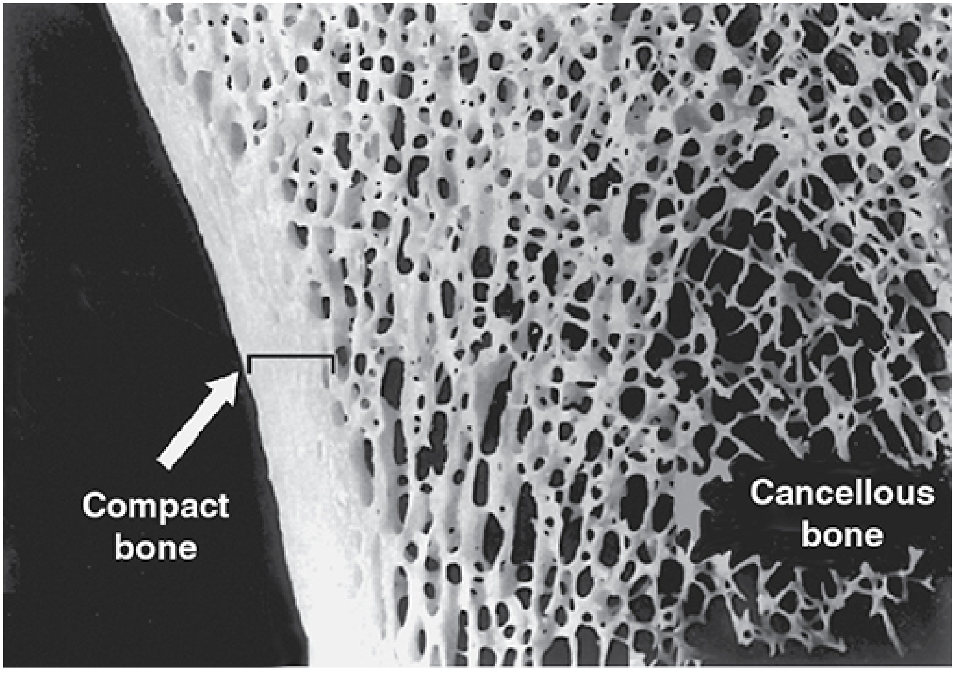
Compact bone (left, dense outer layer) merging into the porous trabecular lattice of cancellous bone (right). - Junqueira's Basic Histology, 17e, Fig. 8-7
Cancellous bone is found at characteristic sites:
-
Epiphyses of long bones - the bulbous ends are largely cancellous with only a thin cortical shell over them
-
Metaphyses - transitional zone between epiphysis and diaphysis contains increasing trabecular bone
-
Diaphysis - a thin layer of cancellous bone lines the inner surface of the cortex, surrounding the central marrow cavity
-
Vertebral bodies - cancellous bone is especially prominent here
-
Short bones (carpals, tarsals) - cancellous core surrounded completely by compact bone
-
Flat bones - the diploë is a thick layer of cancellous bone sandwiched between two plates of compact bone (e.g., skull)
-
Junqueira's Basic Histology, 17e; Medical Physiology (Boron & Boulpaep)
Microstructure - Trabeculae
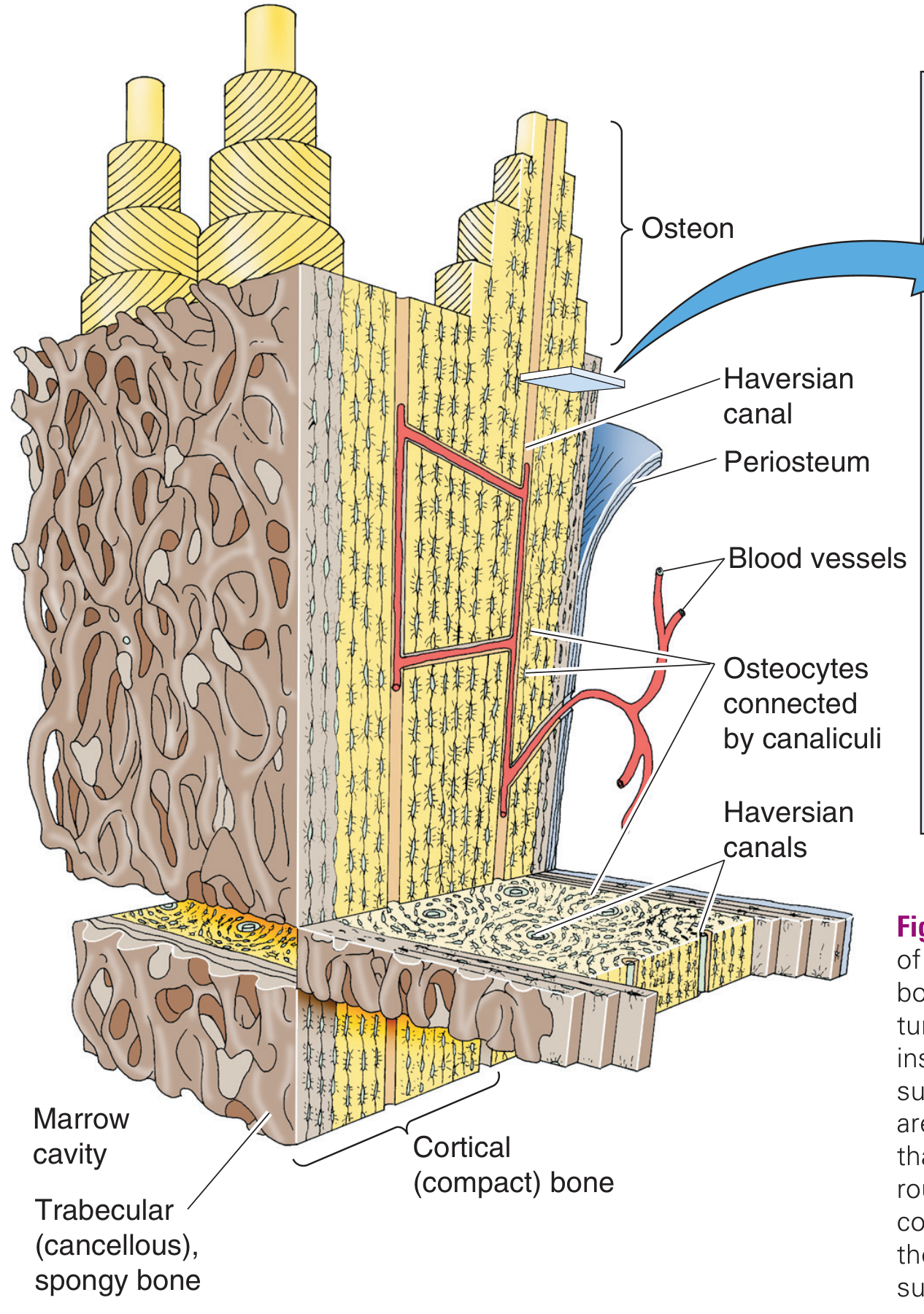
Relationship of trabecular (cancellous/spongy) bone to cortical bone, showing marrow cavity, blood vessels, and cellular components. - Medical Physiology (Boron & Boulpaep), Fig. 52-3
The structural unit of cancellous bone is the trabecula (plural: trabeculae):
-
Thin, interconnected spicules, plates, and rods of bone that form a three-dimensional lattice
-
The spaces between trabeculae are interconnecting cavities filled with bone marrow (red or yellow depending on age/location)
-
Trabeculae extend inward from the cortex into the medullary cavity
-
Their orientation is not random - they align along lines of mechanical stress (Wolff's law), giving the lattice considerable strength without greatly increasing weight
-
Medical Physiology (Boron & Boulpaep); Junqueira's Basic Histology, 17e
Surface Covering - Endosteum
-
All trabecular surfaces are covered by endosteum, a thin cellular layer
-
The endosteum contains osteoprogenitor cells, osteoblasts, and lining cells
-
This is in contrast to the outer surface of compact bone, which is covered by periosteum
-
Because cancellous bone has a much greater surface-area-to-volume ratio than cortical bone, it has far more endosteal surface available for cellular activity
-
Junqueira's Basic Histology, 17e
Cellular Components
The trabeculae and marrow spaces of cancellous bone contain the key bone cells:
| Cell | Location | Function |
|---|---|---|
| Osteoblasts | On trabecular surface (endosteum) | Bone matrix (osteoid) synthesis; mineralization |
| Osteoclasts | On trabecular surface | Bone resorption |
| Osteocytes | Embedded within the mineralized matrix of trabeculae | Mechanosensing; regulate osteoblast/osteoclast activity via canaliculi |
| Lining cells | Quiescent osteoblasts on trabecular surface | Maintain bone surface; respond to stimuli |
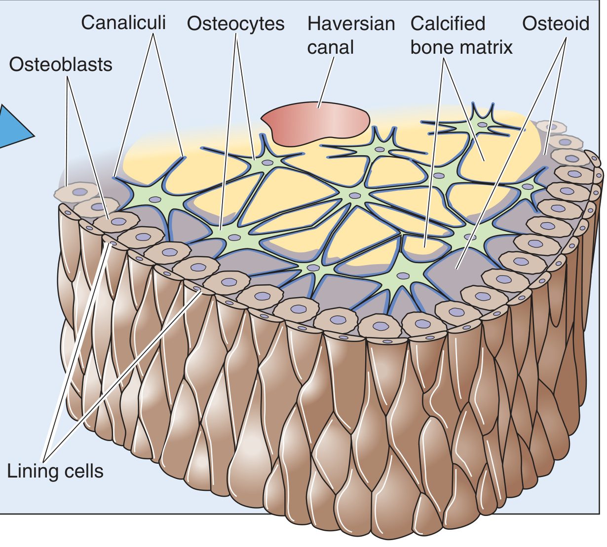
Cellular anatomy of bone tissue - osteocytes embedded in calcified matrix communicate via canaliculi. These same cells populate the trabeculae of cancellous bone. - Medical Physiology (Boron & Boulpaep), Fig. 52-3 inset
Notably, the lacework of bone spicules in cancellous bone is lined in many areas by osteoblasts and osteoclasts - this is where most bone remodeling takes place. Bone turnover rates in cancellous bone are much higher than in cortical bone. - Medical Physiology (Boron & Boulpaep)
Matrix Composition
The extracellular matrix of cancellous trabeculae is identical in composition to compact bone:
- Organic component (osteoid, ~35%): Type I collagen (~90% of osteoid), osteocalcin, osteonectin, and other glycoproteins
- Inorganic component (~65%): Hydroxyapatite crystals [Ca₁₀(PO₄)₆(OH)₂], aligned along collagen fibers
- Type I collagen provides tensile strength; hydroxyapatite provides compressive strength
Histological Organization
At the microscopic level, cancellous bone is composed of lamellar bone in normal adults:
- Trabeculae show parallel bundles of collagen in thin layers (lamellae), with regularly spaced osteocytes between lamellae in lacunae
- Unlike compact bone, cancellous trabeculae are generally too thin to contain Haversian systems (osteons)
- Osteocytes within trabeculae communicate with surface cells and with marrow vasculature directly through canaliculi and short distances through the matrix - no need for a central Haversian canal
In growing bone or fracture healing, trabeculae may initially consist of woven (immature) bone, with randomly arranged collagen and cells, later remodeled into lamellar bone.
- Junqueira's Basic Histology, 17e
Summary Table
| Feature | Cancellous (Trabecular) Bone |
|---|---|
| Synonyms | Spongy bone, trabecular bone, medullary bone |
| Proportion of bone mass | ~20% |
| Structural unit | Trabecula (spicule/plate/rod) |
| Space between trabeculae | Bone marrow (red/yellow) |
| Surface covering | Endosteum |
| Osteons (Haversian systems) | Absent (trabeculae too thin) |
| Main locations | Epiphyses, vertebral bodies, diploë of flat bones |
| Bone turnover rate | High (site of most remodeling) |
| Primary function | Load distribution, hematopoiesis support, mineral reservoir |
Sources: Junqueira's Basic Histology: Text and Atlas, 17e; Medical Physiology (Boron & Boulpaep); Imaging Anatomy: Bones, Joints, Vessels and Nerves
Difference between cortical n cancellous bone
Cortical bone is the dense outer shell (~80% of bone mass) made of tightly packed osteons, while cancellous bone is the inner spongy lattice (~20%) made of trabeculae surrounding marrow spaces. Cortical bone provides strength; cancellous bone handles load distribution and houses marrow.
Searching sources...
Finding Sources
Finding Sources
Reading File
Reading File
Finding Sources
I have all the content needed. Let me fetch the key comparison diagram from the previous session and compile the full answer.
Loading Image
Finding Sources
I now have comprehensive content from multiple authoritative sources. Here is the full comparison:
Cortical Bone vs. Cancellous Bone
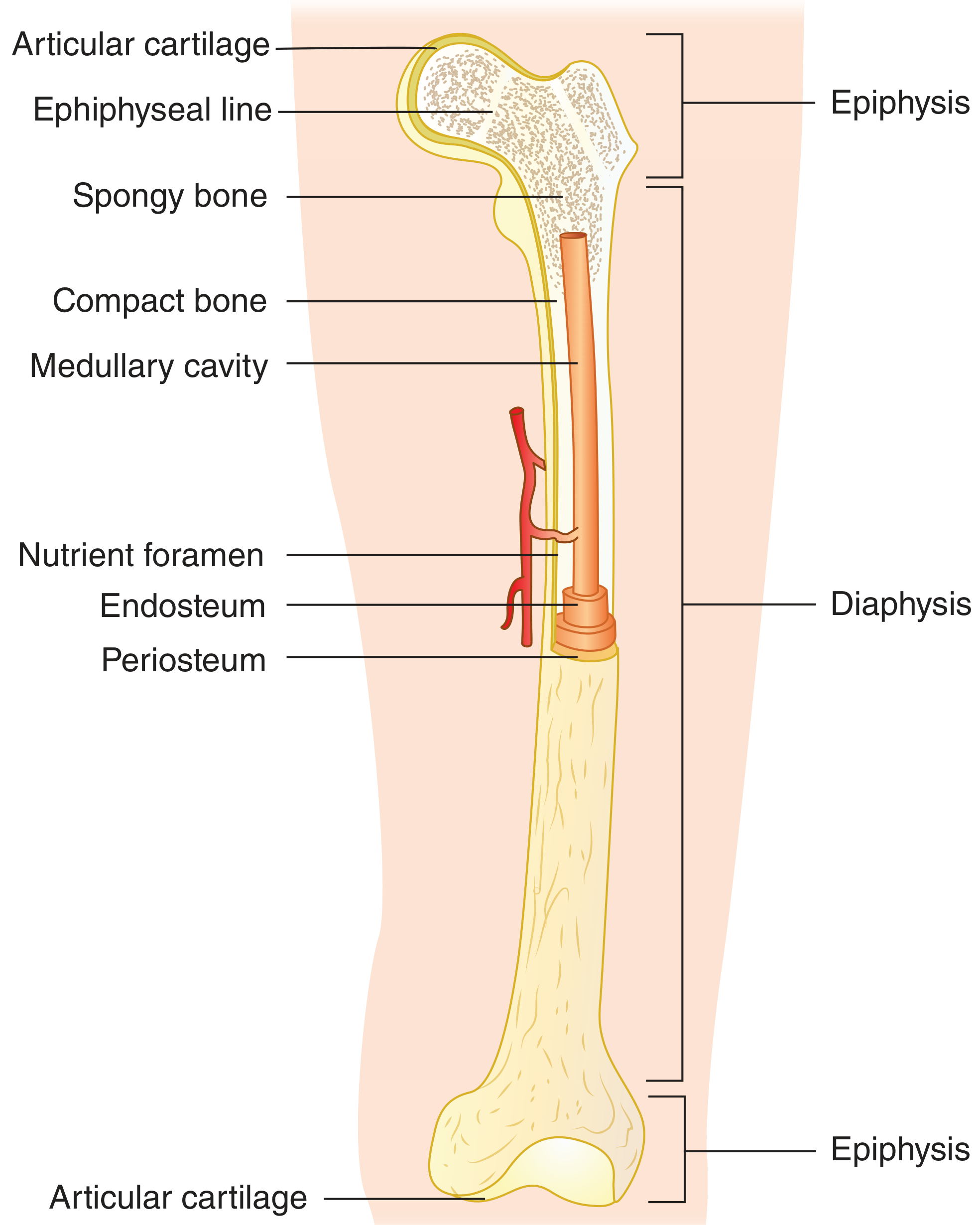
Long bone anatomy showing the spatial relationship of cortical (compact) bone in the diaphysis and cancellous (spongy) bone in the epiphysis. - Firestein & Kelley's Textbook of Rheumatology
Master Comparison Table
| Feature | Cortical (Compact) Bone | Cancellous (Trabecular) Bone |
|---|---|---|
| Synonyms | Compact bone, lamellar bone | Spongy bone, trabecular bone, medullary bone |
| Proportion of total bone mass | ~80% | ~20% |
| Mineralized tissue content | Up to 90% | Only ~20% |
| Structure | Dense, solid with few spaces | Lattice of interconnected trabeculae (plates/rods/spicules) |
| Fundamental unit | Osteon (Haversian system) | Trabecula |
| Haversian canals | Present (central canals within osteons) | Absent (trabeculae too thin) |
| Volkmann's canals | Present (transverse perforating canals) | Absent |
| Surface covering | Periosteum (outer), Endosteum (inner) | Endosteum only |
| Marrow | No marrow within bone tissue | Marrow fills inter-trabecular spaces |
| Porosity | Low (5-10%) | High (75-95%) |
| Vascularity | Lower; relies on Haversian + Volkmann canals | Higher; direct contact with marrow vasculature |
| Bone turnover rate | Low (slow remodeling) | High (major site of remodeling) |
| Remodeling type | Haversian remodeling (cutting cone of osteoclasts) | Surface remodeling by osteoblasts/osteoclasts |
| Primary function | Mechanical support, weight bearing, protection | Load distribution, hematopoiesis, mineral reservoir |
| Main locations | Diaphysis of long bones, outer shell of all bones | Epiphyses, vertebral bodies, flat bone diploë |
1. Structure
Structural comparison: cortical bone (right, with osteons and Haversian canals) vs. trabecular/cancellous bone (left, with marrow-filled spaces). - Medical Physiology (Boron & Boulpaep)
Cortical bone is composed of tightly packed osteons (Haversian systems):
- Each osteon is a cylinder of concentric ring-like lamellae surrounding a central Haversian canal that carries blood vessels and nerves
- Adjacent osteons are connected by transverse Volkmann's (perforating) canals
- Osteocytes sit in lacunae between lamellae and communicate via canaliculi
- Between osteons lie interstitial lamellae (remnants of old osteons)
Cancellous bone has no osteons. It consists of:
-
Thin, interconnected trabeculae (spicules, plates, rods) aligned along mechanical stress lines
-
Inter-trabecular spaces filled with red or yellow bone marrow
-
All trabecular surfaces lined by endosteum bearing osteoblasts and osteoclasts
-
Medical Physiology (Boron & Boulpaep)
2. Mineral Content and Porosity
A key distinction often overlooked:
- Cortical bone: up to 90% mineralized tissue - nearly solid calcified matrix
- Cancellous bone: only ~20% mineralized tissue - the remaining 80% is marrow, blood vessels, and mesenchymal cells
This vast non-mineralized surface area is the basis for cancellous bone's dominant metabolic function - it allows rapid exchange between the bone surface and the circulation. - Firestein & Kelley's Textbook of Rheumatology
3. Surface Area and Remodeling
| Cortical | Cancellous | |
|---|---|---|
| Surface area | Small (dense) | Very large (spongy lattice) |
| Osteoblast/osteoclast activity | Low | High |
| Bone turnover | Slow | Fast |
| Remodeling type | Haversian (internal tunneling by cutting cones) | Endosteal surface remodeling |
Cancellous bone is where most bone remodeling takes place. Its lacework of trabeculae is constantly being synthesized and resorbed. In contrast, cortical bone remodeling via Haversian remodeling is slower; excessive resorption leads to cortical porosity (as seen in osteoporosis). - Rheumatology, 2-Volume Set; Medical Physiology
4. Vascularity
-
Cortical bone: relies on nutrient arteries entering via the nutrient foramen, traveling longitudinally and branching into Haversian and Volkmann's canals
-
Cancellous bone: directly bathed by marrow sinusoids - fenestrated capillaries allow fast exchange of metabolites between circulation and marrow tissue. The metaphysis (rich in cancellous bone) has a higher vascularity than the diaphysis, which is why metaphyseal fractures heal faster than diaphyseal (cortical) fractures
-
Firestein & Kelley's Textbook of Rheumatology; Sabiston Textbook of Surgery
5. Mechanical Role
- Cortical bone: provides axial strength and rigidity for weight bearing; makes up the shaft of long bones; resists bending and torsional forces
- Cancellous bone: provides load distribution - the metaphysis transfers load from the weight-bearing joint surface down to the cortical diaphysis. The trabecular architecture follows Wolff's law - trabeculae align along principal stress lines, providing strength with minimal weight
6. Clinical Relevance
| Clinical Point | Cortical | Cancellous |
|---|---|---|
| Osteoporosis | Increased cortical porosity, cortical thinning | Loss of trabecular number and connectivity |
| Fracture healing | Slower (lower vascularity) | Faster (higher vascularity, more cells) |
| Bone grafting | Structural support (cortical grafts) | Osteogenic/osteoinductive potential (cancellous grafts) |
| Metabolic disease (e.g. hyperparathyroidism) | Relatively spared early | Predominantly affected (high turnover site) |
- Sabiston Textbook of Surgery; Miller's Review of Orthopaedics
Sources: Firestein & Kelley's Textbook of Rheumatology (2022); Medical Physiology (Boron & Boulpaep); Junqueira's Basic Histology, 17e; Sabiston Textbook of Surgery; Miller's Review of Orthopaedics, 9e
This is a shared conversation. Sign in to Orris to start your own chat.