
Breast conservation surgery. To write for 10 marker
breast conserving surgery lumpectomy technique
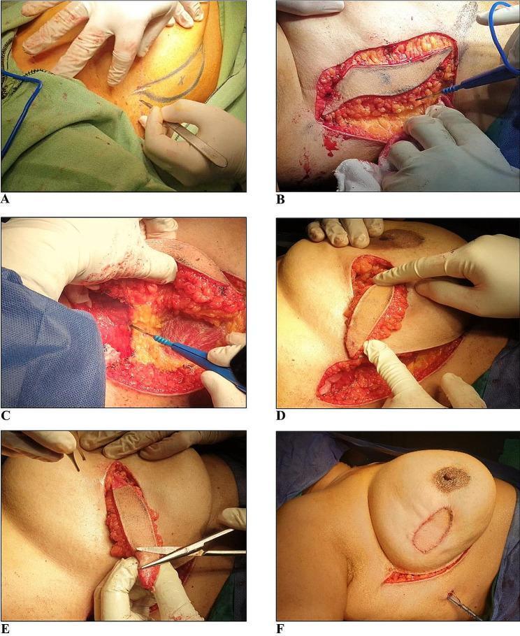
A series of six clinical photographs (A-F) documenting the surgical technique of a lateral intercostal artery perforator (LICAP) flap for oncoplastic breast reconstruction. (A) Pre-incisional view shows marked sickle-shaped margins on the lateral chest wall. (B) Initial incision and dissection of the skin flap. (C) Intraoperative close-up demonstrating the mobilization of the adipocutaneous flap while preserving the vascular perforator within its fatty mesentery. (D) The harvested flap is tunneled subcutaneously and flipped medially to reach the lumpectomy defect in the breast. (E) Preparation of the flap through selective de-epithelialization of the skin edges to ensure a proper fit within the recipient site. (F) Final postoperative view showing the flap fixed within the lumpectomy defect with sutures and the direct primary closure of the lateral donor site. This sequence illustrates a volume-replacement technique used to maintain breast contour and symmetry following breast-conserving surgery (BCS).
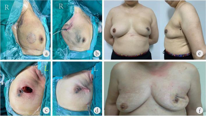
This set of clinical photographs compares the aesthetic outcomes and incision sites of single-port insufflation endoscopic breast-conserving surgery (SIE-BCS) versus conventional breast-conserving surgery (C-BCS). Panels (a, b) show intraoperative SIE-BCS with a single 2.5 cm incision placed within the axillary skin folds, alongside the use of methylene blue for sentinel lymph node and tumor margin identification. Panels (c, d) depict the C-BCS technique, which utilizes two separate incisions: one in the axillary fossa and another directly overlying the breast tumor site. Panels (e, f) provide 12-month postoperative follow-up comparisons. Panel (e) demonstrates a superior aesthetic result following SIE-BCS, with no visible scarring on the breast surface and a well-concealed, faint scar in the axilla (indicated by a black arrow). In contrast, panel (f) shows the outcome of conventional lumpectomy and sentinel lymph node biopsy, characterized by two distinct, visible scars on the breast surface. The images highlight the clinical benefit of single-port endoscopic techniques in minimizing visible scarring and preserving breast aesthetics in cancer treatment.
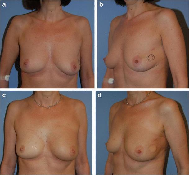
Clinical photographs showing preoperative and postoperative views of a 46-year-old female patient with small breasts and invasive ductal carcinoma (zone VI) in the lateral left breast. Images (a) and (b) provide frontal and oblique preoperative views, illustrating breast hypoplasia with mild ptosis; a surgical marking is visible in the lateral region of the left breast indicating the tumor site. Images (c) and (d) show the three-month postoperative results after breast-conserving surgery (40g lumpectomy) followed by oncoplastic reconstruction. The reconstruction utilized a volume replacement technique via a tunneled lateral thoracodorsal flap with a skin island. The postoperative images demonstrate successful maintenance of breast volume and projection, preservation of the nipple-areola complex (NAC) position, and achievement of good breast symmetry without the need for contralateral surgery. The flap's skin island is visible in the lateral aspect of the left breast in the oblique view (d). These images highlight an effective surgical approach for maintaining aesthetic outcomes in small-breasted patients undergoing oncoplastic breast surgery.
Breast Conservation Surgery (BCS)
Definition
Historical Basis / Key Trials
Indications (Who is suitable for BCS?)
- Single, unifocal tumor - no multicentricity
- Tumor size: T1/T2 tumors (generally ≤4-5 cm) where an acceptable tumor-to-breast volume ratio can be achieved
- Negative/clear surgical margins are achievable
- Patient is suitable for post-operative radiation therapy
- Patient preference - after full counselling on both options
- Neoadjuvant chemotherapy can downstage a previously borderline-sized tumor to allow BCS
Contraindications to BCS
Absolute Contraindications
| Contraindication | Reason |
|---|---|
| Current pregnancy (1st or early 2nd trimester) | Radiation cannot be given during pregnancy |
| Prior irradiation to the same breast/chest wall | Risk of radiation toxicity precludes further RT |
| Multicentric disease (two or more quadrants) | Cannot achieve clear margins with single excision |
| Diffuse malignant-appearing calcifications on mammogram | Suggests widespread intraductal involvement |
| Persistently positive margins despite re-excision | Failure to achieve oncologic clearance |
Relative Contraindications
- Large tumor relative to small breast size (compromises cosmesis) - though advanced oncoplastic techniques may allow BCS in such cases
- Active connective tissue disorders (collagen vascular disease) - risk of poor healing and radiation sensitivity
- Known BRCA1/2 pathogenic mutation (higher risk of ipsilateral and contralateral recurrence, though not an absolute contraindication per se)
Surgical Technique
Pre-operative Steps
- Core needle biopsy for histopathological diagnosis, receptor status (ER/PR/HER2), grade
- Axillary ultrasound to assess nodal status
- Breast MRI - not routine but considered when disease extent is unclear
- Localization for non-palpable lesions: wire-guided localization, radioactive seed localization, or newer techniques (SAVI Scout, radar localization)
- Genetic counselling and BRCA testing where indicated
Operative Steps
- Incision placement - generally over or near the tumor; follow Langer's lines (curvilinear/horizontal in upper quadrants, curvilinear/radial in lower quadrants). A periareolar incision may be used in select cases for cosmesis. Incisions should anticipate possible future mastectomy.
- Excision - the tumor is excised with a cuff of normal breast tissue on all sides. Excision of overlying skin or pectoralis fascia is not required unless the tumor is directly involved.
- Specimen orientation - the specimen must be oriented with sutures/clips in at least two planes (e.g., superior and lateral margins) so the pathologist can assess all six margins.
- Intra-operative specimen radiograph - confirms successful excision of the target lesion (especially non-palpable lesions marked with a clip).
- Cavity shave margins - many surgeons take circumferential shave margins (superior, inferior, medial, lateral, anterior, posterior) from the cavity wall; this reduces re-excision rates by approximately 50%.
- Haemostasis and closure - the breast parenchyma may be re-approximated or the cavity left to fill with seroma; a drain is generally not required.
Axillary Staging (simultaneous with BCS)
- Sentinel lymph node biopsy (SLNB) is the standard for clinically node-negative patients. Blue dye and/or radioisotope (Tc-99m) are injected; the sentinel node(s) are identified and excised for frozen or permanent section.
- Axillary lymph node dissection (ALND) is indicated for clinically/biopsy-proven node-positive disease before surgery, or for more than 2 positive sentinel nodes in certain settings.
- Per the Z0011 trial criteria: ALND can be omitted in patients with 1-2 positive sentinel nodes undergoing BCS with whole-breast radiation and systemic therapy.
Margin Assessment
- Invasive carcinoma: no tumor at the inked margin ("no ink on tumour")
- DCIS: ≥ 2 mm margin
Oncoplastic Surgery
- Better cosmetic outcomes than simple lumpectomy in high-resection-volume cases
- Equivalent oncologic outcomes to standard BCS
- Higher patient-reported outcomes vs mastectomy with reconstruction (BREAST-Q scores)
Adjuvant Therapy After BCS
| Modality | Indication |
|---|---|
| Whole-breast radiation therapy (WBRT) | Mandatory after BCS for invasive cancer; reduces IBTR by ~50%. Typically 40-50 Gy in 15-25 fractions |
| Tumour bed boost | Additional 10-16 Gy to lumpectomy cavity, especially in younger patients or those with high-grade tumours |
| Accelerated partial breast irradiation (APBI) | Selected low-risk patients (older age, small T1, node-negative, ER+); irradiates only the tumour bed |
| Hormonal therapy | 5-10 years of tamoxifen or aromatase inhibitor for ER-positive disease |
| Chemotherapy | For HER2+ or triple-negative tumours, or high Oncotype DX recurrence score |
| Anti-HER2 therapy | Trastuzumab ± pertuzumab for HER2+ disease |
Outcomes
- Local recurrence: 10-year IBTR rates of ~8-10% with BCS + RT vs ~5% with mastectomy; no difference in overall survival.
- Survival: BCS + RT is oncologically equivalent to mastectomy for stage I-II breast cancer.
- Cosmesis: Superior body image and quality of life compared to mastectomy in most patients.
- Risk factors for local recurrence after BCS include: young age, high grade, lymphovascular invasion, positive or close margins, HER2+ or triple-negative biology.
Special Situations
- DCIS: BCS is appropriate for localised DCIS; NSABP B-17 showed RT after lumpectomy reduces IBTR by 52%. NSABP B-24 showed tamoxifen further reduces recurrence in ER+ DCIS.
- Pregnancy: BCS is contraindicated in the first trimester (cannot give RT during pregnancy); feasible in 2nd/3rd trimester if RT is deferred until after delivery.
- Neoadjuvant chemotherapy: Downstages tumour to allow BCS in patients who would otherwise need mastectomy.
Summary Box (for exam recall)
| Feature | Detail |
|---|---|
| Other names | Lumpectomy, WLE, segmental/partial mastectomy, quadrantectomy |
| Key trial | NSABP B-06 - BCS + RT = mastectomy for survival |
| Margin (invasive) | No ink on tumour |
| Margin (DCIS) | ≥ 2 mm |
| RT | Mandatory post-BCS |
| SLNB | Standard axillary staging |
| Absolute contraindications | Pregnancy, prior RT, multicentricity, diffuse Ca++, persistently +ve margins |