
1.Microscopic structure/Histology of a) Hyaline Cartilage***** b) Elastic Cartilage 2. Histology of Bone (Compact bone) 3. Describe microscopic structure of******* a) Elastic Artery(Large sized artery) b) Muscular Artery (Medium sized artery)[Compare both of this]
Reading File
This covers three connected histology units:
-
Cartilage
- Hyaline cartilage: microscopic features, chondrocytes, matrix, perichondrium, sites, and functions
- Elastic cartilage: features and differences from hyaline cartilage
-
Compact bone
- Osteons (Haversian systems), lamellae, canals, osteocytes, lacunae, canaliculi, and periosteum/endosteum
-
Arteries
- Elastic arteries: especially aorta and major branches
- Muscular arteries: distribution arteries
- A direct elastic-versus-muscular artery comparison
High-level overview: Cartilage is an avascular supportive connective tissue whose cells, chondrocytes, lie in lacunae within a firm extracellular matrix. Hyaline cartilage has a glassy matrix with type II collagen not easily seen by light microscopy, whereas elastic cartilage contains conspicuous elastic fibers. Compact bone is organized into vascular osteons with concentric lamellae. Arterial walls have three tunics, but elastic arteries have many elastic lamellae in the tunica media, while muscular arteries have a media dominated by smooth muscle.
Which section should I write as detailed exam notes first?
- Hyaline and elastic cartilage
- Compact bone
- Elastic and muscular arteries with comparison
Write detailed exam notes for all three topics
Reading File
Finding Sources
Finding Sources
Finding Sources
Searching PubMed
"cartilage"[MeSH Terms] AND histology
Searching PubMed
"bone and bones"[MeSH Terms] AND histology
Searching PubMed
"arteries"[MeSH Terms] AND histology
Searching the Web
hyaline elastic cartilage compact bone elastic muscular artery histology OpenStax
Finding Sources
Finding Sources
Searching Images
light micrograph hyaline cartilage elastic cartilage chondrocytes lacunae perichondrium
Searching Images
compact bone histology osteon Haversian canal lamellae micrograph
Reading File
Reading File
Reading File
Histology Exam Notes
1. Cartilage
General features of cartilage
Cartilage is a specialized supporting connective tissue with:
- Cells: chondroblasts and chondrocytes
- Matrix: firm, hydrated extracellular matrix containing collagen fibers, proteoglycans, and glycoproteins
- Lacunae: spaces in the matrix occupied by chondrocytes
- Avascularity: cartilage has no blood vessels, lymphatics, or nerves. Nutrients diffuse through the matrix from the perichondrium or, in articular cartilage, synovial fluid.
- Perichondrium: a connective-tissue covering around most cartilage. It is absent over articular cartilage and at epiphyseal plates.
- Growth:
- Interstitial growth: division of chondrocytes within lacunae and secretion of new matrix from within.
- Appositional growth: addition of new cartilage at the surface by chondroblasts derived from the inner perichondrium.
Cartilage has limited capacity for repair because it is avascular.
A. Hyaline cartilage
Definition
Hyaline cartilage is the commonest variety of cartilage. It has a smooth, translucent, “glassy” appearance due to its homogeneous extracellular matrix.
Sites
- Articular surfaces of synovial joints
- Costal cartilages
- Nasal cartilages
- Thyroid, cricoid, and most arytenoid cartilages of larynx
- Tracheal rings
- Bronchial cartilages
- Epiphyseal growth plates
- Fetal skeleton
Microscopic structure
1. Perichondrium
Present around most hyaline cartilage, except:
- Articular cartilage
- Epiphyseal cartilage plate
It has two layers:
| Layer | Structure and function |
|---|---|
| Outer fibrous layer | Dense irregular connective tissue containing type I collagen fibers, fibroblasts, and blood vessels. It provides protection and nutrition. |
| Inner cellular or chondrogenic layer | Contains chondrogenic cells and chondroblasts. It produces new cartilage by appositional growth. |
2. Cells
Chondroblasts
- Immature cartilage-forming cells.
- Located mainly in the inner layer of perichondrium.
- Oval or spindle-shaped, with basophilic cytoplasm.
- Actively synthesize matrix components, especially type II collagen and proteoglycans.
- Once surrounded by matrix, they become chondrocytes.
Chondrocytes
- Mature cartilage cells.
- Located in spaces called lacunae.
- Round or oval cells with central nuclei.
- May occur singly or in groups.
Isogenous groups or cell nests
- Groups of 2 to 8 chondrocytes derived from division of a single chondrocyte.
- Evidence of interstitial growth.
- Commonly seen in deeper parts of cartilage.
3. Extracellular matrix
The matrix appears homogeneous and glassy in routine H&E sections because its fine type II collagen fibrils are not easily visible.
It contains:
- Type II collagen fibrils: provide tensile strength.
- Proteoglycans: especially aggrecan, rich in chondroitin sulfate and keratan sulfate.
- Glycosaminoglycans: attract water, allowing resistance to compression.
- Adhesive glycoproteins: help bind cells and matrix.
Territorial matrix
- Matrix immediately surrounding lacunae.
- More basophilic and darker-staining.
- Rich in sulfated glycosaminoglycans.
Interterritorial matrix
- Matrix between groups of chondrocytes.
- Paler-staining than territorial matrix.
- Contains relatively more collagen.
Articular cartilage
Articular cartilage is specialized hyaline cartilage covering the articular surfaces of bones in synovial joints.
Features:
- No perichondrium
- Nourished by diffusion from synovial fluid
- Smooth, resilient, low-friction surface
- Distributes mechanical load to underlying bone
- Poor regeneration after injury
Functions
- Provides smooth, low-friction surface at joints.
- Resists compression because of its hydrated proteoglycan-rich matrix.
- Supports the walls of trachea, bronchi, and nose.
- Forms the model for most bones during endochondral ossification.
- Permits longitudinal growth at epiphyseal plates.
Slide identification points
- Glassy, homogeneous matrix
- Chondrocytes in lacunae
- Isogenous groups
- Perichondrium, except in articular cartilage
- Matrix contains no visible fibers in routine H&E stain
Source: Histology: A Text and Atlas, pp. 550 onward. It describes the glassy matrix, lacunae, chondrocytes, matrix composition, and absence of perichondrium on articular cartilage.
B. Elastic cartilage
Definition
Elastic cartilage resembles hyaline cartilage but has abundant elastic fibers in addition to type II collagen. It is more flexible and resilient than hyaline cartilage.
Sites
- Auricle or pinna of ear
- External auditory canal
- Auditory or Eustachian tube
- Epiglottis
- Certain laryngeal cartilages, especially corniculate and cuneiform cartilages
- Vocal folds
Microscopic structure
1. Perichondrium
- Present.
- Similar to the perichondrium of hyaline cartilage.
- Has outer fibrous and inner chondrogenic layers.
2. Chondrocytes
- Present within lacunae.
- Often appear larger and more numerous than in hyaline cartilage.
- May occur in isogenous groups.
- Surrounded by territorial matrix.
3. Matrix
Contains:
- Type II collagen fibrils
- Proteoglycans and glycosaminoglycans
- Dense network of branching elastic fibers and elastic lamellae
Elastic fibers:
- Are often difficult to see with H&E staining.
- Are demonstrated by special stains such as orcein, resorcin-fuchsin, or Verhoeff elastic stain.
- Form a branching network around lacunae and chondrocytes.
Functions
- Provides flexible support.
- Allows repeated bending and return to original shape.
- Maintains patency and shape of structures such as the external ear and epiglottis.
Important point
Unlike hyaline cartilage, elastic cartilage generally does not calcify with aging.
Hyaline cartilage versus elastic cartilage
| Feature | Hyaline cartilage | Elastic cartilage |
|---|---|---|
| Matrix appearance | Glassy, homogeneous | Contains visible elastic fiber network with special stains |
| Main fibers | Type II collagen fibrils | Type II collagen plus abundant elastic fibers |
| Flexibility | Firm, resilient, resists compression | More flexible and elastic |
| Perichondrium | Present except in articular cartilage and epiphyseal plate | Present |
| Calcification with age | May occur | Usually does not occur |
| Major sites | Trachea, bronchi, costal cartilage, nose, articular surfaces | Pinna, epiglottis, auditory tube |
| Function | Support, shock absorption, low-friction joint surface | Flexible support and maintenance of shape |
Source: Histology: A Text and Atlas, Table 7.1 and pp. 550 onward; Junqueira's Basic Histology, cartilage chapter.
2. Histology of Compact Bone
Definition
Compact bone, also called cortical bone, is dense lamellar bone forming the outer cortex of bones and most of the shaft of long bones. Its basic structural unit is the osteon or Haversian system.
Components of compact bone
1. Bone cells
Osteoprogenitor cells
- Stem cells derived from mesenchyme.
- Found in periosteum, endosteum, and bone marrow stroma.
- Differentiate into osteoblasts during bone growth and repair.
Osteoblasts
- Bone-forming cells.
- Found on bone surfaces.
- Cuboidal cells with basophilic cytoplasm and eccentric nuclei.
- Secrete osteoid, the unmineralized organic bone matrix.
- Participate in mineralization.
Osteocytes
- Mature bone cells derived from osteoblasts trapped in matrix.
- Located in lacunae.
- Have flattened cell bodies with long cytoplasmic processes.
- Processes lie in canaliculi and communicate through gap junctions.
- Maintain bone matrix and participate in nutrient exchange and mechanosensation.
Osteoclasts
- Large multinucleated bone-resorbing cells.
- Derived from monocyte-macrophage lineage.
- Lie in shallow resorption depressions called Howship lacunae.
- Have a ruffled border facing the bone surface.
- Important in bone remodeling.
2. Bone matrix
Organic component
Also called osteoid. It consists mainly of:
- Type I collagen fibers
- Proteoglycans
- Glycoproteins, including osteocalcin, osteonectin, and osteopontin
Type I collagen provides tensile strength.
Inorganic component
- Mainly calcium phosphate in the form of hydroxyapatite crystals.
- Provides hardness and resistance to compression.
3. Lamellae
Lamellae are layers of mineralized bone matrix. Collagen fibers in each lamella run parallel to one another, but their direction differs in adjacent lamellae.
This alternating fiber orientation:
- Greatly increases strength
- Resists twisting and bending forces
Types of lamellae:
- Concentric lamellae
- Interstitial lamellae
- Circumferential lamellae
4. Osteon or Haversian system
Definition
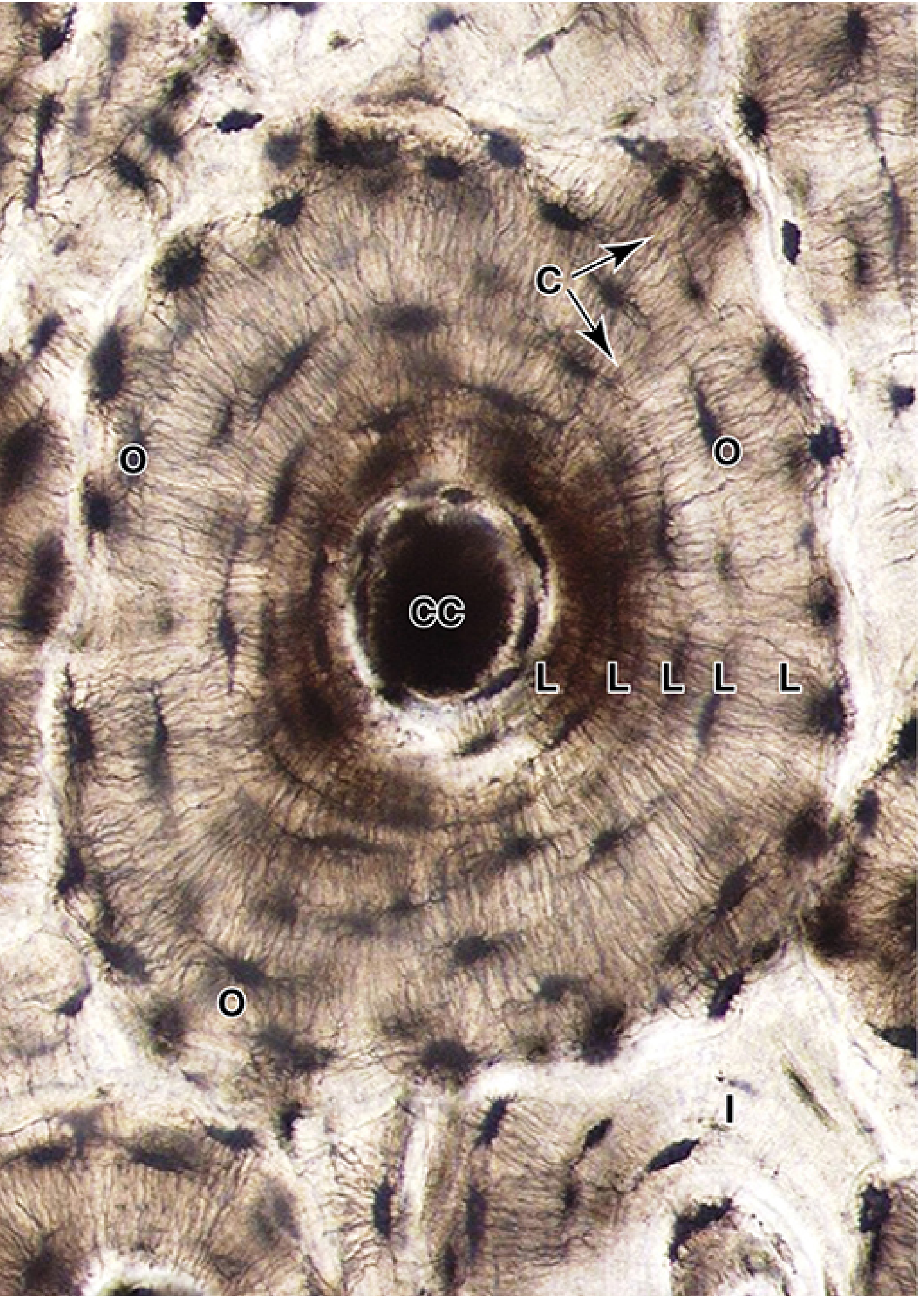
An osteon is a cylindrical structural unit of compact bone. It consists of concentric lamellae arranged around a central Haversian canal.
- Usually 100 to 250 micrometres in diameter.
- Runs longitudinally, parallel to the long axis of the bone.
- Forms most of mature compact bone.
Components of an osteon
A. Haversian canal or central canal
Located at the center of an osteon. It contains:
- Blood vessels
- Nerves
- Loose connective tissue
- Endosteum
B. Concentric lamellae
- 4 to 20 circular layers of bone matrix surrounding the Haversian canal.
- Collagen orientation changes from one lamella to the next.
C. Lacunae
- Small spaces between lamellae.
- Each lacuna contains one osteocyte.
D. Canaliculi
- Fine microscopic channels radiating from lacunae.
- Contain cytoplasmic processes of osteocytes.
- Connect osteocytes with each other and with the central canal.
- Permit diffusion of nutrients, oxygen, metabolites, and signaling molecules.
E. Cement line
- Outer boundary of an osteon.
- Appears as a basophilic or refractile line.
- Contains more noncollagenous proteins and relatively less collagen.
- Marks the boundary of a newly formed osteon.
5. Volkmann canals or perforating canals
- Transverse or oblique canals.
- Connect Haversian canals with one another.
- Connect central canals to blood vessels of periosteum and medullary cavity.
- Carry blood vessels and nerves.
- Not surrounded by concentric lamellae.
Haversian canals versus Volkmann canals
| Feature | Haversian canal | Volkmann canal |
|---|---|---|
| Direction | Longitudinal, parallel to long axis | Transverse or oblique |
| Location | Center of an osteon | Passes between osteons |
| Surrounding lamellae | Surrounded by concentric lamellae | Not surrounded by concentric lamellae |
| Function | Carries vessels and nerves within osteon | Connects Haversian canals with periosteal and medullary vessels |
6. Interstitial lamellae
- Irregular fragments of older osteons between complete osteons.
- Remnants of partially resorbed Haversian systems.
- Result from continuous remodeling of bone.
7. Circumferential lamellae
These are lamellae that encircle the entire bone rather than a single Haversian canal.
Outer circumferential lamellae
- Found just deep to periosteum.
- Extend around the outer surface of bone.
Inner circumferential lamellae
- Found near the medullary cavity.
- Surround the inner surface of compact bone.
8. Periosteum
A connective tissue covering of outer bone surface, except at articular surfaces.
It has two layers:
| Layer | Features |
|---|---|
| Outer fibrous layer | Dense irregular connective tissue with collagen fibers, fibroblasts, vessels, and nerves |
| Inner osteogenic layer | Osteoprogenitor cells and osteoblasts; important in growth and repair |
Sharpey fibers are collagen fibers from periosteum that penetrate bone matrix and anchor periosteum firmly to bone.
9. Endosteum
A thin cellular connective tissue lining:
- Medullary cavity
- Haversian canals
- Volkmann canals
- Trabeculae of spongy bone
Contains osteoprogenitor cells, osteoblasts, and osteoclasts.
Identification of compact bone in ground section
- Numerous circular or oval osteons
- Central Haversian canals
- Concentric lamellae
- Dark lacunae arranged between lamellae
- Fine radiating canaliculi
- Interstitial lamellae between osteons
- Volkmann canals crossing osteons
Source: Junqueira's Basic Histology, pp. 367-370. An osteon consists of concentric lamellae surrounding a central canal containing vessels, nerves, and endosteum; osteocytes occupy lacunae and communicate through canaliculi.
3. Histology of Arteries
General plan of arterial wall
Most arteries have three tunics:
- Tunica intima
- Tunica media
- Tunica adventitia
Arteries are classified mainly by the structure of their tunica media.
| Type | Main feature of tunica media |
|---|---|
| Elastic artery | Many elastic lamellae with smooth muscle cells between them |
| Muscular artery | Predominantly circular smooth muscle cells |
| Arteriole | One or two layers of smooth muscle cells |
A. Elastic artery or large artery
Examples
- Aorta
- Pulmonary trunk and pulmonary arteries
- Brachiocephalic trunk
- Common carotid arteries
- Subclavian arteries
- Common iliac arteries
Function
Elastic arteries receive blood directly from ventricles. Their elastic walls stretch during systole and recoil during diastole.
This:
- Dampens the pulsatile output of the heart
- Maintains blood pressure during diastole
- Ensures relatively continuous forward blood flow
This function is called the Windkessel effect.
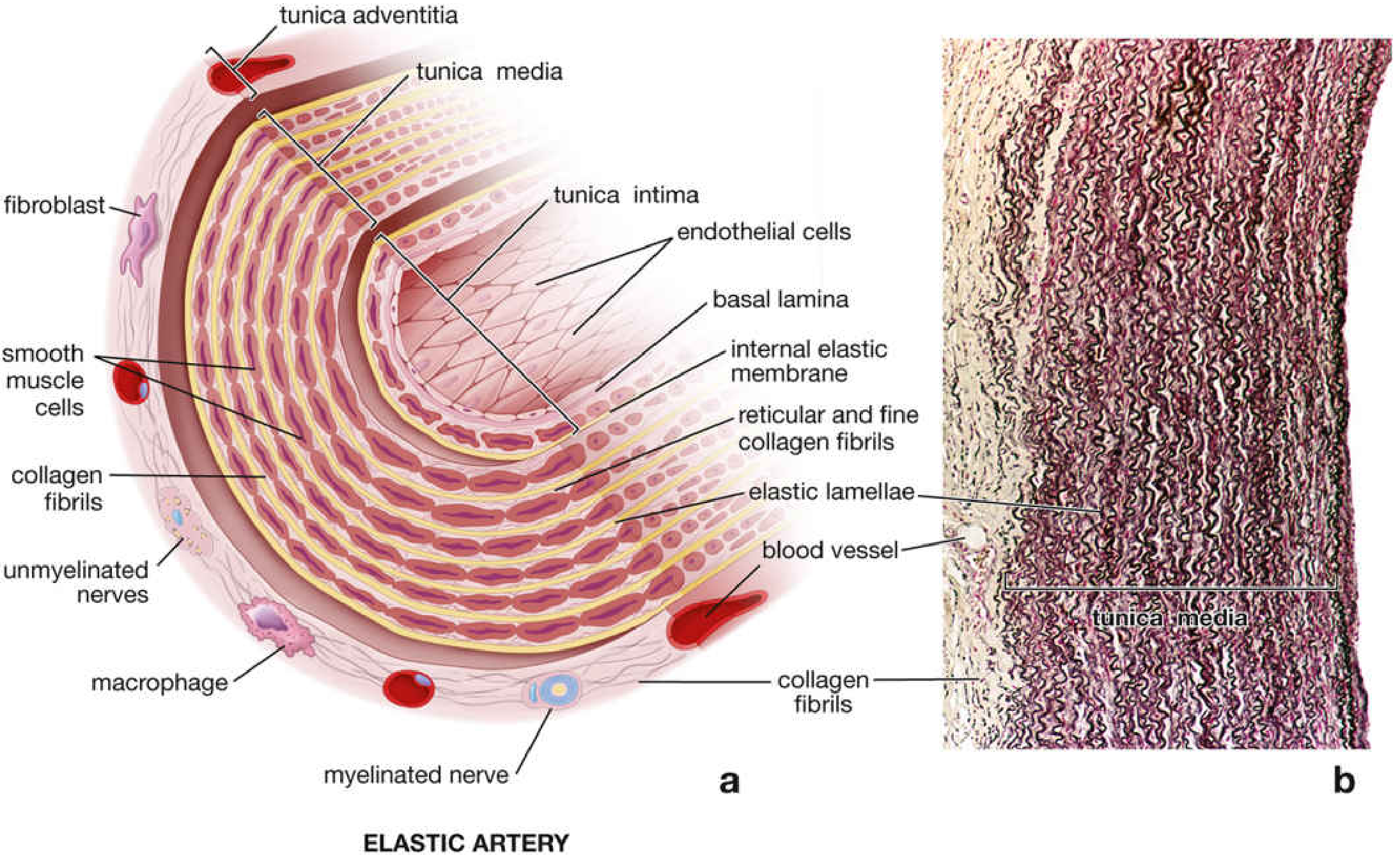
Microscopic structure
1. Tunica intima
The tunica intima is relatively thick and consists of:
- Endothelium: simple squamous epithelium lining lumen
- Basal lamina
- Subendothelial connective tissue containing collagen and elastic fibers
- Some smooth muscle cells may be present in deeper subendothelial region
- Internal elastic lamina is inconspicuous because it blends with the first elastic lamella of tunica media
2. Tunica media
This is the thickest layer.
It consists of:
- Numerous concentric, fenestrated elastic lamellae
- Smooth muscle cells between elastic lamellae
- Collagen fibers
- Ground substance
The elastic lamellae are arranged concentrically around the lumen. They may number 40 to 70 in the aorta.
Functions:
- Stretch during ventricular systole
- Recoil during diastole
- Maintain arterial pressure and continuous blood flow
3. Tunica adventitia
- Relatively thin compared with tunica media.
- Contains collagen fibers, elastic fibers, fibroblasts, nerves, and adipose tissue.
- Contains vasa vasorum, small vessels supplying the outer wall of large arteries.
- Vasa vasorum are especially important because diffusion from lumen cannot adequately supply the thick outer media and adventitia.
- Also contains nervi vasorum, autonomic nerve fibers.
Slide identification points
- Very large lumen
- Thick wall
- Very thick tunica media
- Multiple wavy concentric elastic lamellae
- Internal elastic lamina not clearly distinct
- Adventitia relatively thin
- Vasa vasorum may be seen in adventitia
B. Muscular artery or medium-sized artery
Examples
Most named arteries, such as:
- Radial artery
- Brachial artery
- Femoral artery
- Coronary arteries
- Renal arteries
- Splenic artery
- Mesenteric arteries
- Cerebral arteries
Function
Muscular arteries are called distributing arteries. They control distribution of blood to organs and tissues through contraction or relaxation of smooth muscle in the tunica media.
Microscopic structure
1. Tunica intima
Consists of:
- Endothelium with basal lamina
- Thin subendothelial connective tissue
- A prominent, wavy internal elastic lamina
The internal elastic lamina is especially distinct in contracted vessels. It appears as a wavy, refractile line separating intima from media.
2. Tunica media
This is usually the thickest layer in a muscular artery.
It consists mainly of:
- Multiple layers of circularly arranged smooth muscle cells
- Small amount of collagen and elastic fibers between muscle cells
- Sometimes an external elastic lamina at outer margin
The number of smooth muscle layers varies with vessel size.
Function:
- Vasoconstriction and vasodilation
- Regulation of blood flow to tissues
- Contribution to peripheral vascular resistance and arterial blood pressure
3. Tunica adventitia
- Usually relatively thick.
- Composed of collagen fibers, elastic fibers, fibroblasts, and connective tissue cells.
- Blends with surrounding connective tissue.
- Larger muscular arteries may contain vasa vasorum and nervi vasorum.
- An external elastic lamina may separate media from adventitia, particularly in larger muscular arteries.
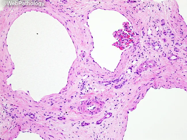
Slide identification points
- Lumen smaller than in an elastic artery of similar wall thickness
- Thick tunica media composed mainly of smooth muscle
- Prominent wavy internal elastic lamina
- External elastic lamina may be visible
- Fewer elastic lamellae than elastic artery
- Adventitia usually well developed
Comparison: Elastic artery versus muscular artery
| Feature | Elastic artery or large artery | Muscular artery or medium-sized artery |
|---|---|---|
| Examples | Aorta, pulmonary trunk, common carotid, subclavian | Radial, femoral, brachial, renal, coronary |
| Main role | Conduct blood from heart and maintain continuous flow | Distribute blood to specific organs |
| Lumen | Very wide | Smaller relative to wall thickness |
| Tunica intima | Thick | Relatively thin |
| Internal elastic lamina | Inconspicuous, merges with elastic lamellae of media | Prominent and wavy |
| Tunica media | Thickest layer; many elastic lamellae with smooth muscle between them | Thickest layer; predominantly circular smooth muscle |
| Elastic lamellae | Numerous, concentric, fenestrated | Few, mainly as internal and sometimes external elastic laminae |
| Smooth muscle | Present between elastic lamellae | Dominant component of media |
| External elastic lamina | Usually indistinct | Often distinct, especially in larger muscular arteries |
| Tunica adventitia | Relatively thin compared with media | Usually well developed and may be as thick as media |
| Vasa vasorum | Prominent in adventitia and outer media | Present in larger muscular arteries, chiefly in adventitia |
| Functional property | High elasticity and recoil | Marked vasoconstriction and vasodilation |
| Effect during systole | Wall expands | Less expansion |
| Effect during diastole | Elastic recoil maintains pressure and blood flow | Smooth muscle tone regulates regional blood flow |
Key exam statement
The most important histological distinction is in the tunica media:
- In an elastic artery, the media contains numerous elastic lamellae.
- In a muscular artery, the media consists mainly of circular smooth muscle cells and has a clearly defined internal elastic lamina.
Source: Histology: A Text and Atlas, vascular system chapter, pp. 1087 onward. Elastic arteries contain multiple elastic lamellae in the media; their internal elastic membrane is not distinct. Muscular arteries have the same three tunics but a media dominated by smooth muscle.
Quick answer
1. Microscopic Structure of Cartilage
General features of cartilage
Cartilage is a specialized supporting connective tissue consisting of:
- Cells: chondroblasts and chondrocytes
- Extracellular matrix: fibers embedded in a firm, hydrated ground substance rich in proteoglycans
- Perichondrium: dense connective tissue surrounding most cartilage
General histological features
- Cartilage is avascular, aneural, and alymphatic.
- Nutrition reaches chondrocytes by diffusion through the matrix from blood vessels in the perichondrium or, in articular cartilage, from synovial fluid.
- Chondrocytes lie in spaces called lacunae.
- Closely placed chondrocytes formed by division of one cell make an isogenous group or cell nest.
- Cartilage grows by:
- Interstitial growth: division of chondrocytes within the matrix.
- Appositional growth: addition of new cartilage at the surface by chondroblasts of the perichondrium.
A. Hyaline Cartilage
Definition
Hyaline cartilage is the commonest type of cartilage. Its matrix appears smooth, homogeneous, and glass-like, hence the term hyaline.
Sites
- Articular surfaces of synovial joints
- Costal cartilages
- Nasal cartilages
- Thyroid, cricoid, and most arytenoid cartilages of larynx
- Tracheal rings
- Bronchial cartilages
- Epiphyseal plates
- Fetal skeleton
Microscopic structure
1. Perichondrium
Present around most hyaline cartilage, except:
- Articular cartilage
- Cartilage of epiphyseal plates
It has two layers:
| Layer | Features |
|---|---|
| Outer fibrous layer | Dense irregular connective tissue, collagen fibers, fibroblasts, and blood vessels |
| Inner chondrogenic layer | Chondrogenic cells and chondroblasts that form new cartilage |
2. Cells
Chondroblasts
- Present mainly beneath the perichondrium.
- Oval or spindle-shaped.
- Basophilic cytoplasm due to abundant rough endoplasmic reticulum.
- Secrete cartilage matrix.
- When enclosed by matrix, they become chondrocytes.
Chondrocytes
- Mature cartilage cells.
- Rounded or oval cells found within lacunae.
- Usually have a central nucleus and basophilic cytoplasm.
- May occur singly or in isogenous groups.
- Maintain and renew the matrix.
3. Matrix
The matrix is abundant and has a glassy appearance in routine H&E staining.
Its main components are:
- Fine type II collagen fibrils
- Proteoglycan aggregates, especially aggrecan
- Glycosaminoglycans, such as chondroitin sulfate and keratan sulfate
- Water
- Adhesive glycoproteins
Type II collagen fibrils are very fine and usually cannot be distinguished with ordinary light microscopy. Therefore, the matrix looks amorphous and homogeneous.
4. Territorial and interterritorial matrix
- Territorial matrix: matrix immediately surrounding the lacunae. It stains more deeply basophilic because it contains more sulfated glycosaminoglycans.
- Interterritorial matrix: matrix between groups of lacunae. It stains relatively lightly.
Functions
- Provides firm but flexible support.
- Resists compression because of its highly hydrated proteoglycan-rich matrix.
- Forms a smooth, low-friction surface at synovial joints.
- Helps distribute pressure and forces over underlying bone.
- Provides a model for endochondral ossification during fetal development and growth of long bones.
- Supports the respiratory passages.
Important points
- Articular cartilage has no perichondrium.
- Articular cartilage receives nutrition from synovial fluid.
- Hyaline cartilage has poor regenerative capacity because it is avascular.
- It may calcify with aging and during endochondral ossification.
Source: Histology: A Text and Atlas with Correlated Cell and Molecular Biology, 7e, pp. 550-551.
B. Elastic Cartilage
Definition
Elastic cartilage resembles hyaline cartilage but contains abundant elastic fibers in its matrix. These fibers provide greater flexibility and recoil.
Sites
- Auricle or pinna of the ear
- External auditory canal
- Auditory or Eustachian tube
- Epiglottis
- Cuneiform cartilages of larynx
- Corniculate cartilages of larynx
- Vocal folds, in some descriptions
Microscopic structure
1. Perichondrium
- Present and similar to that of hyaline cartilage.
- Has outer fibrous and inner chondrogenic layers.
- Provides nutrition and permits appositional growth.
2. Cells
- Chondroblasts occur under the perichondrium.
- Chondrocytes lie in lacunae.
- Chondrocytes may occur singly or in isogenous groups.
- The cells are similar to those in hyaline cartilage.
3. Matrix
The matrix contains:
- Type II collagen fibrils
- Proteoglycans and glycosaminoglycans
- A dense network of branching and anastomosing elastic fibers
- Elastic lamellae in some locations
Elastic fibers are often not prominent in routine H&E sections. They are better demonstrated by special stains such as:
- Orcein
- Resorcin-fuchsin
- Verhoeff stain
Functions
- Provides support while permitting repeated bending.
- Enables elasticity and recoil.
- Maintains the shape of structures such as the auricle and epiglottis.
Important points
- Has perichondrium.
- Does not normally calcify during aging.
- More flexible than hyaline cartilage.
Hyaline Cartilage Versus Elastic Cartilage
| Feature | Hyaline cartilage | Elastic cartilage |
|---|---|---|
| Matrix appearance | Glassy, homogeneous | Similar to hyaline cartilage but with elastic fibers |
| Main fibers | Fine type II collagen fibrils | Type II collagen plus abundant elastic fibers |
| Elastic fibers | Not visible as a network | Numerous, branching, and anastomosing |
| Perichondrium | Present except at articular surfaces and epiphyseal plates | Always present |
| Calcification with age | May occur | Usually does not occur |
| Flexibility | Firm, resilient, resists compression | Highly flexible and elastic |
| Examples | Trachea, bronchi, costal cartilage, articular cartilage | Pinna, epiglottis, Eustachian tube |
| Main function | Support, cushioning, low-friction joint surface | Flexible support and shape maintenance |
2. Histology of Compact Bone
Definition
Compact bone, also called cortical bone, is dense lamellar bone that forms the outer cortex of bones and constitutes most of the shaft of long bones. It is arranged chiefly into osteons or Haversian systems.
Components of bone tissue
Cells of bone
-
Osteoprogenitor cells
- Stem cells that differentiate into osteoblasts.
- Present in periosteum, endosteum, and marrow spaces.
-
Osteoblasts
- Bone-forming cells.
- Synthesize osteoid, the unmineralized organic bone matrix.
- Found on bone surfaces.
- When trapped in matrix, become osteocytes.
-
Osteocytes
- Mature bone cells.
- Present in lacunae between lamellae.
- Maintain the bone matrix.
- Have cytoplasmic processes that pass through canaliculi.
-
Osteoclasts
- Large multinucleated bone-resorbing cells.
- Found in resorption bays called Howship lacunae.
Microscopic structure of compact bone
1. Osteon or Haversian system
The osteon is the structural and functional unit of compact bone.
- It consists of concentric lamellae arranged around a central Haversian canal.
- Osteons are cylindrical structures oriented parallel to the long axis of a long bone.
- Each osteon is usually about 100-250 micrometres in diameter.
- A cement line marks its outer boundary.
2. Haversian canal or central canal
- Located at the center of each osteon.
- Runs longitudinally through the bone.
- Contains:
- Blood vessels
- Nerves
- Loose connective tissue
- Endosteum lining the canal
The central canal provides nutrition to the osteocytes of the osteon.
3. Concentric lamellae
- Circular layers of mineralized matrix surrounding the Haversian canal.
- Usually 4-20 lamellae are present in an osteon.
- Collagen fibers in adjacent lamellae run in different directions.
- This alternating orientation provides great strength and resistance to torsion.
4. Lacunae
- Small spaces between lamellae.
- Each lacuna contains one osteocyte.
- In ground sections, they appear as small dark oval spaces.
5. Canaliculi
- Fine channels radiating from lacunae.
- Contain cytoplasmic processes of osteocytes.
- Connect osteocytes with one another and with the central canal.
- Permit diffusion of nutrients, oxygen, and metabolites through the calcified matrix.
- Osteocyte processes communicate through gap junctions.
6. Cement line
- A thin basophilic line surrounding an osteon.
- Marks the boundary between an osteon and surrounding older bone.
- Contains relatively more noncollagenous proteins and less collagen.
7. Interstitial lamellae
- Irregular lamellae found between complete osteons.
- They are remnants of old osteons that have been partially resorbed during bone remodeling.
8. Circumferential lamellae
These lamellae encircle the whole circumference of the bone.
Outer circumferential lamellae
- Located just beneath the periosteum.
- Extend around the outer surface of bone.
Inner circumferential lamellae
- Located around the medullary cavity.
- Lie beneath the endosteum.
9. Volkmann canals or perforating canals
- Transverse or oblique channels.
- Connect Haversian canals with:
- Periosteal blood vessels
- Medullary blood vessels
- Other Haversian canals
- Carry blood vessels and nerves.
- Unlike Haversian canals, they are not surrounded by concentric lamellae.
10. Periosteum
The periosteum covers the external surface of bone, except at articular surfaces.
It has two layers:
| Layer | Features |
|---|---|
| Outer fibrous layer | Dense connective tissue containing collagen fibers, fibroblasts, blood vessels, and nerves |
| Inner osteogenic layer | Osteoprogenitor cells and osteoblasts; participates in growth and repair |
Sharpey fibers are collagen bundles that anchor the periosteum firmly to bone.
11. Endosteum
- Thin cellular lining of internal bone surfaces.
- Covers:
- Medullary cavity
- Trabeculae of spongy bone
- Haversian canals
- Volkmann canals
- Contains osteoprogenitor cells, osteoblasts, and sometimes osteoclasts.
Identification of compact bone in a ground section
Look for:
- Numerous circular or oval osteons
- Central Haversian canal
- Concentric lamellae
- Dark lacunae
- Fine radiating canaliculi
- Interstitial lamellae between osteons
- Volkmann canals crossing between osteons
- Outer circumferential lamellae near the periosteum
Functions of compact bone
- Provides mechanical strength.
- Protects organs.
- Supports body weight.
- Resists bending and torsional forces.
- Serves as a mineral reservoir, especially calcium and phosphate.
- Contains channels for blood vessels and nerves.
Source: Junqueira's Basic Histology: Text and Atlas, 17e, pp. 367-370.
3. Microscopic Structure of Arteries
General plan of an arterial wall
Arteries have three tunics:
- Tunica intima
- Tunica media
- Tunica adventitia
Tunica intima
- Innermost layer.
- Consists of:
- Endothelium
- Basement membrane
- Subendothelial connective tissue
- Internal elastic lamina, distinct in muscular arteries
Tunica media
- Middle and usually thickest layer.
- Contains smooth muscle cells, elastic fibers or lamellae, collagen, and ground substance.
- Determines the type of artery.
Tunica adventitia
- Outer connective tissue layer.
- Contains collagen fibers, elastic fibers, fibroblasts, nerves, lymphatics, and vasa vasorum in large vessels.
A. Elastic Artery or Large-Sized Artery
Examples
- Aorta
- Pulmonary trunk and pulmonary arteries
- Brachiocephalic trunk
- Common carotid arteries
- Subclavian arteries
- Common iliac arteries
Function
Elastic arteries are conducting arteries. Their main function is to receive blood at high pressure from the heart and convert pulsatile flow into a more continuous flow.
During systole:
- The wall expands as blood is ejected from the ventricles.
During diastole:
- Elastic recoil maintains blood pressure and keeps blood moving forward.
This effect is called the Windkessel effect.
Microscopic structure
1. Tunica intima
- Relatively thick.
- Lined by simple squamous endothelium.
- Has a basement membrane.
- Subendothelial layer contains connective tissue, collagen, elastic fibers, and occasional smooth muscle cells.
- Internal elastic lamina is inconspicuous because it blends with the many elastic lamellae of the media.
2. Tunica media
This is the thickest layer.
It contains:
- Numerous concentric, fenestrated elastic lamellae
- Smooth muscle cells between elastic lamellae
- Collagen fibers
- Ground substance
The elastic lamellae are arranged concentrically and may be numerous in the aorta. They provide distensibility and elastic recoil.
3. Tunica adventitia
- Relatively thinner than the media.
- Contains collagen fibers, elastic fibers, fibroblasts, and connective tissue.
- Contains vasa vasorum, small blood vessels that nourish outer parts of the vessel wall.
- Contains nervi vasorum, autonomic nerve fibers.
- The outer adventitia merges with surrounding connective tissue.
Histological identification
- Very thick wall.
- Very thick tunica media.
- Multiple wavy concentric elastic lamellae.
- Internal elastic lamina not distinct.
- Lumen usually large.
- Vasa vasorum may be visible in adventitia and outer media.
Source: Histology: A Text and Atlas with Correlated Cell and Molecular Biology, 7e, pp. 1087-1089.
B. Muscular Artery or Medium-Sized Artery
Examples
Most named distributing arteries, such as:
- Radial artery
- Femoral artery
- Brachial artery
- Coronary arteries
- Renal artery
- Splenic artery
- Mesenteric arteries
Function
Muscular arteries are distributing arteries. They regulate the distribution of blood to organs by vasoconstriction and vasodilatation.
Microscopic structure
1. Tunica intima
- Endothelium lines the lumen.
- Thin subendothelial connective tissue is present.
- Internal elastic lamina is prominent, wavy, and distinct.
- In contracted arteries, the internal elastic lamina may appear highly folded.
2. Tunica media
- Mainly composed of circularly arranged smooth muscle cells.
- Usually contains approximately 10-40 layers of smooth muscle cells, depending on artery size.
- Elastic fibers, reticular fibers, collagen, and ground substance occur between muscle cells.
- External elastic lamina may be seen at the outer border of the media, particularly in larger muscular arteries.
3. Tunica adventitia
- Relatively thick.
- Made of collagen and elastic fibers arranged longitudinally.
- Contains fibroblasts, nerves, lymphatic vessels, and vasa vasorum in large muscular arteries.
- May be as thick as or thicker than the tunica media in some medium arteries.
Histological identification
- Thick tunica media dominated by smooth muscle.
- Prominent, wavy internal elastic lamina.
- External elastic lamina may be visible.
- Fewer elastic lamellae in media than elastic arteries.
- Relatively round lumen in a well-preserved transverse section.
Comparison: Elastic Artery Versus Muscular Artery
| Feature | Elastic artery | Muscular artery |
|---|---|---|
| Other name | Conducting artery | Distributing artery |
| Size | Large | Medium |
| Examples | Aorta, pulmonary trunk, common carotid, subclavian | Radial, femoral, brachial, coronary, renal |
| Main role | Conducts blood from heart and dampens pulse pressure | Distributes blood to organs and regulates flow |
| Tunica intima | Thick | Comparatively thin |
| Internal elastic lamina | Not distinct, blends with media | Very prominent and wavy |
| Tunica media | Thickest layer, has many elastic lamellae with smooth muscle between them | Mainly circular smooth muscle cells with fewer elastic fibers |
| Main component of media | Elastic lamellae | Smooth muscle |
| External elastic lamina | Usually inconspicuous | Often distinct, especially in larger muscular arteries |
| Tunica adventitia | Relatively thin compared with media | Usually relatively thick |
| Vasa vasorum | Prominent in adventitia and outer media | Present in larger muscular arteries, mainly in adventitia |
| Elastic recoil | Marked | Less marked |
| Vasoconstriction and vasodilatation | Less important function | Major function |
| Histological hallmark | Multiple concentric elastic sheets in media | Prominent internal elastic lamina and smooth-muscle-rich media |
One-line exam answer
Elastic arteries have a media rich in elastic lamellae for pressure buffering and continuous blood flow, whereas muscular arteries have a media rich in smooth muscle for controlling blood distribution to organs.
This is a shared conversation. Sign in to Orris to start your own chat.