
Urticaria
urticaria hives skin wheals
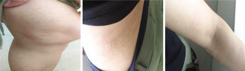
A series of three clinical photographs demonstrating the dermatological resolution of urticaria (hives) across three anatomical regions: the knee joint (A), the lateral waist (B), and the elbow (C). The images show characteristic wheals—edematous, circumscribed, and slightly elevated skin lesions. Panel A displays the knee area with subtle, flesh-colored to slightly erythematous elevations and an uneven skin texture consistent with resolving wheals. Panel B shows the lateral waist with faint, linear, and confluent dermal edema. Panel C depicts the elbow where the lesions have largely flattened, leaving only minimal, faint markings on the skin surface. The progression illustrates the transient nature of urticarial lesions as they gradually diminish in size and elevation. This visual serves as an educational reference for identifying skin morphology in the recovery phase of an acute urticarial reaction, emphasizing the characteristic lack of secondary skin changes (such as scaling or scarring) during the resolution of wheals.
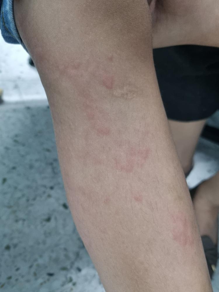
A clinical photograph showing a close-up of a child's lower extremity demonstrating urticaria. The image displays multiple raised, edematous skin lesions known as wheals or hives. These lesions are characterized by pale-to-skin-colored central swellings of varying sizes and irregular shapes, surrounded by a distinct zone of erythema (redness). The distribution is scattered across the leg, illustrating the typical appearance of a hypersensitivity reaction or allergic response. This image serves as an educational reference for identifying cutaneous manifestations of urticaria, focusing on the morphology of superficial dermal edema and associated inflammatory flare.
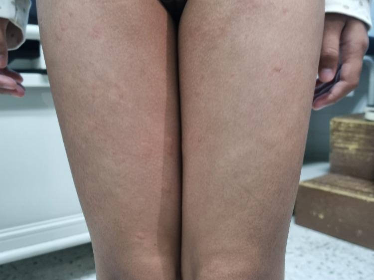
Clinical photograph of a pediatric patient's bilateral lower limbs demonstrating classic urticaria (hives). The image shows multiple discrete and confluent wheals distributed across the thighs. Morphologically, the lesions are characterized by raised, edematous, superficial swellings of varying sizes. The central portions of the wheals appear pale or skin-colored, while the peripheries exhibit varying degrees of erythema. These findings represent a type I hypersensitivity reaction or mast cell degranulation, leading to dermal edema. This visual evidence is a key diagnostic clinical sign for acute urticaria, commonly used in dermatology and pediatric curricula to differentiate transient wheals from other types of papular eruptions or inflammatory skin conditions.
urticaria management guidelines
Urticaria (Hives)
Definition & Epidemiology
- Goldman-Cecil Medicine, p. 4300
- Rosen's Emergency Medicine, p. 2413
Pathophysiology
| Mechanism | Examples |
|---|---|
| IgE-mediated (Type I) | Foods, drugs, insect stings, latex |
| Immune complex/complement | Infections, serum sickness |
| Autoimmune | Functional IgE or IgG autoantibodies against FcεRI or IgE |
| Direct mast cell degranulation | NSAIDs, opioids, radiocontrast media, vancomycin |
| Bradykinin-mediated | ACE inhibitors, hereditary angioedema |
| Vasoactive stimuli | Physical stimuli (cold, pressure, heat, light) |
- Goldman-Cecil Medicine, p. 4301
- Dermatology 2-Volume Set 5e, p. 125
- Harrison's Principles, p. 2851
Classification
1. Acute Urticaria (< 6 weeks)
- Drug reactions (antimicrobials, NSAIDs, opioids, ACE inhibitors, radiocontrast)
- Food reactions (seafood, tree nuts, eggs, peanuts, strawberries)
- Infections: viral (rhinovirus, EBV, hepatitis, coxsackievirus), bacterial, parasitic
- Insect stings/bites
- Transfusion reactions
2. Chronic Urticaria (≥ 6 weeks)
- No identifiable external trigger
- May have autoimmune component (anti-FcεRI or anti-IgE antibodies)
- Angioedema accompanies wheals in ~33-67% of CSU patients
| Subtype | Trigger | Key Feature |
|---|---|---|
| Symptomatic dermographism | Firm stroking of skin | Most common physical urticaria; wheal within 30 min |
| Cold urticaria | Cold exposure | Risk of anaphylaxis with cold water immersion |
| Cholinergic urticaria | Heat, exercise, emotion | Small wheals (1-3 mm) with large surrounding erythema |
| Pressure urticaria | Sustained pressure | Delayed onset 4-8 hours |
| Solar urticaria | UV/visible light | Confined to sun-exposed areas |
| Aquagenic urticaria | Water (any temperature) | May be associated with polycythemia vera |
| Vibratory urticaria | Vibration | Occupational; some familial (ADGRE2 mutations) |
| Exercise-induced anaphylaxis | Exertion ± food | May progress to vascular collapse |
-
Urticarial vasculitis
-
Mastocytosis (cutaneous or systemic)
-
Hereditary angioedema (HAE) - bradykinin-mediated, NO pruritus, NO urticaria
-
CIAS1/NLRP3-associated: familial cold autoinflammatory syndrome (FCAS), Muckle-Wells syndrome
-
Schnitzler's syndrome, Gleich's syndrome
-
Harrison's Principles, p. 2851 (Table 363-1)
-
Rosen's Emergency Medicine, p. 2413
-
Fitzpatrick's Dermatology, p. 718
Clinical Features
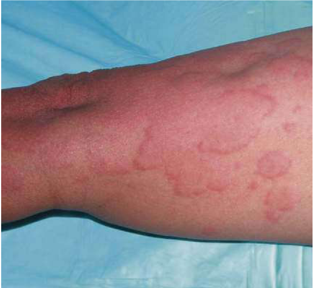
- Circumscribed, raised, pruritic, evanescent areas of edema in the superficial dermis
- Individual wheals last < 24 hours (new lesions continuously develop)
- Pale/white centers with red borders ("wheal and flare")
- May be erythematous or whitish when edema is marked
- Sizes range from a few mm to several cm; may be annular, polycyclic, or map-like
- Fitzpatrick's Dermatology, p. 717
- Harrison's Principles, p. 2851
Differential Diagnosis
- Drug eruption / morbilliform exanthem
- Erythema multiforme (targetoid; fixed > 24 h)
- Urticarial vasculitis
- Angioedema (non-urticarial types)
- Bullous pemphigoid (urticarial phase)
- Polymorphous eruption of pregnancy
- Erythema marginatum
- Systemic mastocytosis
- Sweet's syndrome
Diagnosis
Acute urticaria
- History alone is usually sufficient
- Allergen-specific IgE testing or skin prick testing when a specific trigger is suspected
Chronic urticaria
| Level | Tests |
|---|---|
| Routine (all CSU) | Differential blood count, ESR and/or CRP |
| Extended (guided by history) | Avoidance of suspected triggers; H. pylori testing; thyroid hormones + autoantibodies; autologous serum skin test (ASST) for autoantibodies; allergy skin tests; serum tryptase (if mastocytosis suspected); skin biopsy (if vasculitis suspected) |
- Fitzpatrick's Dermatology (Table 41-1)
- Harrison's Principles, p. 2852
Treatment
Step-Up Approach (EAACI/GALEN Guidelines)
- Cetirizine, fexofenadine, loratadine, desloratadine, levocetirizine
- Preferred over first-generation agents (diphenhydramine, hydroxyzine) due to lack of sedation and anticholinergic side effects
- Can be up-dosed to 4× the standard daily dose if standard dosing is insufficient
- H2 antagonist (famotidine, ranitidine) - modest additional benefit
- Leukotriene receptor antagonist (montelukast 10 mg/day) - particularly useful in aspirin/NSAID-sensitive urticaria
- First-generation antihistamine at night (e.g. hydroxyzine for sedation-aided sleep)
- Omalizumab (anti-IgE monoclonal antibody): approved for CSU in patients ≥12 years
- 300 mg SC every 4 weeks is the standard dose for CSU
- Effective and rapidly acting; approved for chronic inducible urticaria as well
- A 2025 meta-analysis confirms efficacy and safety in pediatric CSU (PMID: 40545961)
- For CSU refractory to omalizumab
- Cyclosporin A (calcineurin inhibitor) - targets T-cells and mast cells
- Hydroxychloroquine, dapsone, or colchicine for urticarial vasculitis
- Not recommended chronically (long-term toxicity)
- Short courses (14-21 days with taper) acceptable for moderate-severe acute urticaria
- Useful in pressure urticaria, vasculitic urticaria, or refractory angioedema
-
Cold urticaria: antihistamines 30-60 min before cold exposure; avoid cold water immersion
-
Cholinergic urticaria: non-sedating antihistamines
-
Solar urticaria: phototherapy for tolerance induction
-
HAE: NOT treated with antihistamines - use C1INH concentrate, icatibant (bradykinin B2 receptor antagonist), ecallantide, or lanadelumab for prophylaxis
-
Harrison's Principles, p. 2852
-
Rosen's Emergency Medicine, p. 2414
-
Dermatology 2-Volume Set 5e, p. 125
-
Goldman-Cecil Medicine, abstract
-
Katzung Pharmacology 16e
Common Causes Summary
| Category | Examples |
|---|---|
| Drugs | Penicillin (most common antibiotic), aspirin/NSAIDs, opioids, ACE inhibitors |
| Foods | Seafood, tree nuts, peanuts, eggs, strawberries, lobster |
| Infections | Rhinovirus, EBV, hepatitis, coxsackievirus, H. pylori, Candida, parasites |
| Physical | Dermatographism, cold, pressure, heat, exercise |
| Systemic diseases | SLE, lymphoma, carcinoma, hyperthyroidism, juvenile RA |
| Inhalants | Pollens, mold, animal dander, dust mites |
| Contact | Latex, topical medications, cosmetics, animal saliva |
| Idiopathic | ~50% of chronic urticaria - no cause found |
Recent Evidence (2024-2026)
- A 2024 systematic review (PMID: 38420865) in JEADV reviewed treatment strategies for acute urticaria, reinforcing antihistamines as first-line
- A 2025 systematic review and meta-analysis (PMID: 40545961) confirmed omalizumab is effective and safe for chronic spontaneous urticaria in children ≥12 years
- A 2025 systematic review (PMID: 40745339) found emerging evidence for vitamin D supplementation as an adjunct in chronic urticaria management