
Necrotizing pancreatitis
I'll read the medical library skill first, then research necrotizing pancreatitis comprehensively.
Reading File
Now I'll run parallel searches across the library and PubMed simultaneously.
Finding Sources
Searching PubMed
"necrotizing pancreatitis"[MeSH Terms] AND management
Searching the Web
necrotizing pancreatitis management guidelines 2024 2025
Good. Now I'll read the key textbook sections and also search for pathophysiology/classification content in parallel.
Reading File
Reading File
Reading File
Finding Sources
Searching PubMed
"necrotizing pancreatitis"[MeSH Terms] AND "step-up approach"
Good content found. Now let me get the pathophysiology and management content from Sleisenger and Fischer's, plus the Yamada section on pathophysiology.
Reading File
Reading File
Finding Sources
Searching PubMed
"acute pancreatitis"[MeSH Terms] AND "necrosectomy"
Excellent. I have found a CT image. Let me fetch it and get Harrison's and Schwartz's content on management.
Loading Image
Reading File
Reading File
Reading File
I now have comprehensive material from multiple textbooks. Let me get the Harrison's management section and the step-up approach content from Schwartz's.
Reading File
Reading File
Reading File
I have rich content from multiple textbooks. Let me fetch the treatment algorithm image and also get the Harrison's section on management of necrosis specifically.
Loading Image
Reading File
Reading File
I now have comprehensive content from 6 textbooks plus an excellent treatment algorithm image. I have enough to write a thorough, well-structured answer.
Necrotizing Pancreatitis
Definition and Classification (Revised Atlanta 2012)
Necrotizing pancreatitis is a severe form of acute pancreatitis characterized by inflammation with necrosis of the pancreatic parenchyma, peripancreatic tissue, or both. It complicates approximately 20% of all acute pancreatitis cases.
Under the 2012 Revised Atlanta Classification, two distinct necrotic collections are recognized:
| Collection | Timing | CT Features | Content |
|---|---|---|---|
| Acute Necrotic Collection (ANC) | <4 weeks | No defined wall, heterogeneous | Fluid + necrosis, intra- and/or extrapancreatic |
| Walled-Off Necrosis (WON) | >4 weeks | Well-defined inflammatory wall, encapsulated | Fluid + necrosis, heterogeneous with possible loculations |
Both collections begin as sterile but may become superinfected - up to 70% of necrotic collections eventually become infected. Infection doubles the mortality rate: from ~10% for sterile necrotizing pancreatitis to ~20% for infected necrosis.
Current Surgical Therapy 14e (Table 2); ROSEN's Emergency Medicine; Fischer's Mastery of Surgery - 8th e
CT Appearances of the Four Peripancreatic Collections
The image below from Fischer's Mastery of Surgery shows the four distinct CT patterns:
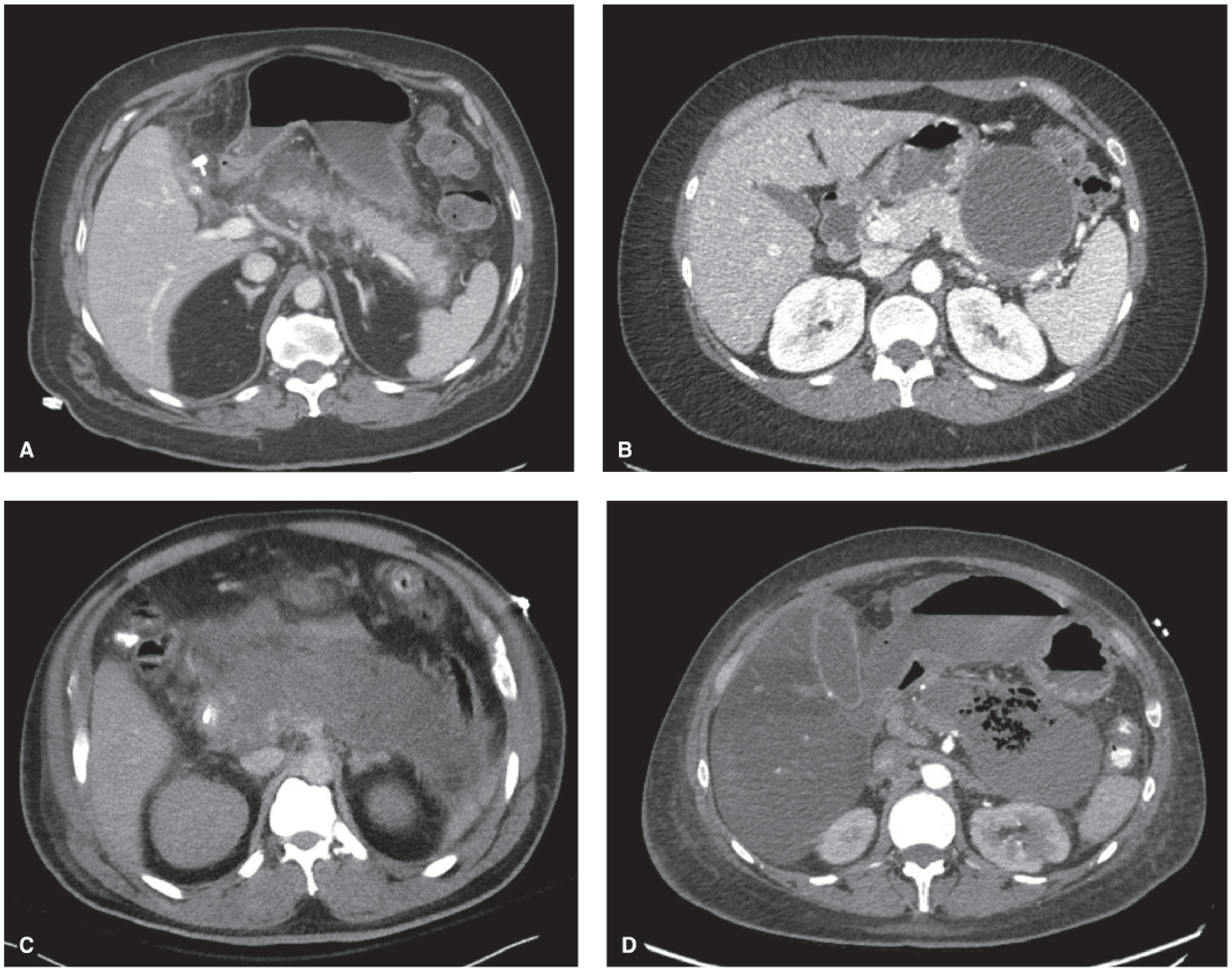
Panel C (ANC): heterogeneous collection without a defined wall, filling the lesser sac. Panel D (WON): heterogeneous collection with a well-defined encapsulating wall, containing gas (emphysematous change) indicating superinfection.
Pathophysiology
The inciting event is premature intracellular activation of digestive enzymes (trypsin) within pancreatic acinar cells. This triggers:
- Local injury: Autodigestion of pancreatic parenchyma and peripancreatic fat, releasing lipase that causes fat necrosis and calcium saponification (contributing to hypocalcemia in ~25% of patients).
- Systemic inflammatory response syndrome (SIRS): Release of TNF-α, IL-1, IL-6. Capillary leakage drives intravascular volume depletion.
- Hemoconcentration: Elevated hematocrit >44% and rising BUN are early markers of severity and predictors of necrosis.
- Organ failure: Respiratory failure (ARDS, PaO₂ <60 mmHg), renal failure (creatinine >2.0 mg/dL), and cardiovascular collapse can occur in the first 48 hours (early phase), driven by SIRS rather than infection.
- Late phase (>2 weeks): Dominated by septic complications from infected necrosis; bacteria reach the necrotic tissue primarily via bacterial translocation from the gut.
Yamada's Textbook of Gastroenterology, 7th e; Harrison's Principles of Internal Medicine 22E
Clinical Presentation and Diagnosis
Symptoms: Severe epigastric pain radiating to the back, nausea, vomiting, fever, tachycardia.
Diagnosis requires 2 of 3 criteria (Harrison's):
- Typical epigastric pain radiating to the back
- Serum lipase or amylase ≥3× upper limit of normal
- Confirmatory imaging on CT/MRI
Lab findings in severe/necrotizing disease:
- WBC 15,000-20,000/μL
- Hematocrit >44% (hemoconcentration)
- BUN >25 mg/dL on admission (strong mortality predictor; BUN AUC 0.918 in PROST trial)
- Hyperglycemia (decreased insulin + increased glucagon + glucocorticoid release)
- Hypocalcemia (~25%)
- Hyperbilirubinemia (~10%)
- ALT >3× ULN strongly suggests gallstone etiology
Imaging:
- Ultrasound: First-line; evaluates gallstones and CBD dilation but poor for necrosis
- Contrast-enhanced CT (CECT): Gold standard for necrotizing pancreatitis. Non-enhancing pancreatic parenchyma on CECT confirms necrosis. Optimal timing is 72-96 hours after onset (to allow demarcation). Gas within the collection = emphysematous/infected necrosis.
- MRI: Better differentiates solid necrotic debris from fluid (important for drainage planning)
Harrison's Principles of Internal Medicine 22E; Schwartz's Principles of Surgery 11th e
Severity Scoring
BISAP Score (within 24 hours - 1 point each):
- BUN >25 mg/dL
- Impaired mental status (GCS <15)
- SIRS present
- Age >60 years
- Pleural effusion on imaging
≥3 points = substantially increased risk for in-hospital mortality.
SOFA Score monitors ongoing organ failure (respiratory, coagulation, liver, cardiovascular, CNS, renal). A score ≥2 in any two systems = multiple organ failure.
Schwartz's Principles of Surgery 11th e; Harrison's 22E
Management
1. Resuscitation and ICU Care
- Aggressive IV fluid resuscitation: Balanced crystalloid (Lactated Ringer's preferred over normal saline). Target: falling hematocrit and BUN over first 12-24 hours, urine output >0.5 mL/kg/hr.
- ICU admission: Patients with persistent SIRS at 24 hours, elevated BISAP ≥3, organ failure, or inadequate response to initial resuscitation.
- Analgesia, bowel rest initially, then early enteral nutrition (nasojejunal feeding preferred - reduces infectious complications, maintains gut barrier integrity).
2. Nutrition
Early enteral nutrition (within 24-48 hours) via nasojejunal tube is preferred over parenteral nutrition. However, the PYTHON trial showed early forced enteral tube feeding did not reduce infection rates compared to on-demand feeding (25% vs 26%).
3. Antibiotics
- Prophylactic antibiotics are NOT recommended - multiple well-designed RCTs show they do not reduce infectious complications, need for surgery, or death. Overuse drives fungal infections and resistant organisms.
- Therapeutic antibiotics ARE indicated for confirmed or strongly suspected infected necrosis. Agents that penetrate pancreatic necrosis include carbapenems, fluoroquinolones, and metronidazole.
- CT-guided fine-needle aspiration (FNA) with Gram stain and culture is used to confirm infection when clinical status is unclear (sensitivity/specificity mid-90% range). Not recommended routinely due to false-negative risk and theoretical risk of introducing infection.
4. Biliary Etiology
- Cholangitis + biliary obstruction: ERCP within 24-48 hours.
- Gallstone pancreatitis without cholangitis: Early ERCP has no benefit (stone usually passes spontaneously). Cholecystectomy during the same admission in mild disease; delayed in severe disease after recovery.
5. Management of Infected/Symptomatic Necrosis - The "Step-Up" Approach
The current paradigm is a minimally invasive step-up approach, deferring intervention as long as possible to allow demarcation (preferably ≥4 weeks). Early surgery carries extremely high perioperative mortality.
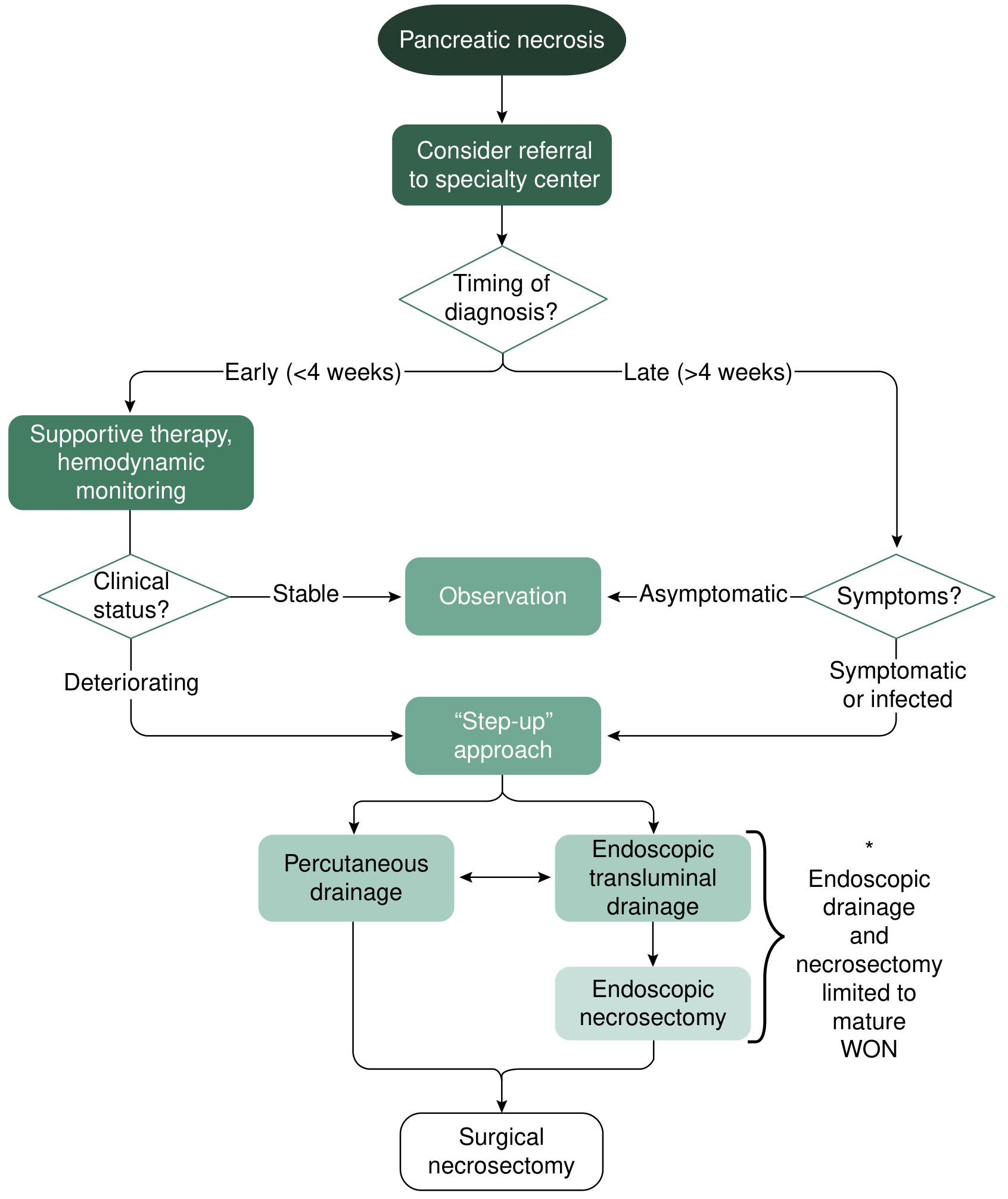
Step 1 - Percutaneous drainage (PD):
- First-line for infected necrosis or ANC with clinical deterioration
- Image-guided by interventional radiology (IR)
- Retroperitoneal access preferred to allow potential VARD
- Serial catheter upsizing + intracatheter irrigation with saline/H₂O₂
- Best for collections extending to retroperitoneum or paracolic gutters
- Up to 50% of patients managed with PD alone, avoiding necrosectomy
- Risk: pancreaticocutaneous fistula if communicating with pancreatic duct
Step 2 - Endoscopic drainage and necrosectomy:
- Best for mature WON within 2 cm of stomach or duodenum
- Requires ≥4 weeks from onset for wall maturation
- EUS-guided cystgastrostomy → balloon dilation → pigtail stents or lumen-apposing metal stents (LAMS)
- Direct endoscopic necrosectomy (DEN): endoscope inserted into collection, mechanical debridement
- PENGUIN trial (Dutch Acute Pancreatitis Study Group): endoscopic necrosectomy vs. surgical necrosectomy - significantly fewer major complications/death, fewer pancreatic fistulas, less new organ failure
- Up to two-thirds of patients avoid operative debridement with endoscopic necrosectomy
Step 3 - Video-Assisted Retroperitoneal Debridement (VARD):
- If percutaneous drainage fails but complete surgical necrosectomy not needed
- The PANTER trial: step-up approach (PD → VARD) vs. primary open surgical necrosectomy - step-up had significantly fewer complications and less new-onset organ failure
Step 4 - Surgical necrosectomy (open or laparoscopic):
- Reserved for failure of all minimally invasive approaches
- Indications per Maingot's Abdominal Operations:
- Diagnostic uncertainty / concurrent intra-abdominal catastrophe
- Infected necrosis documented by FNA or extraluminal gas on CT
- Severe sterile necrosis with persistent organ failure (ideally ≥2 weeks from onset)
- Symptomatic organized pancreatic necrosis ("persistent unwellness")
- Delayed surgery (>4 weeks) significantly better than early surgery - allows demarcation, reduces bleeding risk and collateral organ damage
Current Surgical Therapy 14e; Maingot's Abdominal Operations; Yamada's Textbook of Gastroenterology 7th e; Schwartz's Principles of Surgery 11th e
Complications
| Complication | Details |
|---|---|
| Infected necrosis | Most common cause of death; near-100% mortality without intervention |
| Pancreatic fistula | Communication with pancreatic duct - complicates drainage |
| Pseudoaneurysm | Splenic artery most common; presents as GI bleed or hemosuccus pancreaticus |
| Colonic necrosis | Transverse colon most vulnerable; presents as peritonitis |
| Abdominal compartment syndrome | Massive fluid resuscitation → raised intra-abdominal pressure |
| ARDS | PaO₂ <60 mmHg; ~5-10% of cases |
| Exocrine/endocrine insufficiency | Post-necrotizing pancreatitis diabetes and steatorrhea |
| Chronic pancreatitis | Postnecrotic fibrosis and ductal stricturing |
Prognosis
- Sterile necrotizing pancreatitis: ~10% mortality
- Infected necrotizing pancreatitis: ~20% mortality (effectively 100% without intervention)
- Patients who survive severe necrotizing pancreatitis are at risk for post-necrotizing chronic pancreatitis, as noted in Harrison's (TIGAR-O classification: "Postnecrotic - severe acute pancreatitis")
2025 IAP/APA Guidelines Update: The International Association of Pancreatology 2025 guidelines (PMID 40651900) represent the most current evidence-based recommendations, covering 96 questions across 18 domains including fluid therapy, nutritional support, conservative and interventional treatment of infected necrotizing pancreatitis, discharge criteria, and prevention of recurrence.
This is a shared conversation. Sign in to Orris to start your own chat.