
Types and... Etiology of urinary incontinence. How to approach a case of urinary incontinence? Write in details about... Diagnosis and management of... Stress urinary incontinence. Thirty marks question, PG, ob-gyn.
urinary incontinence types stress urge diagram
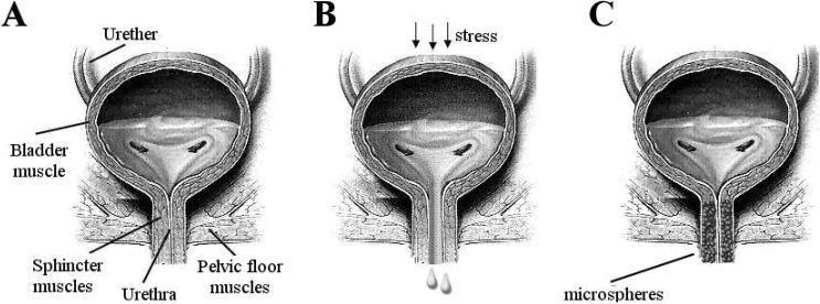
This medical anatomical diagram illustrates the pathophysiology and treatment of stress urinary incontinence (SUI) through three coronal views of the human bladder and urethra (A, B, and C). Diagram A represents normal anatomy and function, showing a healthy bladder muscle, intact ureters, and a closed urethra supported by strong sphincter and pelvic floor muscles. Diagram B depicts the mechanism of stress urinary incontinence, where external 'stress' (indicated by downward arrows) causes involuntary urine leakage through a weakened urethral opening and compromised pelvic floor support. Diagram C demonstrates a therapeutic intervention using urethral bulking agents. It shows the injection of 'microspheres' into the periurethral tissues, which facilitates complete coaptation (closure) of the urethral lumen to prevent leakage. Key labels include the bladder muscle, ureters, urethra, sphincter muscles, and pelvic floor muscles. The illustration is designed for educational use in urology to explain urinary control mechanisms and the clinical application of polymeric microspheres as bulking agents.
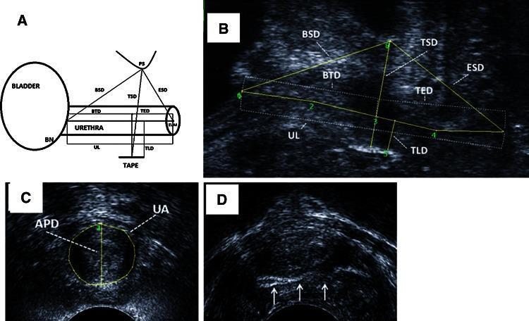
This composite image demonstrates ultrasound-based morphometric evaluation of the female urethra following mid-urethral sling surgery for stress urinary incontinence. Panel A provides a schematic anatomical diagram in the sagittal plane, outlining the relationship between the bladder neck (BN), urethra, pubic symphysis (PS), and the synthetic tape. Panel B is a corresponding sagittal translabial ultrasound image displaying quantitative measurements including urethral length (UL), bladder neck-tape distance (BTD), tape-external urethral meatus distance (TED), and the distance between the tape and pubic symphysis (TSD). Panel C shows an axial ultrasound view at the mid-urethra, where the anterior-posterior diameter (APD) and total urethral area (UA) are delineated. Panel D provides an axial ultrasound view assessing for urethral 'encroachment' or compression by the sling material, indicated by white arrows. These images are used to assess tape positioning, tension, and its impact on urethral anatomy, which are critical factors in surgical outcome and complication analysis in urognecology.
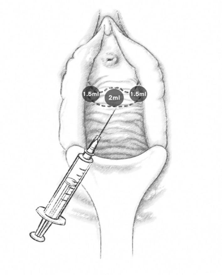
This medical illustration depicts the procedural technique for injecting Platelet-Rich Plasma (PRP) into the anterior vaginal mucosa, typically performed for the treatment of stress urinary incontinence (SUI). The diagram shows a frontal view of the female external genitalia and anterior vaginal wall. The anatomical landmarks include the clitoris, labia majora, and the urethral meatus. The injection protocol is visualized using three targeted sites arranged in a horizontal arc around the mid-urethral region: one central injection point directly inferior to the urethral meatus labeled '2ml' and two lateral injection points labeled '1.5ml' each. A syringe with a needle is shown positioned at an oblique angle, demonstrating the entry into the mucosal tissue. The illustration serves as a clinical guide for medical professionals to visualize the specific volume distribution and spatial arrangement required to provide periurethral support and stimulate tissue repair in the pubourethral ligament area.
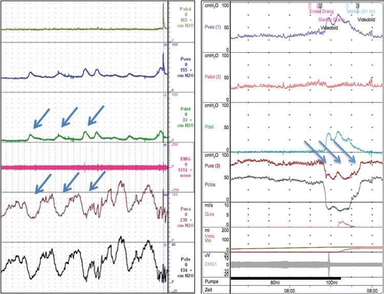
A comparative diagnostic urodynamic study displaying two multicomponent traces: one representing detrusor overactivity (DO) on the left and stress urinary incontinence (SUI) on the right. Each panel tracks several physiological parameters over time (cmH2O), including abdominal pressure (Pabd), vesical pressure (Pves), detrusor pressure (Pdet), urethral pressure (Pura), urethral closure pressure (Pclos), and electromyography (EMG). The left trace (DO) features prominent, rhythmic peaks in the Pdet channel (highlighted by blue arrows), indicating involuntary bladder contractions. These contractions are associated with significant fluctuations in Pura and Pclos, demonstrating urethral instability. The right trace (SUI) shows a sudden drop in Pura and Pclos (blue arrows) during a period of increased intra-abdominal pressure without corresponding involuntary detrusor spikes initially, characteristic of mechanical sphincteric incompetence. This diagnostic imaging is essential for urological evaluation to differentiate between urge and stress incontinence mechanisms by analyzing the relationship between detrusor muscle activity and urethral sphincter integrity.
Urinary Incontinence - Types, Etiology, Approach, and Stress Urinary Incontinence
(30 Marks - PG Ob-Gyn Long Essay)
DEFINITION
PART I: TYPES OF URINARY INCONTINENCE
1. Stress Urinary Incontinence (SUI)
2. Urgency Urinary Incontinence (UUI)
3. Mixed Urinary Incontinence (MUI)
4. Overflow Incontinence
5. Continuous (Total) Incontinence
6. Nocturnal Enuresis
7. Functional Incontinence
8. Extraurethral Incontinence
PART II: ETIOLOGY OF URINARY INCONTINENCE
A. Obstetric / Gynaecologic Causes
- Multiparity and vaginal delivery - strongest risk factor for SUI; vaginal birth confers a twofold increased risk of SUI compared to caesarean delivery. Pelvic floor denervation and levator ani trauma during labor damage pudendal nerve branches and weaken the pubourethral ligaments.
- Prolonged obstructed labor - leads to vesicovaginal fistula (ischaemic necrosis), particularly in resource-poor settings.
- Pelvic organ prolapse - descent of the anterior compartment pulls the urethrovesical junction away from its supporting structures.
- Hysterectomy and pelvic surgery - ureteral/bladder injury can result in ureterovaginal or VVF; disruption of pelvic floor supports contributes to SUI.
- Menopause and oestrogen deficiency - atrophic urethritis reduces mucosal coaptation and urethral closure pressure; advancing age (>50 years) strongly predicts UUI and MUI.
B. Neurological Causes
- Multiple sclerosis, Parkinson's disease, spinal cord injuries - disruption of inhibitory neuronal circuits leads to detrusor overactivity and UUI.
- Stroke, dementia - cortical micturition centre dysfunction.
- Diabetic cystopathy - peripheral neuropathy causes bladder hyposensitivity and acontractile detrusor, leading to overflow incontinence.
- Cauda equina syndrome - denervation of the detrusor and sphincter.
C. Anatomical / Structural Causes
- Urethral diverticulum (post-void dribbling, dysuria, dyspareunia)
- Ectopic ureter (congenital continuous leakage, normal voiding preserved)
- Bladder exstrophy (congenital absence of anterior bladder/urethral wall)
- Pelvic masses (fibroids, ovarian cysts) compressing the bladder
D. Functional and Iatrogenic Causes
- Medications: ACE inhibitors (chronic cough aggravating SUI), alpha-blockers (decreased urethral closure pressure), diuretics (frequency/urgency), anticholinergics (overflow), calcium channel blockers (voiding difficulty), benzodiazepines and alcohol (confusion, diuresis).
- Immobility, constipation (straining increases SUI; faecal impaction causes urgency).
- Urinary tract infections - transient reversible incontinence.
- Obesity - increases intra-abdominal pressure, strong risk factor for all UI subtypes.
- COPD, chronic cough (smoking) - repetitive increase in abdominal pressure.
E. Mnemonic for Transient/Reversible Causes - "DIAPERS"
| Letter | Cause |
|---|---|
| D | Delirium |
| I | Infection (UTI) |
| A | Atrophic urethritis and vaginitis |
| P | Pharmacologic causes |
| P | Psychological causes |
| E | Excessive urine production (hyperglycaemia, hypercalcaemia) |
| R | Restricted mobility |
| S | Stool impaction |
PART III: APPROACH TO A CASE OF URINARY INCONTINENCE
Step 1: History Taking
- Leakage with coughing, sneezing, jumping, running, intercourse → SUI
- Leakage preceded by sudden strong urgency, provoked by running water, change in temperature, inserting key in door → UUI
- Both components present → MUI
- Continuous leakage day and night with otherwise normal voiding → fistula or ectopic ureter
- Duration, severity, and frequency of episodes
- Number of pads used per day (pad usage as a proxy for severity)
- Obstetric history: parity, mode of delivery, birth weight, prolonged labor, instrumental delivery
- Gynaecologic: prior pelvic surgery (hysterectomy, prior anti-incontinence surgery), prolapse, pelvic radiation
- Menopausal status
- Fluid intake (volume, type: caffeine, alcohol, carbonated drinks)
- Associated LUTS: frequency (>8 voids/day), nocturia (>1 void/night), hesitancy, slow stream, incomplete emptying, dysuria, haematuria
- Medical conditions: diabetes, neurological disorders (MS, Parkinson's, stroke), COPD, heart failure, constipation
- Medications (Table 29-4 review - see above)
- Impact on quality of life and social/occupational functioning (validated QOL tools: ICIQ-SF, UDI-6, IIQ-7)
Step 2: Physical Examination
- BMI/obesity assessment
- Mental and cognitive function (dementia → urgency/frequency/enuresis)
- Neurological examination (gait, lower limb reflexes - Parkinson's, MS)
- Cardiovascular - lower limb oedema (nocturnal polyuria from fluid redistribution)
- Pulmonary - evidence of COPD
- Inspect vulva and vagina for atrophic changes, vaginitis, or dermatoses
- Inspect/palpate urethra for tenderness or mass (diverticulum)
- Assess for pelvic organ prolapse (POP-Q staging) - all three compartments
- Evaluate pelvic floor muscle (PFM) tone, strength, and voluntary contraction ability (bidigital palpation)
- Bimanual examination for pelvic/adnexal masses
- Rectal examination for faecal impaction
- Anal wink (S3-S4): gentle perianal skin tap → external anal sphincter contraction
- Bulbocavernosus reflex: gentle clitoral/perianal tap → sphincter contraction
- Absence of these reflexes suggests sacral nerve or pudendal nerve dysfunction
- With a comfortably full bladder, the patient is asked to cough forcefully or perform Valsalva manoeuvre in the lithotomy or standing position
- Instantaneous leakage synchronous with the cough is a positive stress test and supports SUI
- Leakage delayed by a few seconds after the cough suggests detrusor instability
- A sterile, lubricated cotton-tipped swab is inserted into the urethra to the level of the bladder neck
- The patient is asked to Valsalva and the angle of deflection is measured
-
30 degrees from horizontal indicates urethral hypermobility (suggesting urethrovesical junction hypermobility as the mechanism of SUI)
- Note: Current AUA/ACOG guidelines no longer recommend this test routinely in the initial workup
Step 3: Investigations
- Urinalysis (dipstick): Screen for infection (leucocytes, nitrites), haematuria, glycosuria (diabetes), proteinuria
- Midstream urine for MC&S: To exclude or treat UTI before further workup
- Post-Void Residual (PVR) Volume: Measured by bladder scan or in-out catheter immediately after voiding. Normal < 50 mL. Elevated PVR (>200 mL) suggests voiding dysfunction or overflow incontinence.
- Pad Test: Patient weighs pads before and after a defined period (1-hour standardized or 24-48 hour). Quantifies degree of leakage. Does not differentiate SUI from UUI. A 48-hour pad test is more reproducible than a 1-hour test.
- ICIQ-SF (International Consultation on Incontinence Questionnaire - Short Form)
- UDI-6 (Urogenital Distress Inventory)
- IIQ-7 (Incontinence Impact Questionnaire)
- Conservative measures have failed
- The type of incontinence is unclear clinically
- There is recurrence after previous surgical intervention
- Neurological disease is suspected
- Mixed incontinence where the dominant component needs clarification
| Component | What It Assesses |
|---|---|
| Uroflowmetry | Voiding flow rate, pattern, voided volume (normal: >15 mL/s with voided volume >150 mL) |
| Cystometrogram (CMG/filling cystometry) | Bladder compliance, first desire to void, strong desire, capacity; detects uninhibited detrusor contractions |
| Urethral Pressure Profile (UPP) | Urethral closure pressure along length; maximum urethral closure pressure (MUCP); low MUCP (<20 cmH₂O) indicates intrinsic sphincter deficiency (ISD) |
| Valsalva/Cough Leak Point Pressure (VLPP/CLPP) | Pressure at which urine leaks with Valsalva/cough; VLPP <60 cmH₂O = ISD; VLPP >90 cmH₂O = urethral hypermobility |
| Pressure Flow Study (Voiding Cystometrogram) | Detrusor-sphincter coordination during voiding; identifies dysfunctional voiding |
| EMG of external sphincter | Detrusor-sphincter dyssynergia (via anal probe/patch electrodes) |
- Cystoscopy: If haematuria, recurrent UTI, suspected fistula, urethral diverticulum, or prior to complex surgery. Rarely needed for uncomplicated UI.
- Ultrasound (KUB): In recurrent UTI, haematuria, or to assess the upper urinary tract
- MRI pelvis: For suspected urethral diverticulum, complex fistula, neurogenic cases
- Video urodynamics (fluoroscopy + UDS): Reserved for complex or neurogenic cases to assess for vesicoureteral reflux, funneling, bladder neck opening, and cystocele
PART IV: STRESS URINARY INCONTINENCE - DETAILED
Definition
Pathophysiology
Risk Factors for SUI
| Risk Factor | Strength of Evidence |
|---|---|
| Parity / vaginal delivery | Strong (twofold risk vs caesarean) |
| Age <50 years (peaks at 5th decade) | Moderate |
| Obesity (increased IAP) | Strong |
| Pregnancy (50% develop SUI) | Strong |
| Diabetes mellitus | Moderate |
| COPD, chronic cough, smoking | Moderate |
| Race (White > Hispanic > Black) | Moderate |
| Prior pelvic surgery | Moderate |
Diagnosis of SUI
- History of leakage with cough, sneeze, physical activity, intercourse
- Instantaneous leakage on cough stress test with comfortably full bladder
- Absence of urgency as the dominant component
| Feature | SUI | UUI |
|---|---|---|
| Trigger | Cough, sneeze, exercise | Urgency, running water, cold |
| Warning | None (instantaneous) | Strong urgency precedes |
| Amount | Small (drops to splashes) | Moderate to large |
| Nocturia | Not typical | Common |
| Daytime frequency | Normal | Increased (>8/day) |
- Demonstrated leakage with cough or Valsalva on CMG, without a detrusor contraction
- Abdominal leak point pressure (ALPP/VLPP):
- <60 cmH₂O → ISD
- 60-90 cmH₂O → mixed mechanism
-
90 cmH₂O → urethral hypermobility
- Maximum Urethral Closure Pressure (MUCP) <20 cmH₂O → ISD
Management of SUI
A. Conservative Management
- Weight loss: Weight reduction of 5-10% significantly reduces SUI episodes. A landmark RCT (PRIDE trial) showed >50% reduction in incontinence episodes with weight loss.
- Fluid management: Limit intake to ~2 L/day; reduce caffeine, alcohol, carbonated beverages (bladder irritants)
- Treat chronic cough (smoking cessation, COPD management)
- Avoid constipation (high-fibre diet, adequate hydration)
- Avoid heavy lifting where possible
- The cornerstone of conservative management for SUI, recommended as first-line treatment for at least 3 months before considering surgical intervention.
- Based on Arnold Kegel's original description (1948); strengthens levator ani, pubococcygeus, and external urethral sphincter.
- Regimen: Contract pelvic floor muscles for 3-8 seconds; 10-15 contractions per session; 3 sessions per day. Ideally supervised by a pelvic floor physiotherapist.
- Evidence: A 2015 Cochrane review showed that PFMT resulted in fewer UI episodes, improved cure rates, and better QOL compared to controls. Supervised PFMT is more effective than unsupervised.
- Biofeedback (auditory/visual sensory feedback) and electrical stimulation (intravaginal/intrarectal probe) can supplement PFMT, though not proven superior to properly performed PFMT alone.
- "Knack" manoeuvre: Pre-contracting the pelvic floor just before a cough or sneeze significantly reduces leakage.
- Intravaginal pessaries / incontinence pessaries: Incontinence ring or dish pessary supports the anterior vaginal wall and lifts the urethra, recreating the hammock effect. Useful for women who are not surgical candidates or during pregnancy.
- Urethral insert (FemSoft): Occludes the urethral meatus; removed before voiding. Useful for competitive female athletes with activity-specific SUI.
B. Medical (Pharmacological) Management
- A combined serotonin and norepinephrine reuptake inhibitor that increases pudendal nerve firing, enhancing the tone of the rhabdosphincter during the storage phase.
- Dose: 40 mg twice daily
- Reduces UI episodes by approximately 50%
- Side effects: Nausea (most common - 25%, usually transient), dizziness, dry mouth, insomnia, fatigue
- Licensed in the UK and EU for moderate-to-severe SUI in women; considered off-label in several countries including India (not FDA-approved in the US for SUI)
- Used when conservative measures have failed and surgery is contraindicated or declined
- Restores urethral mucosal coaptation and improves the vascular submucosa in post-menopausal women
- Topical (vaginal) oestrogen is preferred over systemic; reduces symptoms of urgency, frequency, and atrophic UI
- Limited evidence for SUI alone; more effective for UUI/mixed UI
- Note: Systemic hormone replacement therapy (HRT) can worsen SUI
- Increases urethral smooth muscle tone; limited evidence and significant cardiovascular side effects; rarely used
C. Surgical Management
i. Midurethral Slings (Gold Standard - First-Line Surgical Treatment)
- A synthetic polypropylene mesh tape is placed under the mid-urethra (not the bladder neck) in a tension-free manner
- Approach: A 1-2 cm incision at the mid-vaginal wall below the urethra; two trocars are passed from below upward behind the pubic bone (retropubic space, Space of Retzius) and exit through two suprapubic skin incisions (bottom-up technique)
- Cystoscopy is performed intraoperatively to confirm bladder integrity
- Long-term efficacy: 17-year follow-up of the original TVT cohort showed ~90% objective and 87% subjective cure
- Complications: Bladder injury (1-5% - detected intraoperatively via cystoscopy), vascular injury, voiding dysfunction, mesh erosion/exposure, de novo OAB
- The tape is passed through the obturator foramen laterally, avoiding the Space of Retzius
- Inside-out (TVT-O, Monarc) or outside-in (ARIS) techniques
- Lower risk of bladder injury and vascular injury compared to TVT
- Higher risk of inner thigh/groin pain and a twofold higher risk of reoperation compared to TVT (based on a large administrative dataset)
- Preferred by some surgeons when retropubic anatomy is distorted (previous pelvic surgery, severe obesity)
- A strip of rectus abdominis fascia (or fascia lata from thigh) is harvested and placed at the bladder neck/proximal urethra
- Particularly favoured for ISD (low VLPP) and recurrent SUI after failed mesh surgery
- Success rate comparable to retropubic colposuspension; higher risk of voiding dysfunction compared to TVT
ii. Retropubic Colposuspension (Burch Urethropexy)
- Burch Colposuspension: Open or laparoscopic; paravaginal sutures on each side of the bladder neck are attached to Cooper's (ileopectineal) ligament, elevating and suspending the bladder neck and proximal urethra
- Long-term (5-year) success rate ~70-80%; comparable to midurethral slings in RCTs
- Lower risk of voiding dysfunction than pubovaginal slings
- Higher risk of subsequent pelvic organ prolapse (posterior compartment) compared to slings, as elevating the anterior compartment transfers strain posteriorly
- Marshall-Marchetti-Krantz (MMK): Similar but sutures are attached to the periosteum of the pubic symphysis; risk of osteitis pubis (1-2%); largely abandoned
- Laparoscopic Burch: Equivalent efficacy to open at 5 years; requires advanced laparoscopic skill; being replaced by midurethral slings in most centres
iii. Periurethral / Transurethral Bulking Agents
- Injectables (e.g., calcium hydroxyapatite - Coaptite; polydimethylsiloxane - Macroplastique; polyacrylamide hydrogel - Bulkamid) are injected periurethrally under cystoscopic guidance to increase urethral coaptation
- Mechanism: Augment the submucosal bulk, narrowing the urethral lumen
- Indications: Women with ISD, elderly or frail patients who cannot tolerate surgery, temporary relief, or those who decline sling surgery
- Success rates are lower (30-50%) and durability is poor; re-injection may be needed
- Minimal invasive; performed as a day-case procedure under local/sedation
- Do not offer for moderate-to-severe SUI as a definitive treatment
iv. Artificial Urinary Sphincter (AUS)
- Mechanical device (AMS 800) placed surgically to occlude the urethra; patient activates a pump to void
- Primarily used in men with post-prostatectomy ISD; rarely used in women
- Indications in women: Severe ISD, failed multiple prior surgeries, neurogenic SUI
- Long-term continence ~80%; high risk of mechanical failure requiring revision
v. Procedures NOT Recommended (NICE Guidelines):
- Anterior colporrhaphy (Kelly plication) - inferior long-term results
- Needle suspension procedures (Stamey, Pereyra, Raz) - poor durability
- Paravaginal defect repair alone - insufficient evidence
- Porcine dermis sling - inadequate data
- MMK procedure - risk of osteitis pubis
Summary Algorithm for SUI Management
Diagnosis of SUI confirmed
        ↓
Conservative: PFMT × 3 months (supervised) + Lifestyle modification
        ↓
Failure or patient declines conservative treatment
        ↓
Counsel regarding surgery
Urodynamics if: unclear diagnosis / prior failed surgery / mixed incontinence
        ↓
Surgical Options:
  • Midurethral sling (TVT/TOT) - Gold Standard
  • Autologous fascial sling (especially if ISD, prior mesh failure)
  • Burch colposuspension (open/laparoscopic)
  • Bulking agents (ISD, elderly/unfit, temporary relief)
KEY POINTS FOR EXAM
- ICS definition: Urinary incontinence = involuntary leakage of urine.
- SUI = leakage with raised IAP, no detrusor contraction; UUI = leakage with urgency; MUI = both.
- Hammock Theory (DeLancey) and Integral Theory (Petros-Ulmsten) explain SUI pathogenesis.
- VLPP <60 cmH₂O = ISD; >90 cmH₂O = urethral hypermobility.
- First-line SUI treatment = supervised PFMT for minimum 3 months.
- Duloxetine = only pharmacological agent for SUI (off-label in many countries).
- Midurethral sling (TVT/TOT) = surgical gold standard; TVT has better long-term efficacy and lower reoperation rate than TOT.
- Burch colposuspension = ~70-80% long-term cure; risks posterior prolapse.
- Bulking agents for ISD/frail patients; lower and less durable cure rates.
- DIAPERS mnemonic = transient causes of UI in elderly.