
I have my MD medicine exams coming up.
What would you like help with?
- Cardiology - heart failure, arrhythmias, ACS, valvular disease
- Respiratory - COPD, asthma, pneumonia, TB, lung cancer
- Nephrology - AKI, CKD, glomerulonephritis, electrolyte disorders
- Gastroenterology - liver disease, IBD, GI bleeding, pancreatitis
- Endocrinology - diabetes, thyroid, adrenal, pituitary disorders
- Neurology - stroke, epilepsy, meningitis, movement disorders
- Infectious Disease - antibiotics, tropical medicine, sepsis
- Hematology/Oncology - anemias, leukemias, coagulation disorders
- Rheumatology - SLE, RA, vasculitis, crystal arthropathies
- Pharmacology - drug mechanisms, side effects, interactions
- Explain concepts and pathophysiology clearly
- High-yield MCQ-style questions with explanations
- Mnemonics and memory aids
- Differentials and clinical reasoning
- Quick revision summaries
- Recent evidence and guidelines
Teach me ecg from basics to advanced for exam as well as daily clinical practice.
ECG Mastery: Basics to Advanced
MODULE 1 - Why ECG? The Big Picture
- Rhythm and rate
- Conduction system integrity
- Chamber size (hypertrophy)
- Ischemia and infarction (location, age, extent)
- Metabolic/electrolyte status
- Drug effects
- Pericardial disease
MODULE 2 - The Cardiac Conduction System
SA Node (right atrium)
↓ (atrial depolarization = P wave)
AV Node (PR delay = AV node slowing)
↓
Bundle of His
↓
Right Bundle Branch + Left Bundle Branch
↙ ↘
Left Anterior Left Posterior
Fascicle Fascicle
↓
Purkinje Fibers → Ventricular myocardium (= QRS)
- SA node fires at 60-100/min (dominant pacemaker)
- AV node fires at 40-60/min if SA fails (junctional escape)
- Purkinje / ventricle fires at 20-40/min (ventricular escape)
- Ventricular repolarization = ST segment + T wave
MODULE 3 - ECG Paper and Calibration
| Parameter | Value |
|---|---|
| Paper speed | 25 mm/s (standard) |
| 1 small square (1 mm) | 0.04 s (40 ms) |
| 1 large square (5 mm) | 0.20 s (200 ms) |
| Standard voltage | 1 mV = 10 mm (2 large squares tall) |
| Half-standard | 1 mV = 5 mm (written on trace) |
MODULE 4 - ECG Waveforms and Intervals

The Waves
| Wave | Represents | Normal |
|---|---|---|
| P wave | Atrial depolarization | <120 ms wide, <2.5 mm tall; upright in II, inverted in aVR |
| QRS complex | Ventricular depolarization | <100-110 ms (2.5 small squares) |
| Q wave | Septal depolarization (normal) or infarction (pathological) | Normal: <0.04 s, <25% of R height |
| ST segment | Plateau of action potential (phase 2) | Isoelectric (flat) |
| T wave | Ventricular repolarization | Upright in I, II, V3-V6; inverted in aVR normal |
| U wave | After-depolarization / Purkinje repolarization | Same direction as T, low amplitude |
The Intervals
| Interval | Normal Range | Meaning |
|---|---|---|
| PR | 120-200 ms (3-5 small squares) | AV conduction time |
| QRS | <100-110 ms | Ventricular conduction |
| QT | <450 ms men, <460 ms women (rate-corrected) | Total ventricular repolarization |
| RR | Depends on rate | Used to calculate heart rate |
- Regular rhythm: HR = 300 ÷ (number of large squares between R-R)
- Or: HR = 1500 ÷ (number of small squares between R-R)
- Irregular: count QRS complexes in 10-second strip × 6
1 large square = 300 bpm | 2 = 150 | 3 = 100 | 4 = 75 | 5 = 60 | 6 = 50
MODULE 5 - The 12 Leads (Understanding the "Camera Angles")
Limb Leads (Frontal Plane)
| Lead | Looks at | Positive Pole Direction |
|---|---|---|
| I | Lateral | 0° (left) |
| II | Inferior | +60° |
| III | Inferior | +120° |
| aVR | Cavity (right shoulder) | -150° |
| aVL | Lateral (left shoulder) | -30° |
| aVF | Inferior (feet) | +90° |
Precordial Leads (Horizontal Plane)
| Lead | Position | Looks at |
|---|---|---|
| V1 | 4th ICS, right sternal border | Septal / RV |
| V2 | 4th ICS, left sternal border | Septal |
| V3 | Between V2 and V4 | Anterior |
| V4 | 5th ICS, midclavicular line | Anterior |
| V5 | Anterior axillary line | Lateral |
| V6 | Midaxillary line | Lateral |
Grouping Leads by Territory
| Territory | Leads | Coronary Artery |
|---|---|---|
| Inferior | II, III, aVF | RCA (right coronary) |
| Anteroseptal | V1, V2, V3, V4 | LAD (left anterior descending) |
| Lateral | I, aVL, V5, V6 | LCx (left circumflex) |
| Posterior | Reciprocal changes in V1, V2 | RCA / LCx |
| Right ventricle | V3R, V4R (right leads) | RCA proximal |
MODULE 6 - The Normal ECG (What to Expect)
Normal Sinus Rhythm Checklist
- Rate: 60-100 bpm
- P wave before every QRS; QRS after every P
- PR interval: 120-200 ms
- P wave: upright in I, II; inverted in aVR
- QRS: <110 ms, normal axis (-30° to +90°)
- R-wave progression in V1-V6 (R grows from V1 to V5)
P Wave Morphology
- Right atrial enlargement (P pulmonale): P wave >2.5 mm tall in II (peaked)
- Left atrial enlargement (P mitrale): P wave >120 ms wide, notched in II; terminal negative deflection in V1 >1 mm² (>1 small square wide AND deep)
QRS Axis
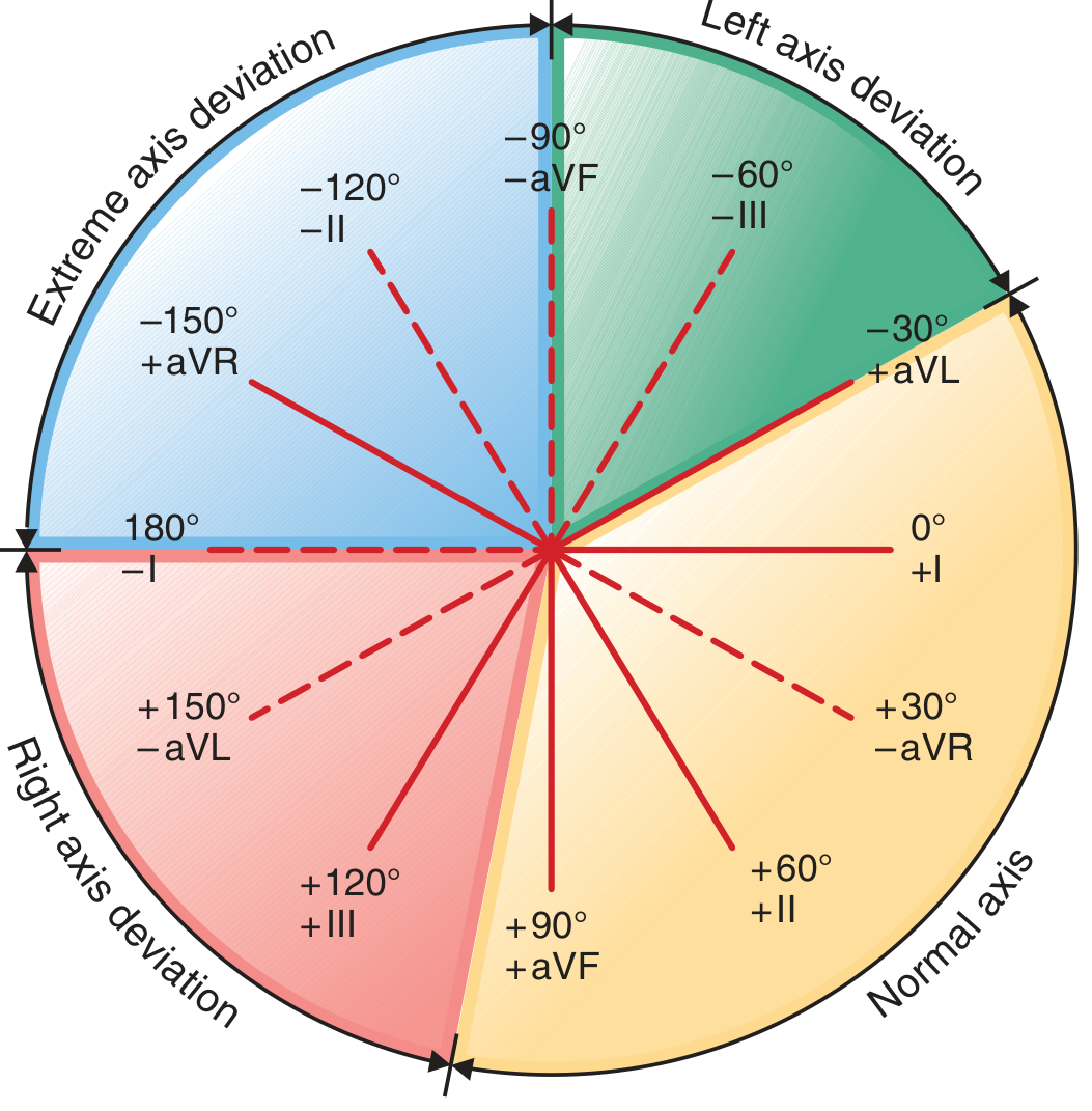
| Axis | Degrees | Leads |
|---|---|---|
| Normal | -30° to +90° | QRS upright in I and aVF |
| Left axis deviation (LAD) | -30° to -90° | QRS upright in I, negative in aVF |
| Right axis deviation (RAD) | +90° to +180° | QRS negative in I, positive in aVF |
| Extreme ("Northwest") axis | -90° to +/-180° | QRS negative in both I and aVF |
- Both upright → Normal
- I up, aVF down → Left axis
- I down, aVF up → Right axis
- Both down → Extreme axis
MODULE 7 - Chamber Enlargement / Hypertrophy
Left Ventricular Hypertrophy (LVH)
- S in V1 + R in V5 or V6 ≥ 35 mm
- R in aVL ≥ 11 mm (Cornell criteria)
- Associated with: LV strain pattern (ST depression + T wave inversion in lateral leads I, aVL, V5, V6)
Right Ventricular Hypertrophy (RVH)
- R ≥ S in V1 (dominant R in V1)
- RAD (axis >+90°)
- rSR' in V1 is NOT RVH - that is RBBB
- ST depression + T wave inversion in V1-V3 (RV strain)
MODULE 8 - Bundle Branch Blocks
Right Bundle Branch Block (RBBB)
- QRS ≥ 120 ms
- RSR' ("M-shaped" or "rabbit ears") in V1
- Wide, slurred S wave in I, V5, V6
- T wave inverted in V1 (secondary change - normal with RBBB)
RBBB = bunny ears in V1 (rSR' = ears of a rabbit)
Left Bundle Branch Block (LBBB)
- QRS ≥ 120 ms
- Broad, notched R (no Q, no S) in I, aVL, V5, V6
- rS or QS (deep S) in V1, V2
- T wave opposite to main QRS deflection (secondary change)
- LBBB: W in V1, M in V6
- RBBB: M in V1, W in V6
Fascicular Blocks (Hemiblocks)
| Block | Axis | QRS duration | Pattern |
|---|---|---|---|
| Left Anterior Fascicular Block (LAFB) | LAD > -45° | Normal or slightly prolonged | Small Q in I, aVL; small R in II, III, aVF |
| Left Posterior Fascicular Block (LPFB) | RAD > +120° | Normal | Small R in I, aVL; small Q in II, III, aVF; must exclude other RAD causes |
MODULE 9 - AV Blocks
First Degree AV Block
- PR interval > 200 ms (>1 large square)
- Every P conducts to QRS (just slowly)
- Usually benign; can be normal in athletes
Second Degree AV Block
- Progressive PR lengthening until a P wave is blocked (QRS dropped)
- "Longer, longer, longer, DROP - then you have a Wenckebach"
- PR after the dropped beat is shortest
- Usually at AV node level; generally benign
- Can be normal in athletes (vagal tone)
- Fixed PR interval; sudden non-conducted P wave (QRS drops without warning)
- Below AV node (His-Purkinje level) - more serious
- Often associated with wide QRS
- High risk of progressing to complete heart block → needs pacemaker
Third Degree (Complete) AV Block
- Complete dissociation: P waves and QRS complexes march independently
- P rate > QRS rate
- QRS escape rhythm: narrow (junctional, 40-60/min) or wide (ventricular, 20-40/min)
- Wide-complex escape = more severe (infranodal block)
- Emergency: Atropine (for narrow-complex), transcutaneous pacing, then permanent pacemaker
Wenckebach = Increasing PR before drop (I = Increasing) Mobitz II = Suddenly drops (II = abrupt, Sudden)
MODULE 10 - Ischemia and Infarction
Spectrum of Ischemic ECG Changes
- Tall, broad, peaked T waves over ischemic zone
- First sign of complete coronary occlusion
- ST elevation in ≥2 contiguous leads
- Threshold: ≥1 mm (most leads), ≥2 mm (V1-V3 men), ≥1.5 mm (V1-V3 women)
- Represents full-thickness (transmural) injury
- Mechanism: epicardial injury current shifts ST vector outward
- Horizontal or downsloping ST depression ≥0.5-1 mm
- Mechanism: ischemia confined to subendocardium; ST vector shifts inward
- Symmetric, deep T wave inversions
- Wellens' syndrome: deep T inversions in V2-V3 = critical proximal LAD stenosis (do NOT stress test - risk of VF)
- Width ≥ 40 ms (1 small square)
- Depth ≥ 25% of R wave height in same lead
- Represent electrically dead (necrotic) myocardium
- Q waves in II, III, aVF = inferior MI; V1-V4 = anterior MI; I, aVL = lateral MI
Localization of STEMI
| Location | ST Elevation In | Reciprocal ST Depression In | Culprit Artery |
|---|---|---|---|
| Inferior | II, III, aVF | I, aVL | RCA (80%), LCx (20%) |
| Anterior | V1-V4 | II, III, aVF (sometimes) | LAD |
| Anteroseptal | V1-V3 | - | LAD proximal |
| Apical/Extensive | V1-V6 | - | LAD proximal (widow maker) |
| Lateral | I, aVL, V5-V6 | V1-V2 | LCx |
| Posterior | Tall R + ST depression V1-V2 | ST elevation in V7-V9 | RCA or LCx |
| Right Ventricular | V1 + V4R elevation | - | Proximal RCA |
Evolution of STEMI Over Time
Minutes: Hyperacute T waves (peaked, tall)
Hours: ST elevation (convex/"tombstone" shape)
6-24h: ST elevation + Q waves appear + T inversion begins
Days: ST returns to baseline + T waves inverted + Q waves persist
Weeks-mos: ST and T normalize; Q waves often persist permanently
MODULE 11 - Arrhythmias
Approach to Any Arrhythmia (systematic)
- Rate: Fast / slow / normal?
- Regular or irregular?
- QRS wide or narrow?
- P waves present? What is the relationship to QRS?
Narrow Complex Tachycardias (QRS < 120 ms)
| Arrhythmia | Rate | Rhythm | P waves | Key feature |
|---|---|---|---|---|
| Sinus tachycardia | 100-160 | Regular | Present, normal | Gradual onset/offset |
| Atrial flutter | 250-350 atrial; 150 ventricular (2:1) | Regular | Sawtooth in II, III, aVF | 150 bpm = think flutter |
| Atrial fibrillation | 100-160 ventricular | Irregularly irregular | No discrete P waves (fibrillatory baseline) | Most common sustained arrhythmia |
| AVNRT | 140-280 | Regular | P buried in QRS or just after | Pseudo-R' in V1, pseudo-S in II |
| AVRT (WPW) | 150-250 | Regular | Retrograde P after QRS | Delta wave in sinus rhythm |
| Atrial tachycardia | 150-250 | Regular | Abnormal P before QRS | Different P morphology |
| Multifocal atrial tachycardia | Variable >100 | Irregular | ≥3 different P wave morphologies | COPD patients |
Wide Complex Tachycardias (QRS ≥ 120 ms) - THE HIGH-STAKES AREA
- VT (ventricular tachycardia) - most dangerous, assume this first
- SVT with aberrant conduction (LBBB/RBBB)
- SVT with pre-excitation (WPW with AF = most dangerous SVT)
- Pacemaker-mediated tachycardia
If in doubt, treat wide complex tachycardia as VT until proven otherwise Never give adenosine or verapamil to pre-excited AF (WPW+AF) - can precipitate VF
- AV dissociation (P waves marching independently) = definitive VT
- Capture beats (narrow QRS among wide) = definitive VT
- Fusion beats = definitive VT
- QRS duration > 160 ms
- Concordance (all precordial leads all-positive or all-negative)
- NW (extreme) axis deviation
Bradyarrhythmias
| Arrhythmia | ECG Feature | Management |
|---|---|---|
| Sinus bradycardia | Regular rhythm <60, P before each QRS | Treat if symptomatic: atropine, pacing |
| Sick Sinus Syndrome | SA dysfunction: Brady-tachy, sinus pauses, sinus arrest | PPM if symptomatic |
| Junctional escape | Narrow QRS, rate 40-60, P absent/inverted/retrograde | Treat underlying cause |
| Ventricular escape | Wide QRS, rate 20-40 | Emergency pacing |
Ventricular Arrhythmias
- Wide (>120 ms), bizarre QRS
- No preceding P wave
- Compensatory pause
- Bigeminy (every other beat), trigeminy (every third)
- ≥3 consecutive wide QRS complexes at ≥100 bpm
- Sustained (>30 s) vs non-sustained (<30 s)
- Monomorphic (uniform QRS) vs polymorphic (varying QRS)
- Polymorphic VT with twisting QRS axis around baseline
- Occurs with long QT (congenital or acquired)
- Triggers: hypokalemia, hypomagnesemia, drugs (class IA/III antiarrhythmics, antipsychotics, antibiotics like azithromycin)
- Treatment: IV magnesium sulfate; correct electrolytes; stop offending drug
- Chaotic, irregular deflections, no organized QRS
- No cardiac output = cardiac arrest
- Immediate defibrillation + CPR
MODULE 12 - Pre-excitation (WPW Syndrome)
- Short PR interval (<120 ms) - bypass of AV node
- Delta wave - slurred upstroke of QRS
- Wide QRS (>110 ms) - fusion of normal and accessory pathway conduction
- ST-T changes secondary to abnormal depolarization
- Positive delta in V1-V3, negative in aVL = left-sided pathway (most common)
- Negative delta in II, III, aVF = posterior/inferior pathway
MODULE 13 - Metabolic and Drug Effects
Hyperkalemia (sequential changes as K+ rises)
| K+ level (approx) | ECG change |
|---|---|
| 5.5-6.5 mEq/L | Peaked ("tented"), narrow T waves |
| 6.5-7.5 mEq/L | Prolonged PR, flattened P waves |
| 7.5-8.0 mEq/L | Wide QRS |
| >8.0 mEq/L | Sine-wave pattern → asystole |
Hypokalemia
- Flattened T waves
- Prominent U waves (U > T wave height = significant)
- Apparent QT prolongation (actually QU prolongation)
- Risk of Torsades
Hypercalcemia
- Shortened QT interval
- Short ST segment or absent ST
Hypocalcemia
- Prolonged QT (prolonged ST segment)
- T wave remains normal
Digoxin
- "Reverse tick" or "Salvador Dali mustache" ST depression (scooping)
- Shortened QT
- T wave flattening or inversion
- Digoxin toxicity: PAT with block, ventricular bigeminy, PVCs, AV blocks
Hypothermia
- Bradycardia
- Osborn (J) wave: positive deflection at J point (most visible in V5, V6)
- Prolonged PR, QRS, QT
- Shivering artifact
MODULE 14 - Pericarditis vs. STEMI
| Feature | Acute Pericarditis | STEMI |
|---|---|---|
| ST elevation | Diffuse (all/most leads) | Regional (contiguous leads) |
| ST morphology | Concave (saddle-shaped) | Convex (domed/tombstone) |
| PR depression | Present (classic) | Absent |
| Reciprocal ST depression | Absent (except aVR) | Present in opposite leads |
| Q waves | No | Develop over hours-days |
| Chest pain position | Pleuritic, worse lying, better leaning forward | Central, radiation to arm/jaw |
MODULE 15 - Pulmonary Embolism ECG
- Deep S wave in Lead I
- Q wave in Lead III
- T wave inversion in Lead III
- Sinus tachycardia (most common sign)
- New RBBB (acute right heart strain)
- RAD
- T wave inversions V1-V4 (RV strain pattern)
- Atrial arrhythmias (AF, flutter)
- P pulmonale
MODULE 16 - The Brugada Pattern
- Coved (convex) ST elevation ≥2 mm in V1-V2
- Down-sloping ST segment followed by T wave inversion
- Spontaneous or drug-induced
MODULE 17 - The 14-Step Systematic ECG Approach (Exam and Clinical)
| Step | What to Assess | What to Look For |
|---|---|---|
| 1 | Calibration & technical | 1 mV = 10 mm, 25 mm/s, correct lead placement |
| 2 | Rhythm | Sinus? Regular? P before every QRS? |
| 3 | Heart rate | 300 rule for regular; count in 10 s for irregular |
| 4 | PR interval | 120-200 ms; short = WPW/junctional; long = AV block |
| 5 | QRS duration | <110 ms; wide = BBB, aberrancy, pacing, hyperK |
| 6 | QT/QTc interval | <450 ms men / <460 ms women |
| 7 | Mean QRS axis | I and aVF method; -30° to +90° normal |
| 8 | P waves | Morphology, size, axis - atrial hypertrophy? |
| 9 | QRS voltages | LVH? Low voltage (pericardial effusion, obesity, COPD)? |
| 10 | R-wave progression | V1 (small r) → V5 (tall R). Poor progression = anterior MI |
| 11 | Q waves | Pathological? Location? Old vs new? |
| 12 | ST segments | Elevation? Depression? Which leads? Shape? |
| 13 | T waves | Tall? Inverted? Symmetric? Asymmetric? |
| 14 | U waves | Prominent (hypokalemia, bradycardia)? |
MODULE 18 - High-Yield Exam Mnemonics
| Mnemonic | What it's for |
|---|---|
| WILLIAM MARROW | BBB: W in V1 = LBBB; M in V1 = RBBB |
| 300-150-100-75-60-50 | Heart rate from R-R large squares |
| I See aVF (Both up = Normal axis) | Quick axis check |
| Longer, longer, longer, DROP | Wenckebach (Mobitz I) |
| DEAD | VT clues: Dissociation, Extreme axis, Absence of P-QRS relationship, Duration >160 ms |
| ABCDE for PE | Arrhythmia, BBB (RBBB), Cor pulmonale (P pulmonale), Deviation (RAD), S1Q3T3 |
| Tented T = hyperK; Flat T + prominent U = hypoK | Electrolytes |
| Concave ST = pericarditis; Convex ST = MI | Distinguishing causes of ST elevation |
| PR depression + ST elevation = pericarditis | Classic pericarditis ECG clue |
MODULE 19 - Quick Clinical Decision Rules
- ST elevation ≥1 mm in ≥2 contiguous leads → Activate cath lab (STEMI protocol) - door to balloon <90 min
- New LBBB + chest pain → Same as STEMI (Sgarbossa criteria)
- ST depression V1-V4 + inferior STEMI → Posterior MI; do V7-V9
- Inferior STEMI + hypotension + clear lungs → Rule out RV MI with V4R
Suggested Study Order for Exams
- Paper calibration + waveform basics (Module 3, 4)
- Leads and normal ECG (Module 5, 6)
- Ischemia/STEMI localization (Module 10) - highest exam yield
- Bundle branch blocks (Module 8)
- AV blocks (Module 9)
- Arrhythmias systematic approach (Module 11)
- Metabolic effects (Module 13)
- Practice 14-step approach on real ECGs (Module 17)
Give me all ecgs for stepwise learning with explanation
ECG Visual Atlas: Stepwise Learning with Real ECGs
LEVEL 1 - The Foundation
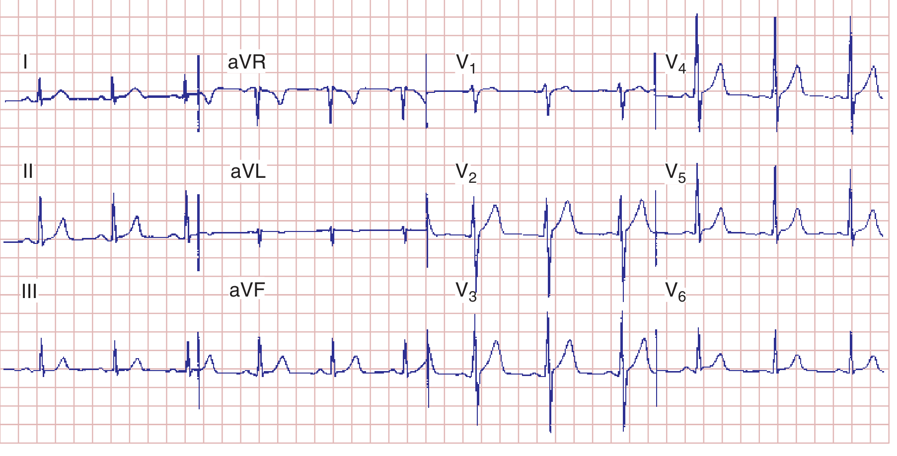
ECG 1: Normal Sinus Rhythm - Your Baseline
| Feature | This ECG | Normal? |
|---|---|---|
| Rate | 75 bpm | ✅ 60-100 |
| Rhythm | Regular, P before every QRS | ✅ Sinus |
| PR interval | 160 ms (4 small squares) | ✅ 120-200 ms |
| QRS duration | 80 ms (2 small squares) | ✅ <110 ms |
| QT interval | 360 ms, QTc ~390 ms | ✅ <450 ms |
| Axis | ~+70° (upright in I and aVF) | ✅ Normal |
| R-wave progression | Small r in V1 → tall R in V5 | ✅ Normal |
ECG 2: Ventricular Depolarization - How the QRS is Born
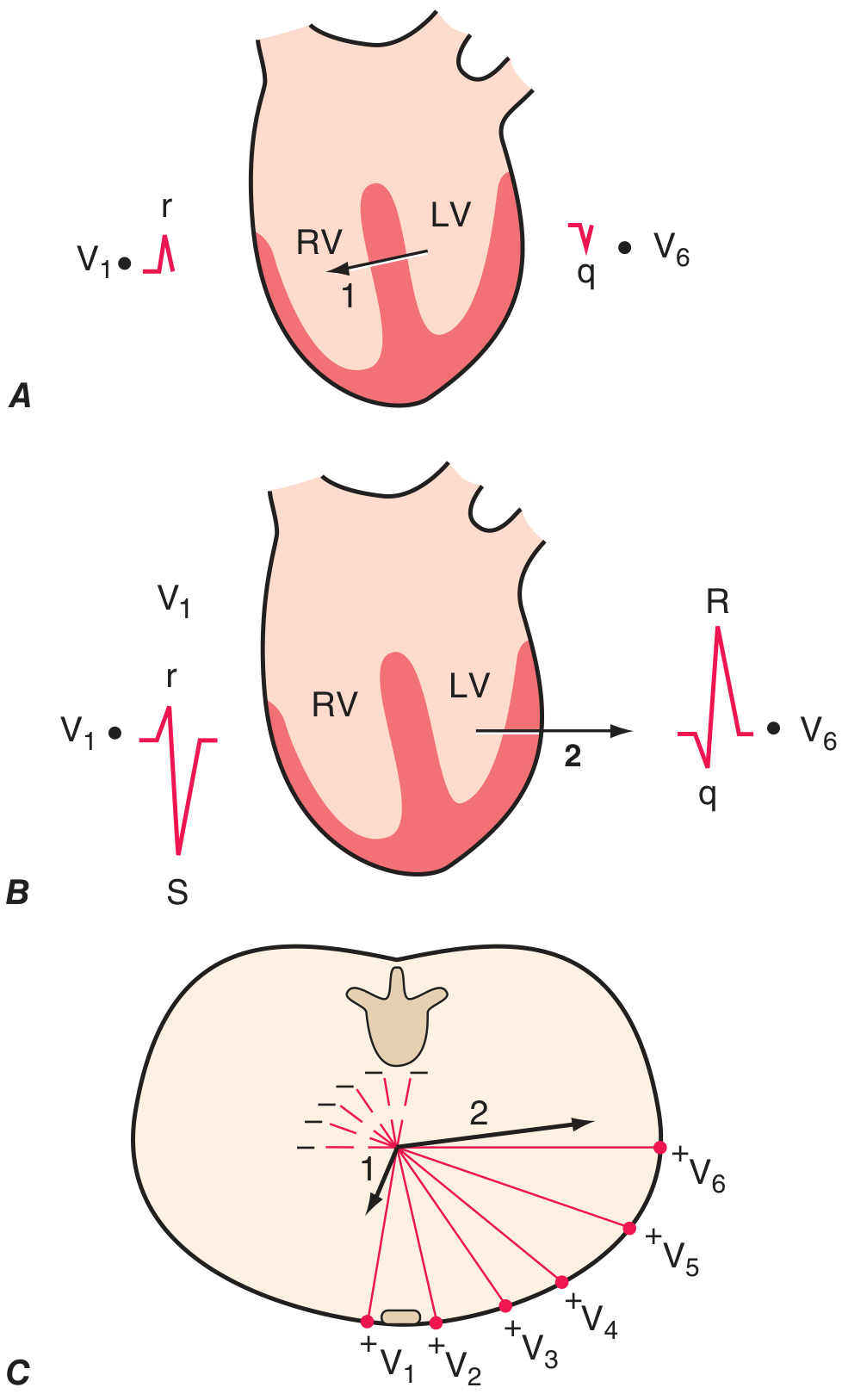
- Phase 1 (septum): Depolarizes left → right. V1 sees it coming → small upward r wave. V6 sees it going away → small downward q wave.
- Phase 2 (ventricles): LV dominates, vector swings left → right. V1 sees it going away → deep S wave. V6 sees it coming → tall R wave.
- V1 = rS (small r, big S)
- V6 = qR (tiny q, big R)
- V1 → V5 = R wave progressively gets bigger (R-wave progression)
LEVEL 2 - Atrial and Ventricular Enlargement
ECG 3: P Wave Morphology - Atrial Abnormalities
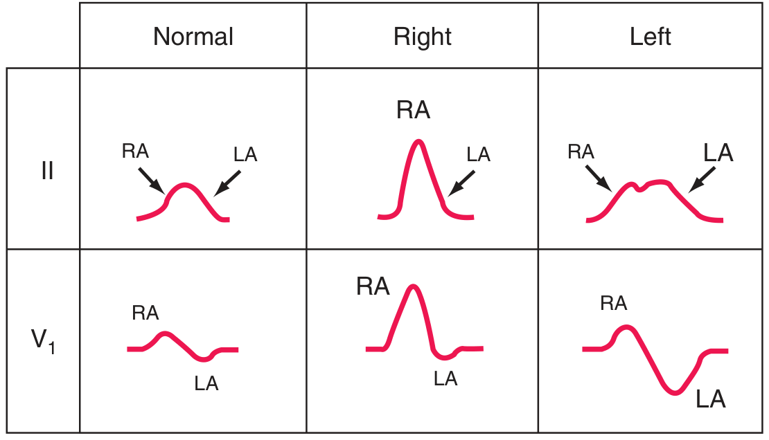
| Lead II | Lead V1 | |
|---|---|---|
| Normal | Small, smooth P (RA then LA humps) | Small biphasic P |
| Right atrial enlargement | Tall (>2.5 mm), peaked P ("P pulmonale") | Large initial positive deflection |
| Left atrial enlargement | Broad (>120 ms), notched P ("P mitrale") | Large terminal negative component >1 mm² |
- RAE (P pulmonale): COPD, pulmonary hypertension, tricuspid stenosis, PE
- LAE (P mitrale): Mitral stenosis, mitral regurgitation, LVH, dilated cardiomyopathy
ECG 4: LVH and RVH Patterns
- S in V1 + R in V5 or V6 ≥ 35 mm
- R in aVL ≥ 11 mm (women); ≥ 28 mm (men) - Cornell criteria
- LV strain pattern: ST depression + asymmetric T inversion in I, aVL, V5, V6
- Dominant R wave in V1 (R ≥ S in V1)
- Right axis deviation (axis > +90°)
- RV strain pattern: T inversion in V1-V3
LEVEL 3 - Ischemia and Infarction (Highest Exam Yield)
ECG 5: The Mechanism of ST Deviation
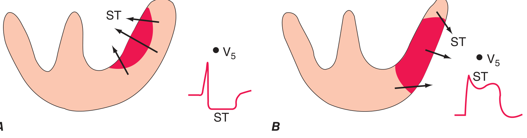
- Panel A (Subendocardial ischemia): Injury current vector points inward → leads overlying the LV see ST depression. This is NSTEMI/unstable angina territory.
- Panel B (Transmural/epicardial ischemia): Injury current vector points outward → leads overlying the zone see ST elevation. This is STEMI territory.
ECG 6: Anterior STEMI (LAD occlusion)
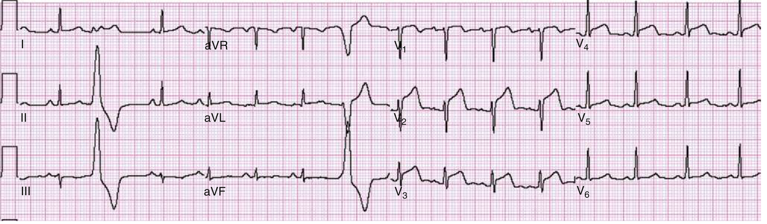
- ST elevation in V1-V4 (anteroseptal leads) - convex/oblique ST morphology
- Note the tall R waves progressing V1-V4, then cut short by the ST change
- This patient had a 90% LAD stenosis - went to cath lab immediately
- Rate: ~70 bpm, sinus
- ST elevation: V1, V2, V3, V4 - ≥2 mm in V1-V3 = diagnostic STEMI
- Reciprocal changes: Look for ST depression in II, III, aVF (inferior leads)
- Q waves: May be developing - marker of established necrosis
- ACTION: Activate cath lab. Door-to-balloon time target: <90 min
ECG 7: Anterolateral STEMI (LAD + circumflex territory)
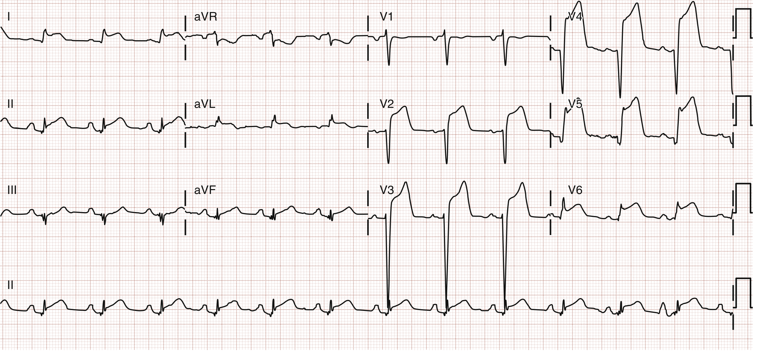
- ST elevation spreading across V2-V6, I, and aVL = extensive anterolateral territory
- This is a large territory MI - "widowmaker" territory (proximal LAD)
- Patient had in-stent thrombosis of previous LAD stent
| Feature | Anterior STEMI (ECG 6) | Anterolateral STEMI (ECG 7) |
|---|---|---|
| ST elevation | V1-V4 only | V2-V6 + I + aVL |
| Territory | Anteroseptal | Extensive anterior + lateral |
| Culprit | Mid-LAD | Proximal LAD or wrapping LAD |
| Severity | Significant | Very large - high risk |
ECG 8: Wellens Syndrome (Critical LAD Stenosis Warning)
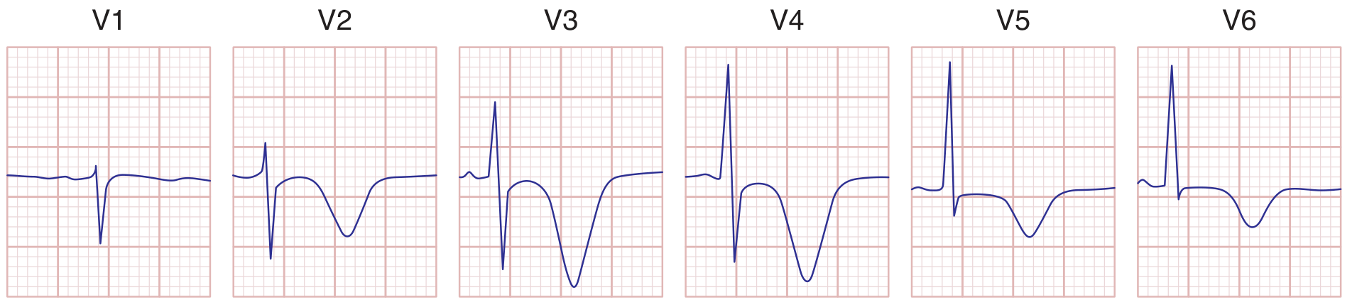
- Deep, symmetric T-wave inversions in V2-V4 (precordial leads)
- Note: no current ST elevation, R waves preserved (no Q waves yet)
- Patient had chest pain that may have resolved
⚠️ DO NOT do a stress test on this patient - can precipitate VF and death. Refer urgently for coronary angiography.
ECG 9: Left Main / Proximal LAD Occlusion - aVR Elevation
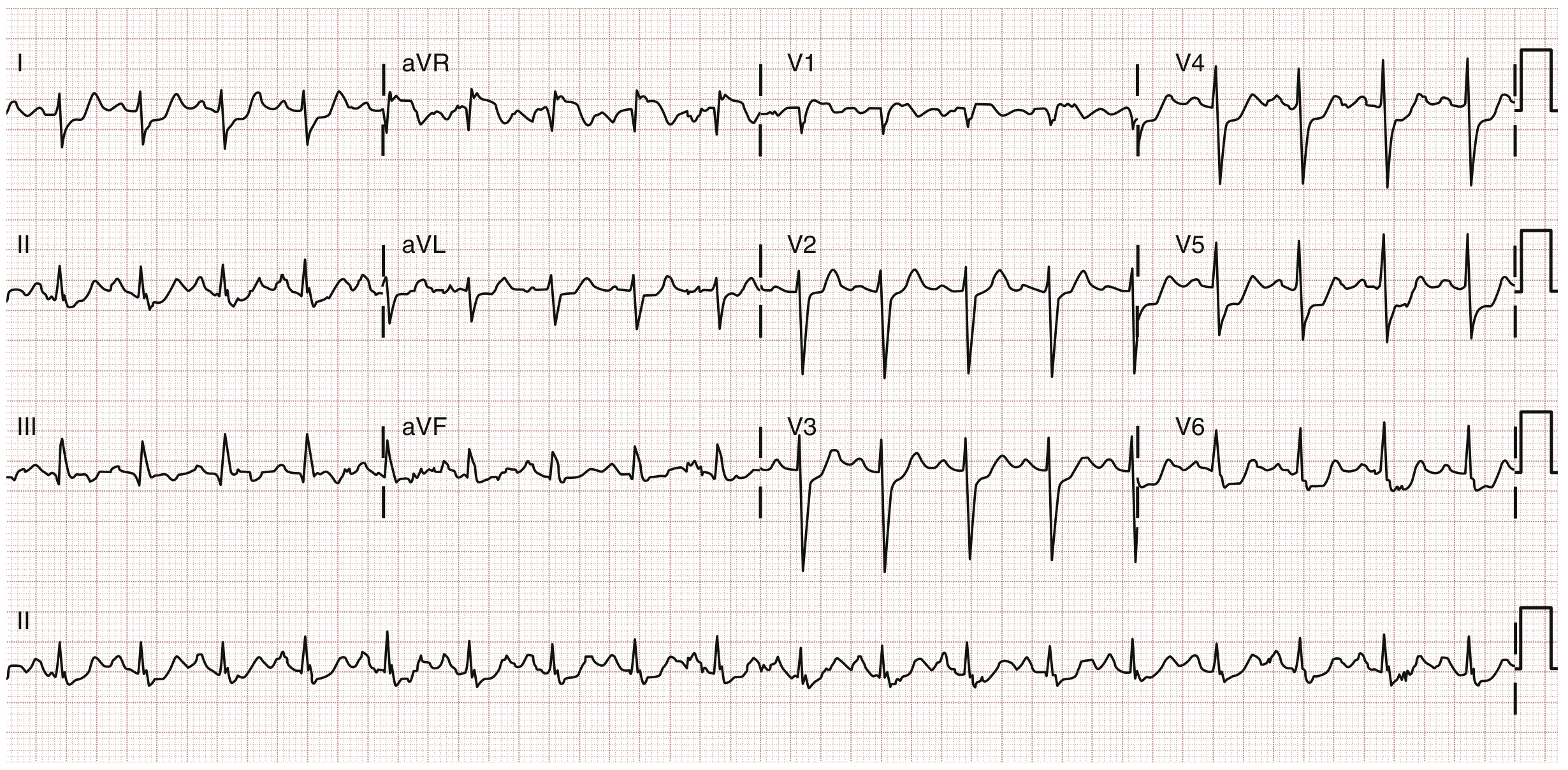
- ST elevation in aVR > 1 mV (the lead pointing at the right shoulder / LV cavity)
- Widespread ST depression in most other leads (I, II, aVL, V4-V6 at minimum)
- This pattern = diffuse subendocardial ischemia from left main occlusion
ST elevation in aVR + widespread ST depression elsewhere = Left main stenosis (78% sensitive, 83% specific) or severe proximal LAD disease
LEVEL 4 - Bundle Branch Blocks
ECG 10: RBBB vs LBBB - The WiLLiaM MaRRow Rule
| V1 | V6 | |
|---|---|---|
| RBBB | M-shaped (rSR') = rabbit ears | W-shaped (wide S) |
| LBBB | W-shaped (QS or rS) | M-shaped (broad notched R) |
- WiLLiam = LBBB has W in V1, M in V6
- MaRRow = RBBB has M in V1, W in V6
- QRS ≥ 120 ms
- rSR' ("rabbit ears") in V1
- Wide slurred S in I, aVL, V5, V6
- T wave inverted in V1-V2 (secondary - normal with RBBB, not ischemia)
- QRS ≥ 120 ms
- Broad monophasic R in I, aVL, V5, V6 (no Q wave, no S wave)
- Deep QS or rS in V1
- T wave inverted in I, aVL, V5, V6 (secondary - normal with LBBB)
- RBBB: Often benign; new RBBB in acute anterior MI = proximal LAD (septal branch involvement)
- LBBB: New LBBB + chest pain = treat as STEMI (modified Sgarbossa criteria)
| Finding | Points |
|---|---|
| ST elevation ≥ 1 mm concordant with QRS (same direction) | 5 |
| ST depression ≥ 1 mm in V1-V3 | 3 |
| ST elevation ≥ 5 mm discordant with QRS | 2 |
LEVEL 5 - AV Blocks (Visual Patterns)
ECG 11-13: The Three AV Blocks
P----P----P----P (every P conducts)
QRS QRS QRS QRS (every P has a QRS)
↕PR: >200ms (stretched but constant)
P P P P P P P
QRS QRS QRS --- QRS QRS ---
PR: ↑ ↑↑ ↑↑↑ drop ↑ ↑↑ drop
- Benign, usually AV node level
- Common in athletes (high vagal tone) and inferior MI
P P P P P P
QRS QRS --- QRS QRS ---
PR: = = = = (CONSTANT then sudden drop)
- Dangerous - below the AV node (His-Purkinje)
- Associated with wide QRS, anterior MI
- Requires pacemaker - high risk of complete block
P P P P P P P P (fast, ~70-80/min)
QRS QRS QRS QRS (slow, 30-50/min)
- Narrow QRS escape = junctional (40-60/min) = AV node still working below block
- Wide QRS escape = ventricular (20-40/min) = dangerous, more hemodynamically unstable
- Emergency: atropine (narrow complex), transcutaneous pacing, urgent permanent pacemaker
LEVEL 6 - Metabolic ECGs (Must Know for Exams)
ECG 14: Hyperkalemia - The Deadly Progression
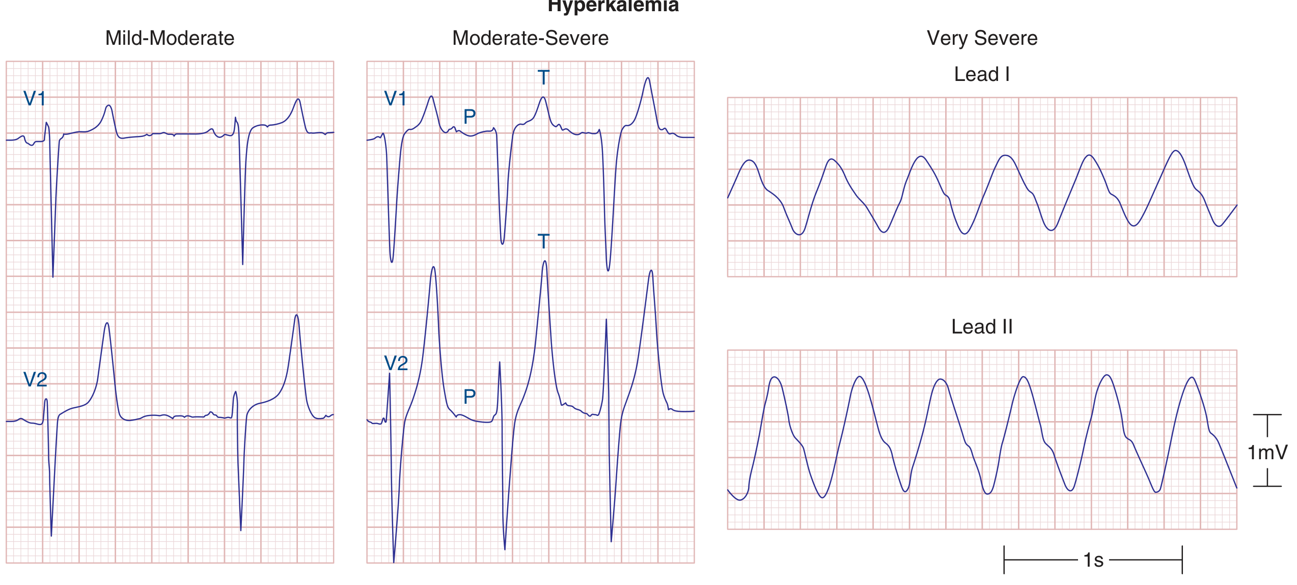
| Panel | K+ Level | ECG Changes |
|---|---|---|
| Mild-Moderate (V1, V2 left panel) | 5.5-6.5 mEq/L | Tall, narrow, peaked ("tented") T waves - classic first sign |
| Moderate-Severe (V1, V2 middle panel) | 6.5-8 mEq/L | Wide QRS, flat/absent P waves, peaked T waves still visible |
| Very Severe (Lead I, II right panel) | >8 mEq/L | Sine wave pattern - imminent asystole |
- Peaked T waves = call for ECG immediately in any CKD/renal patient
- Sine wave = life-threatening emergency - give calcium gluconate NOW
- Treatment sequence: C-BIG-K-D (Calcium, Bicarbonate, Insulin+Glucose, Kayexalate, Dialysis)
ECG 15: Hypokalemia - The U Wave
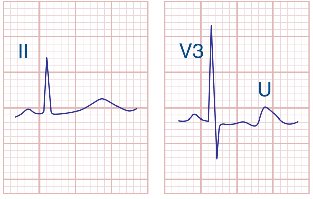
- Lead II (left): Flat T wave, barely visible, slight prolongation of the QT interval
- V3 (right): Prominent U wave labeled - note it is taller than the T wave in this lead
- T wave flattening → T wave inversion
- U wave prominence (U > T = significant hypokalemia)
- Apparent QT prolongation (actually QU prolongation - U wave merging with T)
- Risk: Torsades de Pointes especially if on QT-prolonging drugs
ECG 16: Pericarditis with Cardiac Tamponade - Electrical Alternans
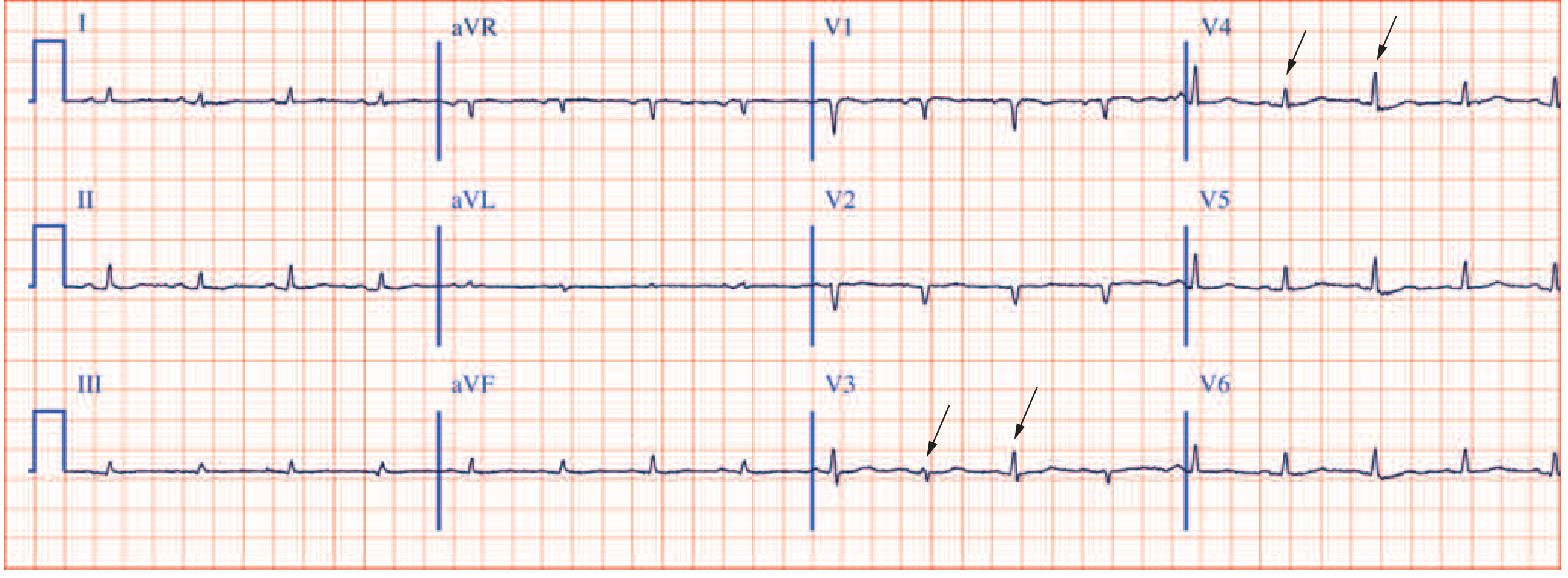
- Sinus tachycardia (compensatory, filling impaired)
- Low QRS voltage (<5 mm all limb leads and/or <10 mm chest leads) - fluid insulating the heart
- Electrical alternans - QRS height alternates every other beat (arrows in V2, V4) - heart swinging inside fluid
- Diffuse saddle-shaped (concave) ST elevation in most leads (not just regional)
- PR segment depression (very specific for pericarditis)
- No reciprocal ST depression (except aVR)
- No Q waves
| Feature | Pericarditis | STEMI |
|---|---|---|
| ST shape | Concave (saddle) | Convex (dome/tombstone) |
| Distribution | Diffuse (all leads) | Regional (2+ contiguous) |
| PR depression | Present | Absent |
| Reciprocal ST depression | Absent | Present |
| Q waves | Never | Develop hours-days |
LEVEL 7 - Clinical Syndromes (Advanced)
ECG 17: Inferior STEMI - RCA Occlusion
- ST elevation in II, III, aVF (inferior leads)
- Reciprocal ST depression in I and aVL (lateral leads)
- III > II elevation = more likely RCA (proximal)
- II > III elevation = more likely LCx
- Right-sided leads (V3R, V4R): If ST elevation here = RV infarction (25-40% of inferior STEMIs)
- RV MI = give fluids (not nitrates!), no diuretics, no morphine
- Posterior leads (V7-V9) or V1-V2: Tall R + ST depression in V1-V2 = posterior extension
- Complete AV block: RCA supplies AV node - inferior MI can cause bradycardia / heart block
ECG 18: WPW (Wolff-Parkinson-White) Pattern
- Short PR (<120 ms) - bypassing the AV node
- Delta wave - slurred initial upstroke of QRS (most visible in lateral leads)
- Wide QRS (>120 ms) - fusion beat (normal + accessory pathway conduction)
- Pseudo-Q waves and ST-T changes - secondary to abnormal depolarization
| Delta wave | Lead | Pathway location |
|---|---|---|
| Positive | V1-V3 | Left-sided (most common, type A) |
| Negative | V1 | Right-sided (type B) |
| Negative | II, III, aVF | Posterior/inferior |
- Orthodromic AVRT: Narrow complex tachycardia (down AV node, up AP) - treat with adenosine
- Antidromic AVRT: Wide complex tachycardia (down AP, up AV node)
- AF with WPW: Irregular wide complex tachycardia - VERY FAST rate (>250 bpm possible) → risk of VF
⚠️ Never give adenosine, digoxin, verapamil, diltiazem in AF+WPW - these block AV node but not accessory pathway, driving all conduction down the fast AP → VF Use: DC cardioversion (if unstable) or IV procainamide/ibutilide
ECG 19: Pulmonary Embolism - S1Q3T3
- Sinus tachycardia - most common, most sensitive
- T-wave inversions V1-V4 - right heart strain pattern (right precordial leads)
- New RBBB or RBBB morphology - acute RV pressure overload
- S1Q3T3 - classic but actually uncommon (<20% of PE cases)
- Right axis deviation
- P pulmonale - peaked P waves from acute RA overload
- Atrial fibrillation or flutter
- S in lead I (deep S = left lateral lead sees delayed RV activation)
- Q in lead III (inferior lead)
- T inversion in lead III (RV strain)
ECG 20: Brugada Pattern
- Coved ST elevation ≥ 2 mm in V1 (and sometimes V2)
- Down-sloping ST segment
- T wave inversion in V1
- Risk of sudden cardiac death from VF (especially at night, during fever)
- Sodium channel mutation (SCN5A - autosomal dominant)
- Unmasked by: fever, cocaine, alcohol, flecainide, procainamide (sodium channel blockers)
- Treatment: ICD (implantable cardioverter defibrillator) for symptomatic patients
- Quinidine for recurrent VF episodes or fever-triggered events
LEVEL 8 - Arrhythmia Recognition (Rhythm Strips)
How to Systematically Approach Any Rhythm Strip
1. RATE → Fast? Slow? Normal?
2. REGULAR? → Measure R-R intervals
3. QRS WIDTH → Narrow (<120ms) or Wide (≥120ms)?
4. P WAVES → Present? Before QRS? Normal morphology?
Arrhythmia Quick Recognition Table
| Arrhythmia | Rate | Regular? | QRS | P waves | Distinguishing Feature |
|---|---|---|---|---|---|
| Sinus tach | 100-160 | Yes | Narrow | Normal, before QRS | Gradual onset, normal P morphology |
| AF | 100-160 ventricular | No (irregularly irregular) | Narrow | Absent (fibrillatory baseline) | Irregularly irregular, no P waves |
| AFL | 150 ventricular | Yes (usually 2:1) | Narrow | Sawtooth in II, III, aVF | Rate 150 = think flutter! |
| AVNRT | 150-250 | Yes | Narrow | Buried in QRS or pseudo-R' in V1 | Abrupt onset/offset, no visible P |
| AVRT (WPW) | 150-250 | Yes | Narrow (or wide) | Retrograde P after QRS | Delta wave in sinus rhythm |
| VT | >100 | Yes | Wide (≥120ms) | Dissociated (march independently) | AV dissociation = definitive VT |
| VF | Chaotic | No | No organized QRS | None | No output - cardiac arrest |
| Torsades | Variable | No | Wide, twisting | None visible | QRS alternates direction around baseline |
| 1° AVB | Any | Yes | Normal | Normal | PR >200ms, all P's conduct |
| Wenckebach | Normal-slow | Grouped beating | Normal | Some P's blocked | Progressive PR then dropped QRS |
| Mobitz II | Normal-slow | Mostly | Normal/Wide | Sudden blocked P | Fixed PR, sudden drop |
| 3° AVB | Slow ventricular | P regular, QRS regular but different rates | Wide or narrow | Dissociated from QRS | P rate > QRS rate, independent |
The Wide Complex Tachycardia Decision Tree
Wide Complex Tachycardia (QRS ≥120ms, Rate >100)
↓
Is patient UNSTABLE? (hypotensive, altered consciousness, pulmonary edema)
YES ──────────────────→ SYNCHRONIZED DC CARDIOVERSION
↓NO
Look for AV Dissociation?
YES ──────────────────→ DEFINITELY VT → Amiodarone or Cardioversion
↓NO
Capture beats or Fusion beats?
YES ──────────────────→ DEFINITELY VT → Amiodarone
↓NO
Brugada morphology criteria? QRS >160ms? Concordance?
YES ──────────────────→ LIKELY VT → Amiodarone
↓NO
Could this be AF + WPW? (irregular wide complex + very fast)
YES ──────────────────→ Procainamide or Cardioversion (NOT adenosine/verapamil)
↓NO
LIKELY SVT with aberrancy → Adenosine trial (have crash cart ready)
Rule: When uncertain, treat wide-complex tachycardia as VT. Adenosine is safe for narrow complex. For wide complex, amiodarone is the safest drug.
LEVEL 9 - Special ECG Patterns (Exam Favourites)
Quick Pattern Recognition Summary
| Pattern | Key ECG Finding | Diagnosis |
|---|---|---|
| Sawtooth waves in II, III, aVF at 300/min | Flutter waves | Atrial flutter |
| Irregularly irregular, no P waves | AF | Atrial fibrillation |
| Short PR + delta wave + wide QRS | Pre-excitation | WPW syndrome |
| Coved ST elevation V1-V2 | Type 1 Brugada | Brugada syndrome |
| Peaked T waves → wide QRS → sine wave | Hyperkalemia | Severe hyperkalemia |
| Prominent U waves, flat T | Hypokalemia | Hypokalemia |
| Diffuse concave ST + PR depression | Pericarditis | Acute pericarditis |
| Regional convex ST elevation | Ischemic injury | STEMI |
| Electrical alternans + low voltage + tachycardia | Tamponade | Cardiac tamponade |
| S1Q3T3 + sinus tachycardia + new RBBB | PE pattern | Pulmonary embolism |
| Deep T inversions V2-V4 + no ST elevation | Wellens | Critical LAD stenosis |
| ST elevation V1-V2 + aVR > ST in other leads | Left main / proximal LAD | Left main disease |
| J wave (Osborn wave) at J point | Hypothermia | Hypothermia |
| Scooping ST depression + short QT | Digoxin effect | Digoxin |
| Prolonged QT + polymorphic VT | Torsades | Long QT syndrome |
Clinical Practice Summary Card
Every ECG you pick up, run through this checklist in 30 seconds:
━━━━━━━━━━━━━━━━━━━━━━━━━━━━━━━━━━━━━━━━
1. RATE → Count (300 rule) / Irregular → count × 6
2. RHYTHM → P before QRS? Regular? Sinus?
3. PR → 120-200ms? Short (WPW)? Long (AVB)?
4. QRS → <110ms? Wide (BBB/VT/pace)?
5. AXIS → I and aVF upright = normal
6. ST SEGMENTS → Elevation (which leads)? Depression?
7. T WAVES → Inverted (where)? Peaked (hyperK)?
8. QT → Prolonged (drugs/electrolytes/long QT)?
9. P WAVES → Peaked (RAE)? Broad (LAE)?
10. U WAVES → Prominent (hypoK)?
11. COMPARE → With previous ECG!
━━━━━━━━━━━━━━━━━━━━━━━━━━━━━━━━━━━━━━━━
PRESS syndrome
posterior reversible encephalopathy syndrome PRES
PRES - Posterior Reversible Encephalopathy Syndrome
Definition
- Acute neurological symptoms (headache, seizures, visual disturbances, altered consciousness)
- Reversible vasogenic edema predominantly in the posterior cerebral regions
- An identifiable precipitating cause (hypertension, drugs, eclampsia, renal disease)
Pathophysiology - Two Competing Theories
Theory 1 - Autoregulatory Failure (Hyperperfusion)
Sudden severe ↑ BP
↓
Cerebral autoregulation overwhelmed
↓
Forced vasodilation (loss of protective vasoconstriction)
↓
↑ Cerebral blood flow → capillary leakage into interstitium
↓
VASOGENIC EDEMA
- Explains posterior predominance: posterior circulation (vertebrobasilar) has less sympathetic innervation than anterior (carotid), making it more vulnerable to autoregulatory breakthrough
- Explains why rate of rise matters more than absolute BP level (eclampsia can cause PRES at relatively lower pressures)
Theory 2 - Endothelial Dysfunction (Toxic/Autoimmune)
Calcineurin inhibitors, chemotherapy, autoimmune states
↓
Direct endothelial injury → Blood-brain barrier breakdown
↓
Vasogenic edema (without high BP)
- Explains PRES occurring at normal or mildly elevated BP (e.g., cyclosporine toxicity)
- Explains PRES in sepsis, TTP, autoimmune diseases
Harrison's: "Both of these processes likely play some role in each of these disorders." - Harrison's Principles of Internal Medicine 22E, Ch. 318
Causes / Precipitating Factors
1. Hypertensive Emergencies (Most Common)
- Malignant / accelerated hypertension (diastolic usually >125 mmHg)
- Hypertensive emergency from any cause: renal artery stenosis, acute glomerulonephritis, pheochromocytoma, Cushing's syndrome
2. Obstetric
- Eclampsia - most important single cause in young women
- HELLP syndrome (Hemolysis, Elevated Liver enzymes, Low Platelets)
- Pre-eclampsia with severe features
3. Immunosuppressive / Cytotoxic Drugs
| Drug Category | Examples |
|---|---|
| Calcineurin inhibitors | Cyclosporine, tacrolimus (most commonly implicated) |
| mTOR inhibitors | Sirolimus, everolimus |
| Chemotherapy | Cisplatin, bevacizumab, rituximab, methotrexate (IT or HD) |
| Biologic agents | Anti-VEGF agents, CAR-T therapy |
| Others | Erythropoietin (via BP + volume expansion) |
Important: "Many of the substances implicated can cause this syndrome even at low doses or after years of treatment. Therefore, normal serum levels do not exclude them as inciting agents." - Harrison's 22E
4. Renal Disease
- End-stage kidney disease (ESKD) on dialysis
- Acute glomerulonephritis
- Thrombotic microangiopathy (TMA), HUS, TTP
5. Autoimmune / Inflammatory
- Systemic lupus erythematosus
- Vasculitis (PAN, granulomatosis with polyangiitis)
- Scleroderma (hypertensive crisis)
6. Other
- Post-carotid endarterectomy hyperperfusion syndrome
- Sepsis / multi-organ dysfunction
- Cocaine, amphetamines, sympathomimetics
Clinical Features - The Four Cardinal Symptoms
| Symptom | Frequency | Notes |
|---|---|---|
| Headache | 50-80% | Diffuse, subacute, non-specific in character; ranges from mild to severe |
| Seizures | 60-90% | Often the presenting complaint; can be focal or generalized; non-convulsive seizures possible - low threshold for EEG |
| Visual disturbances | 20-40% | Cortical blindness, hemianopia, visual hallucinations, blurred vision - due to occipital lobe involvement |
| Altered consciousness | 25-50% | Confusion → stupor → coma in severe cases |
- Focal neurological deficits (hemiparesis, aphasia) - reflect atypical or extensive PRES
- Papilledema and retinal haemorrhages (especially in hypertensive PRES)
- Cerebellar signs if posterior fossa involved
- Balint syndrome (simultanagnosia, optic ataxia, gaze apraxia) - rare, severe occipital involvement
Adams & Victor's Neurology: "The neurologic syndrome is usually dominated by symptoms referable to the occipital and adjacent parietal region."
Critical point: "The rapidity of rise, rather than the absolute value of pressure, is the most important risk factor." - Harrison's 22E
Imaging - The Key to Diagnosis
MRI Brain (Gold Standard)
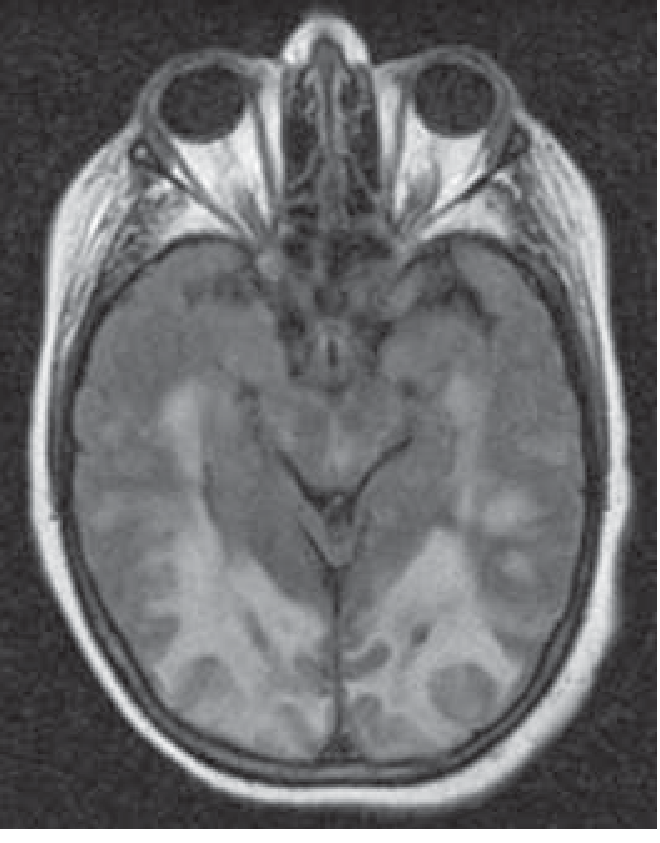
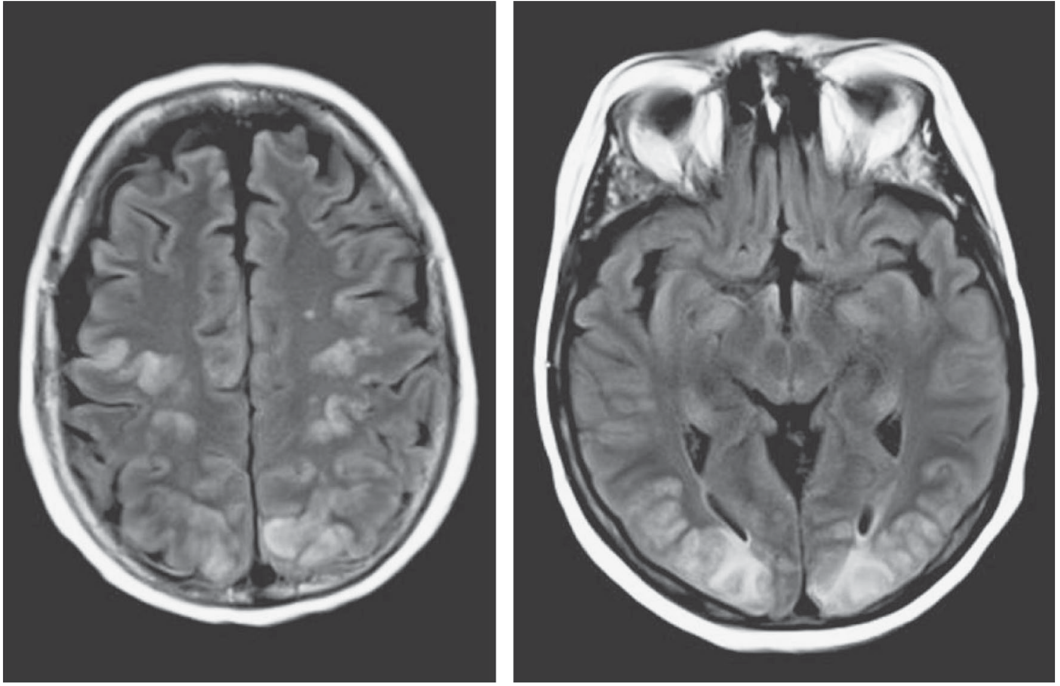
| Sequence | Finding |
|---|---|
| T2/FLAIR | Bilateral hyperintensity (bright signal) in posterior regions - the hallmark |
| DWI | Isointense or hypointense (increased diffusivity on ADC map) = vasogenic edema (distinguishes from cytotoxic edema of stroke) |
| ADC map | High ADC (bright) = free water movement = vasogenic edema |
| T1 post-contrast | Usually no enhancement; patchy enhancement if BBB severely disrupted |
| SWI/GRE | Microhaemorrhages in complicated cases |
Distribution of Edema
- Frontal lobes
- Temporal lobes
- Cerebellum and brainstem
- Deep structures (basal ganglia, thalami) - rare
- Unilateral (rare)
Grainger & Allison's Radiology: "MRI demonstrates bilateral cortico-subcortical T2 confluent hyperintensities, predominantly in the watershed zones of parietal and occipital lobes... DWI demonstrates iso- or hypointense areas within the lesions, reflecting increased diffusivity and, therefore, vasogenic oedema."
CT Brain
- Less sensitive than MRI
- May show patchy hypodensity in posterior regions
- Used when MRI unavailable; can support diagnosis but not exclude it
MRA/CTA
- May show segmental vasospasm or vessel caliber changes (overlap with RCVS)
- Not required for diagnosis but obtained in atypical cases
DWI Signal - The Critical Distinction
| PRES | Ischaemic Stroke | |
|---|---|---|
| DWI | Iso or hypointense (bright on ADC) | Bright (restricted diffusion) |
| ADC | High (bright) | Low (dark) |
| Edema type | Vasogenic | Cytotoxic |
| Reversibility | Yes (with treatment) | No (infarction permanent) |
Diagnosis
- Compatible clinical syndrome (seizures, headache, visual disturbance, encephalopathy)
- Characteristic MRI findings (posterior vasogenic edema)
- Identifiable precipitant
- Exclusion of alternative diagnoses
| Investigation | Purpose |
|---|---|
| MRI brain (FLAIR + DWI + ADC) | Confirm diagnosis, exclude stroke |
| BP measurement (bilateral arms) | Quantify hypertension |
| Urine dipstick + 24h protein | Screen for eclampsia/nephropathy |
| FBC, LFTs, LDH, blood film | Exclude TTP, HELLP |
| Drug levels (cyclosporine, tacrolimus) | Note: toxicity occurs even at normal levels |
| EEG | If seizures, to detect non-convulsive status epilepticus |
| Urine βhCG | Exclude pregnancy / eclampsia |
| Renal function panel | CKD, ESKD contribution |
| Urine toxicology | Cocaine, amphetamines |
Treatment
1. Treat the Underlying Cause (Most Important)
- Hypertensive emergency: Controlled BP reduction
- Eclampsia: IV magnesium sulfate + antihypertensives + delivery of fetus
- Drug-induced: Stop the offending agent (cyclosporine, tacrolimus → switch to alternative immunosuppressant; rituximab, bevacizumab → discontinue)
- ESKD/fluid overload: Dialysis with careful volume removal
2. Blood Pressure Management
Adams & Victor's: "A typical target is a pressure of 150/100 mmHg or a 20 percent reduction in mean pressure."
| Agent | Dose / Notes |
|---|---|
| Labetalol | IV boluses or infusion - first-line in most settings |
| Nicardipine | IV infusion - titratable, preferred in eclampsia |
| Hydralazine | IV - used in obstetric settings |
| Sodium nitroprusside | IV - potent, titratable but cyanide risk with prolonged use |
| Magnesium sulfate | Specifically for eclampsia - also anticonvulsant effect |
3. Seizure Management
- First-line: Benzodiazepines (lorazepam IV) for acute seizure control
- Maintenance: Levetiracetam (preferred - fewer interactions, no hepatic metabolism)
- Non-convulsive seizures: Screen with EEG; treat similarly
- Duration of anticonvulsants: Usually short-term - can discontinue once PRES resolved and no recurrent seizures (typically weeks, not indefinitely)
4. Monitoring
- Repeat MRI in 4-8 weeks to document radiological resolution
- BP monitoring long-term
- If drug-induced: discuss alternative immunosuppression with transplant/oncology team
Prognosis
- Majority resolve completely within days to weeks with prompt treatment (clinical and radiological)
- Irreversible neurological deficits in ~10-15% - usually from:
- Delayed diagnosis
- Progression to cytotoxic edema / infarction
- Intracranial haemorrhage (microhemorrhages or lobar bleeding)
- Mortality ~5-15% in severe cases, especially if associated with malignant hypertension, sepsis, or major organ failure
- Recurrence: Can occur if underlying cause not addressed
PRES vs Key Differentials
| Condition | Key Differentiating Feature |
|---|---|
| Ischaemic stroke | DWI restriction (bright); vascular territory distribution; no posterior predominance |
| Venous sinus thrombosis | DWI restriction; often haemorrhagic; MRV shows sinus occlusion |
| Encephalitis | CSF pleocytosis; fever; temporal lobe involvement (herpes); positive viral PCR |
| RCVS (reversible cerebral vasoconstriction) | Thunderclap headache; normal MRI or SAH; multifocal vasospasm on angiography; no parenchymal edema initially |
| Vasculitis | Multifocal infarcts; CSF inflammation; systemic features; vessel wall enhancement on MRI |
| CJD / prion disease | DWI in cortical ribboning or basal ganglia; not posterior predominant; CSF 14-3-3 |
| Hypertensive haemorrhage | Blood on CT/MRI; no edema pattern |
High-Yield Exam Summary
| Feature | Key Point |
|---|---|
| Full name | Posterior Reversible Encephalopathy Syndrome |
| Pathophysiology | Vasogenic edema from autoregulatory failure OR endothelial dysfunction |
| Classic causes | Hypertensive emergency, eclampsia, cyclosporine/tacrolimus, CKD |
| Triad | Headache + Seizures + Visual disturbance ± encephalopathy |
| MRI hallmark | Bilateral posterior T2/FLAIR hyperintensity NOT restricted on DWI |
| DWI pattern | Iso/hypointense (ADC high) = vasogenic edema - distinguishes from stroke |
| Most common precipitant | Hypertension; but BP may be normal in drug-induced PRES |
| Most diagnostic mistak | Ordering CT only (less sensitive); mistaking for posterior stroke |
| Treatment | Remove cause + controlled BP reduction (20% MAP reduction) + seizure management |
| Prognosis | Usually reversible; ~15% not fully reversible; mortality ~5-15% severe cases |
| Overlap syndrome | RCVS (may coexist with PRES in up to 10-15%) |
Recent Evidence
Renal tubular acidosis
renal tubular acidosis
Renal Tubular Acidosis (RTA) - Complete Guide
Overview and Framework
↓ HCO₃⁻ + ↑ Cl⁻ + Normal Anion Gap = RTA (or GI HCO₃⁻ loss)
Normal Renal Acid-Base Physiology (Foundation)
Proximal Tubule (reabsorbs ~85-90% of filtered HCO₃⁻)
Tubular lumen Proximal tubule cell Blood
HCO₃⁻ + H⁺ ──CA──→ H₂CO₃ → H₂O + CO₂ Na⁺ + HCO₃⁻ ──→ blood
↓ diffuses in
CO₂ + H₂O ──CA──→ H₂CO₃ → H⁺ + HCO₃⁻
↓ Na⁺/H⁺ exchanger ↓ NBC1 cotransporter
H⁺ secreted into lumen HCO₃⁻ exits to blood
- Key transporters: Apical Na⁺/H⁺ exchanger (NHE3), apical H⁺-ATPase; basolateral Na⁺/HCO₃⁻ cotransporter (NBC1 / SLC4A4)
- Net result: HCO₃⁻ reabsorption, NO net acid excretion (urine pH unchanged)
Distal Tubule / Collecting Duct (secretes net acid, reabsorbs remaining 5-10% HCO₃⁻)
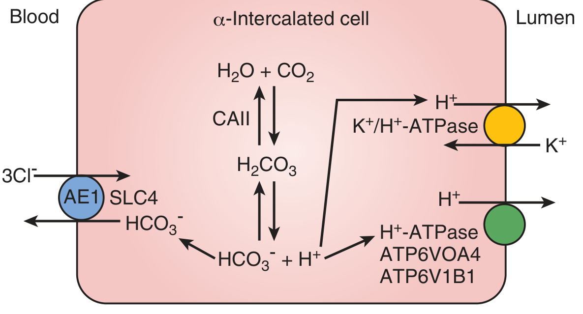
- Apical H⁺-ATPase (ATP6V0A4, ATP6V1B1 subunits) → secretes H⁺ into lumen
- Apical H⁺/K⁺-ATPase → secretes H⁺, reabsorbs K⁺
- Basolateral AE1 (SLC4A1) → exports HCO₃⁻ to blood in exchange for Cl⁻
- Intracellular carbonic anhydrase II (CA II) → generates H⁺ + HCO₃⁻
- Can reduce urine pH to as low as 4.5 (1000:1 H⁺ gradient)
- H⁺ is buffered in urine by titratable acids (phosphate) and ammonia → NH₄⁺
Classification of RTA
| Type | Name | Primary Defect | Serum K⁺ | Urine pH |
|---|---|---|---|---|
| Type 1 | Distal RTA | ↓ H⁺ secretion in collecting duct | ↓ Low (hypokalemia) | >5.5 (cannot acidify) |
| Type 2 | Proximal RTA | ↓ HCO₃⁻ reabsorption in proximal tubule | ↓ Low (hypokalemia) | Variable (<5.5 at low serum HCO₃⁻; >5.5 after alkali) |
| Type 3 | Mixed | Combined defect (CA II mutation) | Low | Variable |
| Type 4 | Hypoaldosteronism RTA | ↓ NH₄⁺ excretion, ↓ distal Na⁺ reabsorption | ↑ High (hyperkalemia) | <5.5 (can acidify) |
TYPE 1 - Distal RTA (Classical RTA)
Mechanism
- Secretory defect: Reduced H⁺-ATPase activity (most common) - can't pump enough H⁺
- Gradient defect: H⁺ leaks back from lumen to cell (amphotericin B - creates pores)
- Rate-dependent: Adequate pump but insufficient capacity at high acid loads
Causes
| Inheritance | Gene | Protein | Features |
|---|---|---|---|
| Autosomal recessive | ATP6V1B1 | H⁺-ATPase B1 subunit | + Sensorineural deafness |
| Autosomal recessive | ATP6V0A4 | H⁺-ATPase A4 subunit | Without deafness |
| Autosomal dominant | SLC4A1 | AE1 (Cl⁻/HCO₃⁻ exchanger) | Milder, often incomplete |
| Autosomal recessive | CA2 | Carbonic anhydrase II | Type 3 RTA + osteopetrosis + cerebral calcification |
- Autoimmune: Sjögren syndrome (most common cause in adults - can cause profound hypokalemia with weakness and respiratory arrest), SLE, rheumatoid arthritis, hypergammaglobulinaemia
- Drugs/toxins: Amphotericin B (gradient defect - creates membrane pores), lithium, ifosfamide, toluene (glue sniffing)
- Structural: Chronic obstructive uropathy, renal medullary sponge kidney, pyelonephritis
- Metabolic: Hypercalciuria, hyperparathyroidism, Wilson's disease, Fabry disease
- Other: Multiple myeloma, sickle cell disease
Clinical Features
- Hyperchloremic, normal AG metabolic acidosis
- Hypokalemia (often severe) - due to:
- Loss of electrogenic H⁺ secretion → enhanced K⁺ secretion to maintain electroneutrality
- Loss of H⁺/K⁺-ATPase reabsorptive activity
- Compensatory hyperaldosteronism (itself causes K⁺ wasting)
- Urine pH persistently >5.5 despite acidosis (cannot acidify below 5.5)
- Mechanism:
- Metabolic acidosis → bone demineralization → hypercalciuria (calcium release from bone buffer)
- Acidosis → impaired tubular citrate reabsorption → hypocitraturia (citrate is the most potent stone inhibitor)
- Alkaline urine (pH >5.5) promotes calcium phosphate precipitation (brushite, apatite stones)
- Hypocitraturia is the most critical stone-forming factor in Type 1 RTA
- Stone type: Calcium phosphate (not calcium oxalate)
- Bone disease (rickets in children, osteomalacia in adults) from chronic acidosis
- Growth retardation in children
- Polyuria, polydipsia (from hypercalciuria-induced NDI)
- Muscle weakness (hypokalemia)
- Respiratory paralysis (severe hypokalemia - notably in Sjögren's) ⚠️
- Autosomal recessive form + sensorineural hearing loss (ATP6V1B1 mutation)
Diagnostic Tests
UAG = [Na⁺ + K⁺]urine - [Cl⁻]urine
| Result | Meaning |
|---|---|
| Positive UAG | Low urine NH₄⁺ → renal cause (RTA) |
| Negative UAG | High urine NH₄⁺ → GI cause (diarrhea) |
- Give 0.1 g/kg NH₄Cl orally
- Normal: urine pH drops to <5.5 within 6 hours
- Type 1 RTA: fails to drop urine pH below 5.5
- Maximizes distal Na⁺ delivery and aldosterone → should lower urine pH <5.3 in normals
- Type 1 RTA: fails
TYPE 2 - Proximal RTA
Mechanism
Normal: PCT reabsorbs HCO₃⁻ when serum level ≥ 24 mEq/L
Type 2 RTA: threshold lowered to ~15-18 mEq/L
Phase 1 (serum HCO₃⁻ above threshold):
→ massive HCO₃⁻ spills into urine → urine pH >5.5 (alkaline)
Phase 2 (serum HCO₃⁻ falls below new threshold):
→ no more spilling → distal nephron can acidify → urine pH <5.5 (acid)
Causes
- Autosomal recessive: SLC4A4 mutation → NBC1 (basolateral Na⁺/HCO₃⁻ cotransporter) defect + ocular abnormalities (band keratopathy, glaucoma, cataract)
| Cause | Notes |
|---|---|
| Wilson's disease | Copper deposition - classic association |
| Cystinosis | Most common hereditary cause in children |
| Galactosaemia, fructose intolerance | Metabolic |
| Multiple myeloma | Light chain deposition |
| Tenofovir | Most important drug cause currently (HIV treatment) |
| Ifosfamide | Alkylating agent - proximal tubule toxicity |
| Aristolochic acid | Herbal remedy (Aristolochia) |
| Cidofovir, adefovir | Acyclic nucleoside phosphonates |
| Heavy metals | Lead, cadmium, mercury |
| Acetazolamide, topiramate | Carbonic anhydrase inhibitors (drug-induced Type 2 RTA) |
Brenner & Rector: "Fanconi syndrome is usually drug-associated; currently, important causes include aristolochic acid, ifosfamide, and acyclic nucleoside phosphonates (e.g., tenofovir, cidofovir, adefovir)."
Clinical Features
- Hyperchloremic normal AG metabolic acidosis
- Hypokalemia - worsens with alkali treatment (NaHCO₃ → ↑ distal Na⁺ and HCO₃⁻ delivery → ↑ K⁺ wasting)
- No nephrocalcinosis / rare nephrolithiasis (citrate excretion relatively normal, unlike Type 1)
- Rickets/osteomalacia (mainly from phosphate wasting in Fanconi syndrome)
- Growth retardation in children
- Features of Fanconi syndrome if present: glycosuria, aminoaciduria, phosphaturia, hypouricaemia
- With carbonic anhydrase inhibitors (acetazolamide): glaucoma treatment context
Diagnostic Tests
- Measure when serum HCO₃⁻ > 20 mEq/L (after alkali supplementation)
- Type 2 RTA: FEHCO₃ > 10-15% (massive bicarbonate wasting)
- Type 1 RTA: FEHCO₃ < 5% (distal nephron can recapture it)
Formula: FEHCO₃ = (Urine HCO₃⁻ × Plasma Cr) / (Plasma HCO₃⁻ × Urine Cr) × 100
TYPE 3 - Mixed RTA (Rare)
- Cause: Autosomal recessive mutations in carbonic anhydrase II (CA II) gene
- Features proximal + distal defects combined
- Classic triad: RTA + osteopetrosis + cerebral calcification
- Also: mental retardation, fractures (from osteopetrosis)
- Very rare - mainly academic/exam question
TYPE 4 - Hyperkalemic RTA (Most Common Type in Adults)
Mechanism
- ↓ Na⁺ reabsorption in collecting duct → ↓ lumen electronegativity → ↓ K⁺ secretion → hyperkalemia
- Hyperkalemia itself suppresses renal ammonia (NH₃) synthesis and excretion → ↓ NH₄⁺ excretion → H⁺ accumulates → metabolic acidosis
↓ Aldosterone → ↓ collecting duct Na⁺ reabsorption
↓
↓ Lumen electronegativity → ↓ K⁺ secretion → HYPERKALEMIA
↓
Hyperkalemia → ↓ renal NH₃ synthesis → ↓ NH₄⁺ buffering of H⁺
↓
H⁺ retention → HYPERCHLOREMIC METABOLIC ACIDOSIS
Causes
- Diabetic nephropathy - most common overall cause of Type 4 RTA
- Chronic interstitial nephritis
- NSAID use (suppress renin via prostaglandin inhibition)
- Calcineurin inhibitors (cyclosporine, tacrolimus)
- HIV nephropathy
- Addison's disease
- Bilateral adrenalectomy
| Drug | Mechanism |
|---|---|
| ACE inhibitors, ARBs | ↓ Angiotensin II → ↓ aldosterone synthesis |
| Potassium-sparing diuretics (spironolactone, eplerenone) | Aldosterone receptor antagonists |
| Heparin, LMWH | Direct inhibition of aldosterone synthesis |
| Trimethoprim, pentamidine | Block ENaC channels (aldosterone resistance at tubule) |
| Amiloride, triamterene | Block ENaC channels |
| Calcineurin inhibitors | Suppress renin-angiotensin-aldosterone axis |
| NSAIDs | ↓ Prostaglandins → ↓ renin → ↓ aldosterone |
- PHA type I: mutations in ENaC (autosomal recessive - severe) or mineralocorticoid receptor (autosomal dominant)
- PHA type II (Gordon's syndrome): gain-of-function WNK kinase mutations → hyperkalemia + hypertension + Type 4 RTA
Clinical Features
- Hyperkalemia (disproportionate to degree of acidosis) - the defining feature
- Mild metabolic acidosis (serum HCO₃⁻ 17-22 mEq/L, rarely severe)
- Urine pH <5.5 (distal acidification intact, but NH₄⁺ excretion is low)
- Mild-moderate CKD usually present
- No nephrocalcinosis, no nephrolithiasis
- Risk of cardiac arrhythmias from hyperkalemia
Harrison's: "In generalized distal RTA (type 4 RTA), hyperkalemia is disproportionate to the accompanying reduction in GFR."
Diagnostic Test
- Low renin + low aldosterone = hyporeninemic hypoaldosteronism (DM, NSAIDs)
- Low renin + high aldosterone = PHA (aldosterone resistance)
- High renin + low aldosterone = primary adrenal insufficiency Trans-tubular potassium gradient (TTKG): <6 in hypoaldosteronism or resistance
Master Comparison Table
| Feature | Type 1 (Distal) | Type 2 (Proximal) | Type 4 (Hyperkalemic) |
|---|---|---|---|
| Defect site | Collecting duct | Proximal tubule | Collecting duct |
| Primary defect | ↓ H⁺-ATPase | ↓ HCO₃⁻ reabsorption | ↓ Aldosterone / resistance |
| Serum HCO₃⁻ | Very low (<10) | Moderate (↓12-20) | Mildly low (17-22) |
| Serum K⁺ | ↓ Hypokalemia | ↓ Hypokalemia | ↑ Hyperkalemia |
| Urine pH | Always >5.5 | <5.5 (at steady state); >5.5 after alkali | <5.5 (can acidify) |
| Urine anion gap | Positive | Positive | Positive |
| FEHCO₃ | <5% | >10-15% (after alkali) | <5% |
| Nephrocalcinosis | Yes (common) | No | No |
| Nephrolithiasis | Yes (up to 70%) - Ca phosphate | Rare | Rare |
| Bone disease | Yes (osteomalacia/rickets) | Yes (rickets - phosphate wasting) | No |
| Classic causes | Sjögren's, amphotericin B, hereditary | Tenofovir, ifosfamide, Fanconi, myeloma | DM nephropathy, ACEi/ARB, K⁺-sparing diuretics |
| Response to alkali | Works (with K⁺ supplement) | Worsens hypokalemia (need K⁺ + alkali) | Treat hyperkalemia + aldosterone |
| NH₄⁺ excretion | Low (pump failure) | Low in acidosis | Very low (main problem) |
| Hearing loss | ATP6V1B1 mutation | Ocular (SLC4A4 mutation) | Not applicable |
Key Diagnostic Algorithm
Normal Anion Gap Metabolic Acidosis
↓
Stool/GI losses?
YES: Diarrhea → Urine AG negative (high NH₄⁺ excretion)
NO → RTA likely → Urine AG POSITIVE (low NH₄⁺)
↓
Check serum K⁺
┌─────────────────────────────┐
│ K⁺ LOW (hypokalemia) │ │ K⁺ HIGH (hyperkalemia) │
│ │ │ │
Check urine pH → TYPE 4 RTA
│ │ (aldosterone deficiency/
Urine pH >5.5 Urine pH <5.5 resistance)
at all times with acidosis
(even in acidosis) but rises with
↓ alkali dosing
TYPE 1 RTA ↓
(Distal) Measure FEHCO₃ during HCO₃⁻ therapy
FEHCO₃ >10-15% → TYPE 2 RTA (Proximal)
Urine pH Quick Rule
| Condition | Urine pH |
|---|---|
| Normal with acidosis | <5.5 (kidneys acidify maximally) |
| Type 1 RTA | >5.5 always (even in acidosis) - can NEVER acidify |
| Type 2 RTA | <5.5 at steady state; >5.5 after NaHCO₃ (bimodal) |
| Type 4 RTA | <5.5 (pump intact, just insufficient NH₄⁺) |
| Diarrhea (GI loss) | <5.5 (kidneys compensate correctly) |
| Urinary tract infection with urease-producing organisms | Falsely alkaline (splits urea → NH₃) |
Treatment
Type 1 (Distal) RTA
| Treatment | Dose | Notes |
|---|---|---|
| Potassium citrate | 1-2 mEq/kg/day (divided doses) | Preferred - provides K⁺ + alkali + citrate (inhibits stones) |
| Sodium bicarbonate | 1-3 mEq/kg/day | If K⁺ already replete |
| Shohl's solution (Na citrate + citric acid) | Oral | Alternative alkali source |
| Treat underlying cause | (e.g., stop amphotericin, treat Sjögren's) |
Type 2 (Proximal) RTA
| Treatment | Notes |
|---|---|
| Potassium citrate (preferred) or K⁺-Cl⁻ | Must precede or accompany alkali to prevent alkali-induced hypokalemia |
| Large doses of alkali required | Much more than Type 1 - proximal wasting is massive (up to 10-20 mEq/kg/day) |
| Thiazide diuretics (paradoxical benefit) | ↓ ECF volume → ↑ proximal reabsorption (including HCO₃⁻) - reduces alkali requirement |
| Treat Fanconi cause | Phosphate + Vitamin D for rickets; stop tenofovir; treat Wilson's, cystinosis |
| Vitamin D + phosphate | For associated rickets/osteomalacia from phosphate wasting |
Harrison's: "Therapy with NaHCO₃ will enhance delivery of HCO₃⁻ to the distal nephron and enhance renal potassium secretion, thereby causing hypokalemia. For this reason, potassium supplementation is often added to alkali therapy."
Type 4 RTA
| Treatment | Notes |
|---|---|
| Dietary K⁺ restriction | First step |
| Loop diuretics (furosemide) | ↑ distal K⁺ excretion, ↑ urine flow |
| Fludrocortisone (mineralocorticoid) | For true aldosterone deficiency (primary/hyporeninemic) |
| Stop offending drugs | ACE-I, ARB, K⁺-sparing diuretics, NSAIDs, heparin, trimethoprim |
| Sodium bicarbonate | Correct acidosis once K⁺ controlled |
| Treat hyperkalemia | As per standard protocol if acute/severe |
| Dialysis | If refractory in ESKD |
Note: Fludrocortisone may cause fluid retention and hypertension; use cautiously in elderly and DM patients.
High-Yield Mnemonics
Sjögren's, Amphotericin B, Drugs (lithium, toluene), Hypercalciuria, Autoimmune (SLE), Lithiasis (nephrocalcinosis), Toxins
Wilson's, Cystinosis, Tenofovir/ifosfamide, Myeloma, Ifosfamide, Aristolochic acid
Hyperkalemia, Hyporeninism, Hypoaldosteronism, Hyperchloremia - and it's the most High-frequency type in clinical practice
Exam Scenarios
Urine examination in renal diseases
Urine Examination in Renal Diseases - Complete Guide
Why Urine Examination Matters
- Distinguish pre-renal, intra-renal, and post-renal AKI
- Point to the specific type of glomerular disease
- Diagnose ATN, interstitial nephritis, pyelonephritis
- Quantify proteinuria severity
- Monitor disease progression
- Guide whether a renal biopsy is needed
Goldman-Cecil: "In a normal person, the urine contains no red blood cells, urinary albumin excretion is <30 mg/day, and total urinary protein excretion is <140 mg/day."
Structure of Urine Examination
URINE EXAMINATION
├── 1. MACROSCOPIC (Appearance, Colour, Odour)
├── 2. PHYSICAL (Volume, Specific Gravity, Osmolality, pH)
├── 3. DIPSTICK (Chemical)
│ ├── Protein, Blood, Glucose, Ketones
│ ├── Bilirubin, Urobilinogen
│ ├── Nitrite, Leukocyte esterase
│ └── pH, Specific gravity
├── 4. MICROSCOPY (Sediment)
│ ├── Cells (RBCs, WBCs, Epithelial cells)
│ ├── Casts (Hyaline, Granular, RBC, WBC, Tubular, Waxy, Fatty)
│ ├── Crystals
│ └── Organisms
└── 5. QUANTITATIVE TESTS
├── 24-hour urine protein / creatinine ratio
├── Albumin:Creatinine ratio (ACR)
├── Urine electrolytes (Na, K, Cl)
├── Urine osmolality
└── FENa, Urea nitrogen
PART 1 - Macroscopic Appearance
Colour
| Urine Colour | Cause | Clinical Context |
|---|---|---|
| Pale yellow / straw | Dilute urine | Normal, diabetes insipidus, diuretics |
| Deep yellow / amber | Concentrated urine | Volume depletion, fever |
| Red / pink | Haematuria (RBCs present) | Glomerulonephritis, stones, tumour, UTI |
| Red / pink (no RBCs on microscopy) | Haemoglobinuria or myoglobinuria | Haemolysis (haemoglobin), rhabdomyolysis (myoglobin) |
| Cola / tea / dark brown | Myoglobinuria, haemoglobinuria, bilirubin | Rhabdomyolysis, severe haemolysis, liver disease |
| Orange | Concentrated bilirubin, rifampicin | Jaundice, anti-TB drugs |
| Green / blue-green | Biliverdin, Pseudomonas infection, propofol | Rare |
| Milky white / turbid | Pyuria (pus), chyluria, phosphaturia | UTI, lymphatic fistula |
| Frothy | Heavy proteinuria | Nephrotic syndrome |
Odour
| Odour | Cause |
|---|---|
| Ammonia | UTI with urease-producing organisms |
| Fruity / sweet | Ketonuria (DKA, starvation) |
| Mousy / musty | Phenylketonuria |
| Maple syrup | Maple syrup urine disease |
| Fishy | Trimethylaminuria |
| Foul / putrid | Bacterial UTI |
PART 2 - Physical Properties
Volume
| Category | Volume (24h) | Causes |
|---|---|---|
| Polyuria | >3000 mL | DI (central/nephrogenic), DM, diuretics, hypercalcaemia, psychogenic polydipsia, CKD (loss of concentrating ability) |
| Oliguria | <400 mL | Pre-renal AKI, ATN, obstruction |
| Anuria | <100 mL | Complete obstruction, bilateral cortical necrosis, severe ATN, rapidly progressive GN |
Specific Gravity (SG)
| SG | Meaning |
|---|---|
| 1.001-1.003 | Very dilute (free water excretion or DI) |
| 1.010 | Isosthenuria (urine = plasma osmolality) - tubular dysfunction |
| 1.015-1.030 | Normal concentration |
| >1.020 | Concentrated urine (volume depletion, SIADH) |
| Fixed at 1.010 | Lost concentrating ability - CKD or ATN |
- Pre-renal AKI → SG >1.020 (concentrated, kidneys trying to retain water)
- ATN → SG ~1.010 (isosthenuria, tubular injury = can't concentrate)
- Diabetes insipidus → SG 1.001-1.005
Urine pH
| pH | Cause |
|---|---|
| Acidic (<5.5) | Acidaemia, high protein diet, volume depletion, uric acid stones |
| Alkaline (>6.5) | Alkalosis, vegetarian diet, UTI (urease organisms), RTA Type 1 (urine ALWAYS alkaline despite acidaemia) |
| Persistently alkaline despite systemic acidosis | Type 1 RTA (cannot acidify) |
Urine Osmolality
| Osmolality | Context | Interpretation |
|---|---|---|
| >500 mOsm/kg | Pre-renal AKI, SIADH | Tubules concentrating normally |
| 250-350 mOsm/kg | ATN, CKD | Isosthenuria (tubular damage) |
| <100 mOsm/kg | DI, psychogenic polydipsia | Maximal dilution (ADH absent or ignored) |
PART 3 - Dipstick Analysis
Protein
| Dipstick Result | Approximate Protein | Interpretation |
|---|---|---|
| Trace | 10-20 mg/dL | May be normal (concentrated urine) |
| 1+ | ~30 mg/dL | Mild |
| 2+ | ~100 mg/dL | Significant |
| 3+ | ~300 mg/dL | Severe |
| 4+ | >1000 mg/dL | Nephrotic range possible |
- Detects albumin only - misses Bence-Jones protein (immunoglobulin light chains) in myeloma → need sulphosalicylic acid (SSA) test or urine protein electrophoresis
- False positive: concentrated urine, alkaline urine (pH >8), contamination with antiseptics
- False negative: dilute urine, non-albumin proteinuria
Blood (Haemoglobin)
- Detects intact RBCs, free haemoglobin, and myoglobin
- Positive dipstick + no RBCs on microscopy = haemoglobinuria (haemolysis) or myoglobinuria (rhabdomyolysis)
- Distinguish haemoglobin vs myoglobin by plasma colour:
- Haemolysis → pink plasma (free Hb)
- Rhabdomyolysis → clear plasma (myoglobin is rapidly cleared from plasma)
Other Dipstick Parameters
| Parameter | Positive Meaning | Clinical Significance |
|---|---|---|
| Glucose | Glucose in urine | DM (blood glucose > renal threshold ~180 mg/dL), Fanconi syndrome (glucosuria at normal blood glucose = proximal tubule defect) |
| Ketones | Acetoacetate | DKA, starvation, alcoholism, prolonged fasting |
| Bilirubin | Conjugated bilirubin | Hepatocellular disease, obstructive jaundice |
| Urobilinogen | ↑ = haemolysis or hepatitis; ↓/absent = biliary obstruction | Liver disease, haemolytic anaemia |
| Nitrites | Gram-negative bacteria (convert nitrate → nitrite) | UTI (E. coli, Klebsiella, Proteus) - NOT Enterococcus, Staphylococcus |
| Leukocyte esterase | PMN leukocytes | UTI, interstitial nephritis, sterile pyuria |
| pH | See above | Acid-base status, RTA, stones |
| SG | Concentration | Volume status |
PART 4 - Urine Microscopy (The Most Important Part)
How to Perform
- Collect fresh midstream urine (examine within 1-2 hours - cells degenerate rapidly)
- Centrifuge 12 mL at 1500-2000 rpm for 5 minutes (450g)
- Decant supernatant, resuspend pellet in ~0.5 mL remaining urine
- Place drop on slide with coverslip
- View with subdued light; scan at ×100 first, then ×400 for detail
- Count cells per HPF (high-power field ×400); casts per LPF (low-power field ×100)
- Phase contrast microscopy preferred for dysmorphic RBCs
CELLULAR ELEMENTS
1. Red Blood Cells (Erythrocytes)
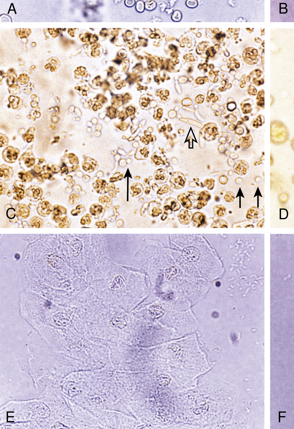
| Feature | Dysmorphic RBCs | Non-dysmorphic (Isomorphic) RBCs |
|---|---|---|
| Shape | Irregular, spiculated, blebs, membrane folds ("acanthocytes") | Uniform biconcave disks |
| Origin | Glomerulus (forced through GBM under pressure → deformed) | Collecting system, ureter, bladder, urethra |
| Implies | Glomerular disease | Urological cause (stones, tumour, trauma, UTI) |
| Sensitivity | ~87.5% for glomerular disease when dysmorphic | 85% for urological disease when isomorphic |
| Gold standard method | Phase contrast microscopy |
NKF Primer: "In one study, up to 85% of patients with nondysmorphic microscopic hematuria had a urologic disorder, whereas 87.5% of those with dysmorphic hematuria had glomerular disease."
2. White Blood Cells (Leukocytes)
| WBC Type | Appearance | Significance |
|---|---|---|
| Neutrophils (PMNs) | ~12 μm, multilobed nucleus | UTI, interstitial nephritis, glomerulonephritis |
| Glitter cells | Swollen PMNs with Brownian motion granules | Dilute or hypotonic urine during infection |
| Eosinophils | Require Hansel stain or Wright stain | Allergic interstitial nephritis (drug-induced), atheroembolic disease |
| Lymphocytes | Small, round | Viral nephritis, renal transplant rejection |
3. Renal Tubular Epithelial (RTE) Cells
- 12-20 μm, larger than PMNs, oval or egg-shaped
- A few are normal
- Many RTE cells = tubular injury → ATN, interstitial nephritis
- Lipid-laden RTE cells = oval fat bodies (nephrotic syndrome)
CASTS - The Most Diagnostically Powerful Finding
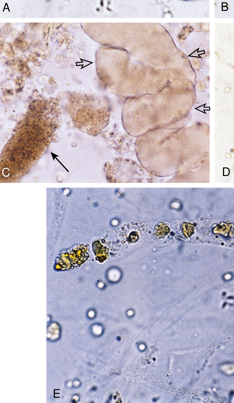
Cast Types - Detailed
- Protein alone (Tamm-Horsfall matrix only)
- Nearly transparent, low refractive index - need subdued light to see
- Non-specific - seen in: concentrated urine of normal individuals, dehydration, post-exercise, fever, diuretic use, early CKD
- Presence alone has little diagnostic value
- Fine or coarsely granular material (degenerated proteins or cells)
- Non-specific but almost always pathological
- Found in: ATN, glomerulonephritis, interstitial nephritis, volume depletion after exercise
- "Muddy brown" granular casts = the classic finding of ATN (from degenerated tubular cell debris)
- Fresh: brown/red, intact erythrocytes visible in matrix
- Degraded: pink-orange granular (hard to distinguish from coarsely granular)
- Pathognomonic of glomerulonephritis / intraparenchymal bleeding
- Also rare in: anticoagulant nephropathy, tubulointerstitial disease
- Context: always accompanied by dysmorphic haematuria, proteinuria, and granular casts ("the company they keep")
- PMNs embedded in hyaline matrix
- Pathognomonic of:
- Pyelonephritis (distinguishes from lower UTI which never causes WBC casts)
- Acute interstitial nephritis (drug-induced, etc.)
- Also in: proliferative glomerulonephritis, lupus nephritis
- Sloughed tubular cells in hyaline matrix
- Classic early finding in ATN (tubular cell necrosis and sloughing)
- Also: severe interstitial nephritis
- Hyaline material with HIGH refractive index - appears waxy, brittle, with fissures at edges
- Form in dilated, atrophic tubules = marker of chronic parenchymal disease / advanced CKD
- Also in: post-infectious GN, amyloidosis, any severe chronic kidney disease
- "Broad" = dilated tubules = end-stage renal disease
- Oval fat bodies and free lipid droplets in cast matrix
- Characteristic of nephrotic syndrome with lipiduria
- "Maltese cross" = cholesterol esters rotating polarized light (seen under polarized light)
- Oval fat bodies = tubular cells that have reabsorbed luminal fat
- Haemoglobin casts: haemolysis
- Myoglobin casts: rhabdomyolysis (brown pigment, no RBCs)
- Bile casts: severe jaundice
Cast Summary Table
| Cast Type | Key Feature | Disease |
|---|---|---|
| Hyaline | Colourless, translucent | Non-specific (normal in concentrated urine) |
| Muddy brown granular | Brown, granular | ATN (classic) |
| RBC cast | Red/brown, intact RBCs | Glomerulonephritis (pathognomonic) |
| WBC cast | WBCs in matrix | Pyelonephritis, AIN |
| Tubular cell cast | Tubular cells in matrix | ATN (early), AIN |
| Waxy / Broad | Waxy, fissured, wide | Advanced CKD (dilated atrophic tubules) |
| Fatty cast + oval fat bodies + Maltese cross | Polarized light birefringence | Nephrotic syndrome |
| Granular cast (non-muddy) | Granular | Non-specific: GN, AIN, ATN |
PART 5 - Crystals in Urine
| Crystal | Shape | pH Conditions | Clinical Significance |
|---|---|---|---|
| Calcium oxalate (monohydrate) | "Envelope" or dumbbell-shaped | Acidic | Hyperoxaluria, ethylene glycol poisoning, IBD |
| Calcium oxalate (dihydrate) | Envelope/bipyramidal | Acidic | Oxalate stone formers |
| Uric acid | Rhomboid, rosettes, diamond shapes | Acidic (pH <5.5) | Gout, uric acid stones, tumour lysis syndrome |
| Triple phosphate (struvite) | "Coffin lid" | Alkaline (pH >7) | Infection with urease organisms (Proteus), struvite stones |
| Calcium phosphate (brushite) | Flat plates, rosettes | Alkaline | Type 1 RTA stones |
| Cystine | Hexagonal (classic), colourless | Acidic | Cystinuria (pathognomonic) |
| Bilirubin | Orange-brown needles | - | Liver disease |
| Sulfonamide | "Sheaves of wheat" | Acidic | Sulfa drug toxicity |
| Calcium carbonate | Yellow spheres | Alkaline | Non-pathological (horse/rabbit urine) |
| Ammonium biurate | "Thorny apple" | Alkaline | Old urine specimens |
| Cholesterol | Notched squares | - | Nephrotic syndrome |
PART 6 - Proteinuria - Quantification and Types
Types of Proteinuria by Mechanism
| Type | Protein | Mechanism | Diseases | Daily Protein |
|---|---|---|---|---|
| Glomerular | Albumin (large molecular weight) | Damaged glomerular filtration barrier (loss of size/charge selectivity) | Nephrotic syndrome (MCD, FSGS, MN, DM), GN | 500 mg - >20 g/day |
| Tubular | β₂-microglobulin, retinol-binding protein, α₁-microglobulin (low MW) | Damaged proximal tubule - fails to reabsorb filtered small proteins | AIN, ATN, Fanconi syndrome, toxins | 1-2 g/day |
| Overflow | Bence-Jones protein (light chains), myoglobin, haemoglobin | Overproduction exceeds tubular reabsorption threshold | Multiple myeloma, rhabdomyolysis, haemolysis | Variable |
| Secretory | Tamm-Horsfall protein | Normal secretion by tubules | Benign; excessive in some conditions | Minimal |
Quantification Methods
- Most practical (random urine sample)
- Ratio of urine protein (mg/dL) to urine creatinine (mg/dL)
- Approximates 24-hour protein in g/day
| PCR | Approximate 24h Protein |
|---|---|
| <0.2 | <200 mg/day (normal) |
| 0.2-3.5 | 200 mg - 3.5 g (subnephrotic) |
| >3.5 | >3.5 g (nephrotic range) |
- Preferred for screening and monitoring diabetic/hypertensive nephropathy
| ACR | Category |
|---|---|
| <3 mg/mmol (<30 mg/g) | Normal |
| 3-30 mg/mmol (30-300 mg/g) | Microalbuminuria (moderately increased) |
| >30 mg/mmol (>300 mg/g) | Macroalbuminuria (severely increased) |
- Gold standard for proteinuria quantification
- Inconvenient; relies on adequate collection (check creatinine excretion ~15-20 mg/kg/day to assess completeness)
Significance of Proteinuria Levels
| Protein Level | Implication |
|---|---|
| <150 mg/day | Normal |
| 150 mg - 1 g/day | Benign orthostatic proteinuria, AIN, mild GN, hypertensive nephrosclerosis |
| 1-3.5 g/day | Significant GN (IgA, lupus, focal GN), tubulointerstitial disease |
| >3.5 g/day | Nephrotic syndrome - MCD, FSGS, MN, DN, amyloidosis |
- Protein present when upright, absent when lying flat
- Common in adolescents (10-15% of young males)
- Benign - confirm by comparing first morning (recumbent) sample vs daytime sample
PART 7 - Urine Electrolytes and Indices
Urine Sodium (UNa)
| UNa | Pre-renal AKI | ATN (Intrinsic) |
|---|---|---|
| Value | <20 mEq/L | >40 mEq/L |
| Reason | Kidneys maximally retain Na⁺ (intact tubules) | Tubular injury → can't retain Na⁺ |
Fractional Excretion of Sodium (FENa)
FENa = (UNa × PCr) / (PNa × UCr) × 100
| FENa | Interpretation |
|---|---|
| <1% | Pre-renal AKI (tubules working, conserving Na⁺) |
| >2% | ATN or intrinsic renal (tubular damage, Na⁺ wasting) |
| 1-2% | Indeterminate / overlap |
- False low (<1%) FENa in ATN: Contrast nephropathy, myoglobinuria (rhabdomyolysis), haemoglobinuria, early obstruction, some GN
- False high (>1%) FENa in pre-renal: Diuretic use (use FEUrea instead), CKD with salt wasting, adrenal insufficiency
Fractional Excretion of Urea (FEUrea)
FEUrea = (UUrea × PCr) / (PUrea × UCr) × 100
- More useful when diuretics have been given
- FEUrea <35% = pre-renal
- FEUrea >50% = intrinsic renal / ATN
Urine:Plasma Ratios in AKI
| Index | Pre-renal | ATN |
|---|---|---|
| Urine osmolality | >500 mOsm/kg | <350 mOsm/kg |
| Urine Na⁺ | <20 mEq/L | >40 mEq/L |
| U/P creatinine ratio | >40 | <20 |
| U/P osmolality | >1.5 | <1.1 |
| FENa | <1% | >2% |
| Urine specific gravity | >1.020 | ~1.010 |
| Urine sediment | Normal / hyaline casts | Muddy brown granular + tubular cell casts |
Urine Anion Gap (UAG) - for Normal AG Acidosis
UAG = [Na⁺ + K⁺]urine - [Cl⁻]urine
| UAG | NH₄⁺ excretion | Cause of acidosis |
|---|---|---|
| Negative (Cl⁻ > Na⁺+K⁺) | High (appropriate) | GI losses (diarrhoea) |
| Positive (Na⁺+K⁺ > Cl⁻) | Low (inappropriate) | RTA (renal tubular cause) |
PART 8 - Urine Findings by Disease (The "Urinary Fingerprints")
Comprehensive Disease-Urine Pattern Table
| Disease | Colour/Appearance | Protein | Haematuria | Casts | Other |
|---|---|---|---|---|---|
| Nephrotic syndrome | Frothy, pale | Massive (>3.5 g/day) | Minimal | Fatty casts, oval fat bodies, waxy casts | Maltese cross under polarised light; lipiduria |
| Nephritic syndrome / proliferative GN | Dark, smoky | Moderate (1-3 g/day) | Macroscopic or microscopic (dysmorphic) | RBC casts (pathognomonic) | Granular casts, WBC casts |
| IgA nephropathy | Cola-coloured (synpharyngitic) | Variable | Episodic gross haematuria (follows URTI by 24-48h) | RBC casts | Dysmorphic RBCs, proteinuria |
| ATN | Muddy brown | Mild (tubular) | Minimal | Muddy brown granular + tubular cell casts | FENa >2%, SG ~1.010 |
| Acute Interstitial Nephritis (AIN) | Normal/turbid | Mild | Microscopic | WBC casts, tubular cell casts | Eosinophiluria (Hansel stain) - 60-70% sens., 85-90% spec. |
| Pyelonephritis | Turbid, foul odour | Mild | Microscopic | WBC casts (distinguishes from cystitis), granular | Bacteria, WBCs, nitrites, LE dipstick positive |
| Pre-renal AKI | Concentrated, amber | None / trace | None | Hyaline casts only | SG >1.020, UNa <20, FENa <1% |
| Diabetic nephropathy | Frothy (late) | Microalbuminuria → macroalbuminuria → nephrotic | Absent (haematuria = think other diagnosis) | Hyaline; Kimmelstiel-Wilson nodules on biopsy | ACR monitoring essential |
| Minimal change disease | Frothy | Massive (selective - mainly albumin) | Absent | Fatty casts, oval fat bodies | Highly selective proteinuria (IgG: transferrin ratio <0.1) |
| Membranous nephropathy | Frothy | Heavy (non-selective) | Absent/minimal | Fatty, waxy casts | Non-selective proteinuria (IgG: transferrin ratio >0.2) |
| Rapidly Progressive GN (RPGN) | Dark, smoky | Significant | Gross haematuria + dysmorphic | RBC casts | ANCA, anti-GBM - urgent biopsy |
| Lupus nephritis | Variable | Variable | Variable | "Telescoped sediment" - all cast types | Waxy + RBC + WBC + granular casts simultaneously |
| Rhabdomyolysis | Dark brown (tea/cola) | Positive dipstick | Dipstick positive, no RBCs on microscopy | Pigmented granular casts | Myoglobin (positive dipstick for blood, NO RBCs) |
| Myeloma kidney | Variable | Large (but dipstick negative!) | Absent | Waxy casts | Bence-Jones protein (SSA+, dipstick−) |
| Haemolytic uraemic syndrome (HUS) | Haemoglobinuria | Variable | Haematuria | Variable | Schistocytes on blood film; dipstick positive for blood |
| Renal stones | Haematuria | Absent | Gross haematuria (painful) | None | Crystals (oxalate, uric acid, cystine by type) |
| Bladder/renal tumour | Haematuria (painless) | Absent/mild | Painless macroscopic haematuria (isomorphic RBCs) | None | Urine cytology; cystoscopy |
| Type 1 RTA | Alkaline | Mild | Microscopic | Hyaline | Urine pH always >5.5; hypercalciuria; hypocitraturia; nephrocalcinosis |
| Nephrocalcinosis | Normal | Variable | Microscopic | Variable | Calcium phosphate crystals; linked with Type 1 RTA |
| Cystinuria | May form stones | Absent | Haematuria if stones | None | Hexagonal cystine crystals (pathognomonic) |
| Alport syndrome | Smoky haematuria | Variable | Persistent microscopic haematuria | RBC casts | Family history + deafness + ocular abnormalities |
The "Telescoped Sediment" (Lupus Nephritis)
- RBC casts
- WBC casts
- Granular casts
- Waxy casts
- Fatty casts
PART 9 - Key Exam Algorithms
Algorithm 1: Approach to Proteinuria
Proteinuria detected on dipstick
↓
Confirm with spot PCR or 24h collection
↓
Is it orthostatic? (First morning void = recumbent)
If yes → absent in first morning urine → orthostatic proteinuria (benign)
↓ No
Quantify: <150 mg/day = normal
150mg-3.5g = subnephrotic → work up GN, DM, HTN
>3.5 g/day = NEPHROTIC RANGE
↓
Check sediment:
RBC casts + haematuria → Nephritic component → GN (biopsy likely)
Fatty casts + oval fat bodies → Pure nephrotic (MCD, MN, FSGS)
Both → Mixed nephrotic-nephritic (lupus, MPGN)
Algorithm 2: Approach to Haematuria
Haematuria (>3-5 RBCs/HPF or positive dipstick)
↓
Microscopy: RBCs present?
No RBCs → Haemoglobinuria or Myoglobinuria
Check plasma: pink = Hb; clear = myoglobin
↓ RBCs present
Dysmorphic RBCs? (>20% dysmorphic or acanthocytes >5%)
YES → Glomerular origin → Check protein, casts, BP, creatinine
→ GN work up (ANCA, anti-GBM, complement, ASOT, ANA, anti-dsDNA)
NO → Urological origin → Urology referral
→ (Stones, tumour, infection, trauma)
↓
RBC casts?
YES → Glomerulonephritis / RPGN → Urgent nephrology
NO → Isolated haematuria → Consider IgA nephropathy, thin GBM disease
Algorithm 3: AKI Differentiation by Urinalysis
AKI detected (↑ creatinine)
↓
Urinalysis + microscopy + FENa
↓
┌────────────────┬─────────────────┬─────────────────┐
│ Pre-renal │ ATN │ Interstitial │
│ SG >1.020 │ SG ~1.010 │ Nephritis │
│ UNa <20 │ UNa >40 │ │
│ FENa <1% │ FENa >2% │ FENa variable │
│ Normal sediment│ Muddy brown │ WBC casts │
│ (Hyaline casts)│ granular casts │ Eosinophiluria │
│ │ Tubular cell │ Mild proteinuria │
│ │ casts │ │
└────────────────┴─────────────────┴─────────────────┘
↓
RBC casts → Glomerulonephritis → RPGN?
→ Urgent nephrology + biopsy
Quick Summary Mnemonics
- Hyaline - non-specific
- Granular (muddy brown) - ATN
- RBC cast - GN (glomerulonephritis)
- WBC cast - pyelonephritis / AIN
- Tubular cell cast - ATN (early)
- Fatty cast - Nephrotic syndrome
Pre-renal = Concentrated (SG >1.020, UNa <20, FENa <1%) = Tubules working hard ATN = Diluted/Fixed (SG ~1.010, UNa >40, FENa >2%) = Tubules broken
Ischaemic nephropathy
ischaemic nephropathy renal artery stenosis
Ischaemic Nephropathy - Complete Guide
Definition and Terminology
- Progressive loss of renal function caused by significant reduction in renal perfusion
- Due to renal artery stenosis (RAS) or occlusive disease of the main renal artery or its branches
- Resulting in chronic ischaemia of the renal parenchyma
Brenner & Rector: "Renal artery stenosis is much more common than renovascular hypertension. Not all stenoses are associated with hypertension."
Epidemiology
- Atherosclerotic RAS (ARAS) is identified in 6.8% of individuals >65 years in population-based US surveys (>60% occlusion by Doppler)
- Among patients undergoing coronary angiography: ARAS found in 14-20%
- Among patients with peripheral vascular disease: RAS in 30-50%
- Among patients with abdominal aortic aneurysm: RAS in 33%
- Among patients presenting with unexplained ESKD: possible ARAS contribution in up to 40%
- Prevalence of high-grade RAS in hypertensive patients with azotemia rises from 3.2% in the 6th decade to >25% in the 8th decade
- ARAS accounts for ~85% of all RAS; fibromuscular dysplasia (FMD) accounts for ~10-15%
Causes of Renal Artery Stenosis
1. Atherosclerotic RAS (ARAS) - 85%
- Plaque located at the proximal renal artery (ostium and proximal 1-2 cm) - often an extension of aortic plaque
- Usually bilateral in 30-40% of cases (or unilateral in a solitary kidney)
- Strongly associated with: hypertension, hyperlipidaemia, smoking, DM, CAD, PVD, aortic aneurysm
- Progressive - natural history studies show progression in ~50% over 5 years; ~10-15% develop complete occlusion
- Linked to very high cardiovascular mortality (the kidney disease is a marker of systemic vascular burden)
2. Fibromuscular Dysplasia (FMD) - 10-15%
- Affects mid-portion and distal renal artery (not the ostium)
- Noninflammatory disorder of vessel wall layers
- Medial fibroplasia = most common subtype (85% of FMD) → classic "string of beads" appearance (alternating stenoses and microaneurysms)
- Also affects cerebral arteries in 25% (screen for intracranial aneurysms)
- Rarely causes major loss of renal function
- Responds very well to balloon angioplasty alone (no stent needed)
- Smoking is a risk factor for progression
3. Other Causes (Rare)
| Cause | Notes |
|---|---|
| Takayasu arteritis | Young women, large vessel vasculitis; bilateral RAS common |
| Giant cell arteritis | Elderly; may involve renal arteries |
| Renal artery dissection | Spontaneous or traumatic; acute presentation |
| Aortic coarctation | Congenital; affects renal perfusion |
| Renal artery aneurysm | May compress adjacent artery |
| Iatrogenic | Aortic stent grafts impinging on renal ostia |
| Extrinsic compression | Retroperitoneal fibrosis, tumour, haematoma |
| Neurofibromatosis type 1 | Children; renal artery dysplasia |
| Radiation nephritis | Post-renal radiation |
Pathophysiology
The Goldblatt Model
Two-kidney, one-clip model (2K1C) - equivalent to UNILATERAL RAS:
RAS (one kidney) → ↓ renal perfusion pressure
↓
Activation of Juxtaglomerular Apparatus → ↑ RENIN secretion
↓
Renin → Angiotensin I → ACE → ANGIOTENSIN II
↓ ↓
Vasoconstriction ALDOSTERONE (adrenals)
↓ ↓
↑ SVR Na⁺ + H₂O retention
↓ ↓
HYPERTENSION (renin-dependent)
Contralateral kidney excretes excess volume (natriuresis)
One-kidney, one-clip model (1K1C) - equivalent to BILATERAL RAS or solitary kidney:
Both kidneys ischaemic → ↑ RAAS + volume retention (no contralateral escape)
↓
VOLUME-DEPENDENT HYPERTENSION
↓
Flash pulmonary oedema episodes (volume overload without adequate natriuresis)
↓
Azotaemia (bilateral reduced GFR)
Goldman-Cecil: "Bilateral critical renal artery stenosis is a volume-dependent form of hypertension that, unlike unilateral renal artery stenosis, can cause azotemia."
Mechanisms of Renal Parenchymal Injury in Ischaemic Nephropathy
↓ Renal perfusion pressure (stenosis >70-80%)
↓
Autoregulation overwhelmed:
┌──────────────────────────────────────┐
│ ↓ Glomerular perfusion pressure │
│ ↓ GFR (filtration fraction changes) │
│ Tubular ischaemia │
└──────────────────────────────────────┘
↓
Chronic tubular hypoxia → Tubular atrophy
↓
Interstitial fibrosis (TGF-β, Ang II-mediated)
↓
Glomerulosclerosis (ischaemic glomeruli: wrinkled GBM,
collapsed capillaries, periglomerular fibrosis)
↓
Cortical loss → Small, shrunken, scarred kidney
- Angiotensin II direct toxicity: Ang II causes efferent arteriolar constriction, mesangial proliferation, TGF-β upregulation → fibrosis
- Oxidative stress: Reduced NO production, excess reactive oxygen species
- Inflammatory infiltrate: Macrophages, T cells infiltrate ischaemic parenchyma → cytokine-mediated injury
- Atheroemboli: Cholesterol crystals from unstable plaques embolise into interlobar vessels → additional ischaemia ("atheroembolic renal disease" - distinct but often coexistent)
- Reperfusion injury: When stenosis worsens or revascularisation attempted - excess free radicals, proximal tubular necrosis, "no-reflow phenomenon"
Why the Posterior Circulation Matters
Clinical Presentations
1. Renovascular Hypertension
- Accelerating, resistant (refractory to ≥3 drugs), or severe hypertension
- Onset of hypertension at unusual age (very young or very old)
- Previously well-controlled hypertension that suddenly worsens
- Hypertension onset following ACE inhibitor initiation (bilateral RAS unmasked)
2. Flash Pulmonary Oedema (Pickering Syndrome)
- Recurrent acute pulmonary oedema without clearly precipitating cardiac cause
- Classic in bilateral RAS or RAS to solitary kidney
- Volume-dependent hypertension → acute fluid overload → pulmonary oedema
- ECG: no acute MI; LV function may be relatively preserved
- Highly characteristic: should always prompt evaluation for bilateral RAS
- Can resolve dramatically with revascularisation
3. Ischaemic Nephropathy
- Progressive unexplained CKD in a patient with diffuse atherosclerosis
- Worsening renal function after starting ACE inhibitor or ARB (≥20-30% rise in creatinine) → strongly suggests bilateral RAS
- Asymmetric kidney sizes on imaging (one kidney >1.5 cm smaller than the other)
- Kidneys without proteinuria or haematuria (sediment typically bland)
4. Acute Renal Artery Occlusion
- Sudden flank pain ± haematuria ± nausea/vomiting
- Sudden onset anuria (if bilateral or solitary kidney)
- Causes: thrombosis on ARAS plaque, embolism (AF, endocarditis), dissection, trauma
5. Incidental Finding
- ARAS found on vascular imaging done for another reason (e.g., aortic aneurysm CT)
Clinical Clues / "Red Flags" for RAS
| Clinical Feature | Significance |
|---|---|
| Hypertension onset <30 years (especially women) | FMD |
| Hypertension onset >55 years with diffuse atherosclerosis | ARAS |
| Refractory hypertension (resistant to ≥3 drugs including diuretic) | RAS in 20% of referrals |
| Abrupt ↑ in creatinine after ACE-I or ARB | Bilateral RAS (efferent arteriole dilatation → ↓GFR) |
| Asymmetric kidney sizes on USS (>1.5 cm difference) | Unilateral RAS |
| Flash pulmonary oedema (recurrent, no cardiac cause) | Bilateral RAS |
| Unexplained progressive CKD in elderly vascular patient | ARAS |
| Epigastric / flank bruit | RAS (sensitivity 40%, specificity 90%) |
| Hypokalaemia with hypertension | Secondary hyperaldosteronism from ARAS |
| Worsening renal function with diuretics | Volume depletion unmasking bilateral RAS |
| Co-existing CAD, PVD, aortic aneurysm | Screen for ARAS |
Imaging - The Diagnostic Approach
Angiographic Imaging
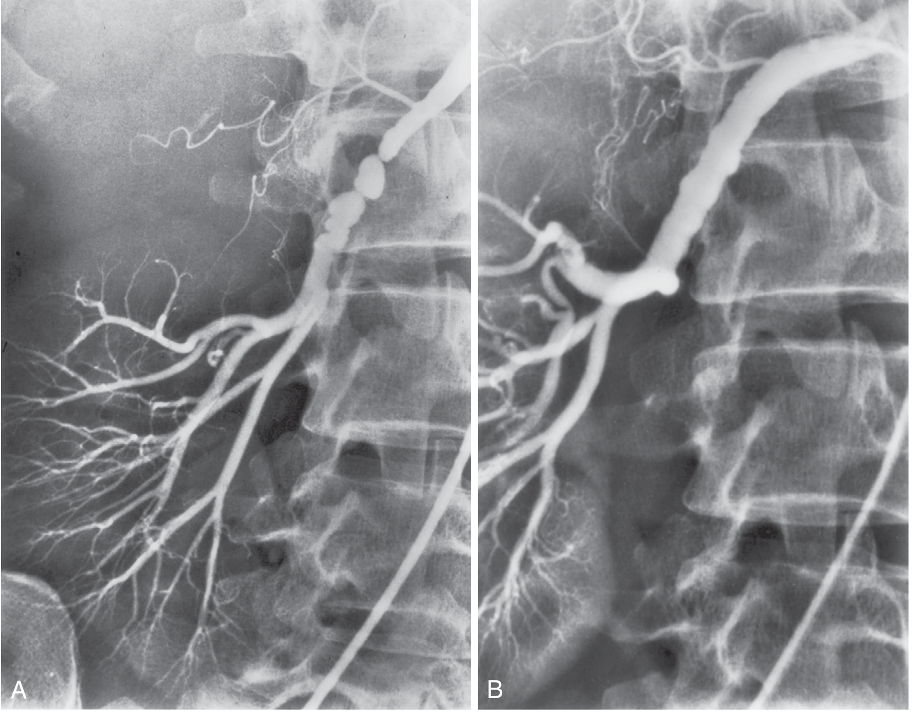
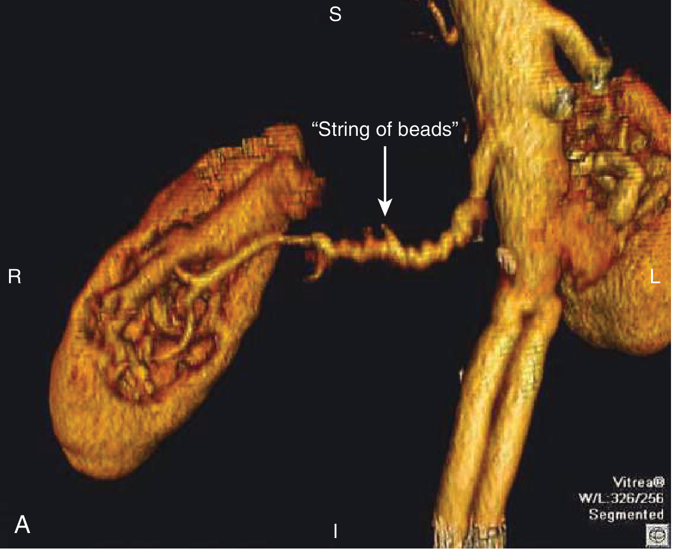
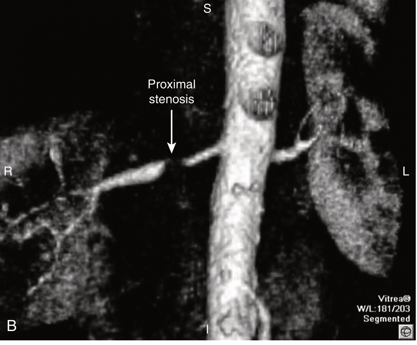
Diagnostic Modalities Compared
| Modality | Sensitivity | Specificity | Advantages | Limitations |
|---|---|---|---|---|
| Renal Duplex USS | 85-95% | 92-97% | First-line; cheap; non-invasive; provides functional data (peak systolic velocity >200 cm/s, RAR >3.5) | Operator-dependent; limited by obesity, bowel gas; misses accessory arteries |
| CT Angiography (CTA) | 96-98% | 94-98% | Fast; 3D reconstruction; detects ostial lesions well; shows aorta | Iodinated contrast (nephrotoxicity); radiation; may miss distal FMD |
| MR Angiography (MRA) | 90-97% | 85-95% | No radiation; no iodinated contrast | Gadolinium contraindicated in GFR <30 (nephrogenic systemic fibrosis); overestimates stenosis; pacemakers |
| Captopril-enhanced renal scintigraphy | 85% | 85% | Functional (shows which kidney is ischaemic); RAAS-dependent | Unreliable with bilateral disease or CKD; rarely used now |
| Digital Subtraction Angiography (DSA) | Gold standard | Gold standard | Definitive; allows simultaneous intervention | Invasive; iodinated contrast; risk of atheroemboli; reserved for when intervention planned |
Goldman-Cecil: "Current practice favors limiting invasive arteriography to carrying out endovascular intervention (e.g., stenting and/or angioplasty)."
Functional Tests (Physiological Significance)
| Test | Principle | Significance |
|---|---|---|
| Peripheral plasma renin activity (PRA) | ↑ in renin-dependent renovascular HTN | Sensitivity ~55-60%, specificity poor alone |
| Captopril-stimulated PRA | Exaggerated PRA rise after ACE inhibition | Sensitivity ~75%; useful in unilateral RAS |
| Renal vein renin ratio | Affected:unaffected kidney renin >1.5:1 | Predicts benefit of revascularisation; requires invasive sampling |
| Split renal function (isotope GFR) | Quantifies GFR contribution per kidney | Guides revascularisation vs nephrectomy decision |
| Translesional pressure gradient | >20 mmHg = haemodynamically significant | Used during angiography; important for selecting which lesions to treat |
Pathology (Kidney Biopsy / Nephrectomy Findings)
- Small, shrunken kidney on the stenotic side
- Finely granular surface (ischaemic cortical scarring)
- May show large cortical scars (from atheroemboli)
| Lesion | Description |
|---|---|
| Ischaemic glomerulosclerosis | Wrinkled, collapsed GBM; retracted glomerular tuft; periglomerular fibrosis - pathognomonic |
| Tubular atrophy | Simplified tubular epithelium, thickened tubular basement membranes |
| Interstitial fibrosis | Progressive in chronic ischaemia |
| Arteriolosclerosis | Hyaline thickening of arterioles (from hypertension) |
| Atheroemboli | Cholesterol clefts (ghost-like spaces) within vessels - pathognomonic of cholesterol embolism; surrounded by giant cells |
The Renin-Angiotensin System in RAS
UNILATERAL RAS:
↓ Perfusion pressure → ↑ Renin → ↑ Ang II → HTN
Contralateral kidney: pressure natriuresis compensates → volume normal
RAAS-dependent hypertension → ACE-I/ARB very effective for BP
BUT: ACE-I may worsen stenotic kidney GFR (removes efferent constriction)
BILATERAL RAS (or solitary kidney with RAS):
Both kidneys ischaemic → ↑ Renin + volume retention (no contralateral escape)
ACE-I or ARB → removes efferent arteriolar tone in BOTH kidneys
→ Precipitous ↓ GFR → ACUTE KIDNEY INJURY ⚠️
- A rise in creatinine >20-30% after starting ACE-I/ARB is pathognomonic of bilateral RAS or RAS to solitary kidney
- This is not an absolute contraindication to ACE-I/ARB - a monitored trial is acceptable
- But close monitoring of creatinine and K⁺ is mandatory
- If creatinine rises >30% from baseline = stop ACE-I/ARB and investigate for bilateral RAS
Management
Goals of Therapy (Brenner's Framework)
- Blood pressure control - prevent cardiovascular morbidity and mortality
- Preservation of renal function - prevent progression to ESKD
- Volume regulation - prevent flash pulmonary oedema
- Prevent vascular progression - anti-atherosclerotic therapy
Medical Therapy (Cornerstone of Management for ARAS)
| Drug Class | Agent | Indication / Notes |
|---|---|---|
| ACE inhibitor / ARB | Ramipril, perindopril / losartan, irbesartan | Excellent BP control in unilateral RAS; use with caution + monitoring in bilateral RAS; reduce creatinine by >30% → stop and investigate |
| Calcium channel blocker | Amlodipine | Safe in bilateral RAS; good BP control; does not depend on RAAS |
| Beta-blocker | Carvedilol, bisoprolol | Useful adjunct; particularly if concurrent CAD or heart failure |
| Loop diuretic | Furosemide | For bilateral RAS with volume overload; reduces pulmonary oedema episodes |
| High-intensity statin | Atorvastatin 40-80 mg | Slows progression of ARAS; anti-inflammatory; reduces cardiovascular mortality (mandatory in all ARAS) |
| Antiplatelet | Aspirin 75-100 mg | Reduces cardiovascular events in diffuse atherosclerosis |
| Glycaemic control | Insulin / oral agents | DM accelerates ARAS progression |
| Smoking cessation | Critical - smoking accelerates ARAS and FMD progression |
The CORAL and ASTRAL Trials - Landmark Evidence
- 806 patients with ARAS randomised to revascularisation + medical vs medical alone
- Result: No difference in BP control, renal function, hospitalisation, or mortality
- Criticism: 40% of enrolled lesions were not haemodynamically significant (<60% stenosis); excluded patients clinicians thought would definitely benefit
- 947 patients with ARAS >60% randomised to renal artery stenting + medical vs medical alone
- All patients received aggressive medical therapy (irbesartan ± amlodipine, statin, antiplatelet)
- Result: No difference in the composite renal/cardiovascular endpoint
- Criticism: Average stenosis 67%; well-preserved baseline GFR; excluded patients with recent flash pulmonary oedema or rapidly deteriorating renal function; may have underpowered for truly high-risk patients
Goldman-Cecil: "For atherosclerotic renal artery stenosis, renal artery revascularization may reduce the intensity of required medical therapy but does not lead to better control."
Revascularisation - When to Consider
| Clinical Scenario | Evidence for Revascularisation | Recommendation |
|---|---|---|
| FMD with hypertension | Very good - angioplasty resolves HTN in ~45% | Angioplasty strongly recommended (no stent) |
| Refractory / resistant hypertension with haemodynamically significant ARAS | Reasonable evidence | Consider revascularisation if truly refractory to ≥3 drugs |
| Flash pulmonary oedema (recurrent) with bilateral ARAS | Best evidence for stenting | Revascularisation strongly indicated |
| Rapidly deteriorating renal function with bilateral ARAS or ARAS to solitary kidney | Logical but limited RCT evidence (excluded from CORAL/ASTRAL) | Consider revascularisation |
| Creatinine rise with ACE-I/ARB indicating bilateral RAS | Reasonable | Revascularisation if medication cannot be managed otherwise |
| Stable ARAS, controlled BP, stable CKD | No benefit shown (CORAL, ASTRAL) | Medical therapy alone |
| Complete renal artery occlusion, atrophic kidney | No benefit (irreversible) | Medical therapy; consider nephrectomy if contributing to HTN |
- Atherosclerotic ostial lesions require stenting (angioplasty alone has >50% restenosis rate due to elastic recoil of aortic plaque)
- Non-ostial ARAS and FMD → angioplasty alone preferred
- Procedural complication rate ~5.2% (cholesterol embolisation, dissection, branch occlusion)
FMD vs ARAS - Comparison
| Feature | FMD | Atherosclerotic RAS |
|---|---|---|
| Age | 15-55 years | >55 years |
| Sex | Female predominance | Male predominance |
| Location | Mid/distal renal artery | Proximal / ostial |
| Angiographic appearance | "String of beads" (microaneurysms + stenoses) | Smooth proximal stenosis ± calcification |
| Bilateral | 30-35% | 30-40% |
| Other vessels | Carotid/cerebral (25%), iliac | Coronary, aorta, iliac (diffuse atherosclerosis) |
| Renal function loss | Rare | Common (ischaemic nephropathy) |
| Pathology | Non-inflammatory medial fibroplasia | Atherosclerotic plaque; cholesterol crystals |
| Aetiology | Unknown; smoking worsens | Cardiovascular risk factors |
| Treatment | Angioplasty alone (no stent) - 45% cure | Stent + aggressive medical therapy |
| Response to revascularisation | Excellent (~45% cure of HTN) | Modest BP benefit; rarely cures |
| Natural history | Usually stable; rarely causes ESKD | Progressive; major cause of ischaemic ESKD |
Prognosis and Natural History of ARAS
- Progression: ~50% show progression on serial imaging over 5 years
- Occlusion rate: ~10-15% progress to complete occlusion
- Renal atrophy: Kidney length decreases ~0.7 cm/year on the stenotic side without treatment
- Cardiovascular mortality is the dominant outcome - not ESKD
- 5-year CV mortality: ~20-30% in ARAS patients
- ARAS patients have 3-4x higher rate of myocardial infarction than matched hypertensives without RAS
- ESKD: Significant bilateral ARAS contributes to ~5-12% of incident ESKD in patients >50 years (often underdiagnosed)
Atheroembolic Renal Disease (Related but Distinct)
- Cholesterol crystals embolise from aortic/renal artery plaques into small renal vessels
- Triggers: Catheter manipulation, aortic surgery, anticoagulation (dislodges friable plaque)
- Clinical: Acute or subacute renal failure, often after angiography or cardiac catheterisation
- Systemic features: Livedo reticularis, blue toe syndrome, eosinophilia, ↑ ESR, ↓ complement
- Urine: May show eosinophiluria, haematuria, mild proteinuria
- Biopsy: Cholesterol clefts (biconvex needle-shaped spaces after cholesterol dissolved) in small vessels, surrounded by giant cell reaction
- No specific treatment; statin therapy; supportive; often irreversible
High-Yield Exam Summary
| Feature | Key Point |
|---|---|
| Most common cause | Atherosclerotic RAS (85%) - ostial/proximal lesion |
| Second cause | FMD (10-15%) - mid-vessel, "string of beads", young women |
| Mechanism | ↓ Renal perfusion → ↑ RAAS → renovascular HTN + ischaemic nephropathy |
| Unilateral RAS | Renin-dependent HTN; contralateral kidney compensates; no azotaemia |
| Bilateral RAS | Volume-dependent HTN; azotaemia; flash pulmonary oedema |
| ACE-I/ARB clue | ≥20-30% creatinine rise after starting = bilateral RAS until proven otherwise |
| Flash pulmonary oedema | Recurrent episodes without cardiac cause = bilateral RAS (Pickering syndrome) |
| First-line imaging | Renal duplex USS; confirm with CTA or MRA |
| Gold standard imaging | DSA (reserved for when intervention planned) |
| FMD treatment | Balloon angioplasty alone (no stent) - ~45% cure |
| ARAS treatment | Aggressive medical therapy (RAAS blockade + statin + antiplatelet) first; selective stenting for refractory cases |
| CORAL trial lesson | Stenting + medical = medical alone for stable ARAS; BUT excluded high-risk patients (flash pulmonary oedema, rapidly declining GFR) |
| Pathology | Ischaemic glomerulosclerosis: wrinkled GBM, periglomerular fibrosis |
| Prognosis dominated by | Cardiovascular mortality (not ESKD) |
| Atheroemboli distinction | Post-procedure cholesterol crystals → acute/subacute renal failure + blue toes + livedo reticularis |
Clinical Approach Algorithm
Suspected Renovascular Disease
↓
Clinical clues: resistant HTN / flash pulmonary oedema /
progressive CKD in atherosclerotic patient /
creatinine rise with ACE-I / asymmetric kidneys
↓
RENAL DUPLEX ULTRASOUND (first-line)
Peak systolic velocity >200 cm/s; RAR >3.5
Kidney size asymmetry
↓
Confirm: CTA (preferred) or MRA (if GFR adequate)
↓
RAS confirmed?
↓ YES
Haemodynamically significant? (>70-75% stenosis, or gradient >20 mmHg)
↓ YES
Unilateral vs Bilateral?
↓
┌──────────────────────────────────┬─────────────────────────────────┐
│ UNILATERAL ARAS, stable │ BILATERAL ARAS or high-risk: │
│ → Medical therapy: │ Flash pulmonary oedema / │
│ ACE-I/ARB + CCB + statin + │ Rapidly ↓ GFR / Refractory HTN │
│ antiplatelet + risk factor │ → Consider STENTING │
│ modification │ + aggressive medical therapy │
│ Monitor creatinine closely │ │
└──────────────────────────────────┴─────────────────────────────────┘
↓
FMD?
→ BALLOON ANGIOPLASTY ALONE (no stent)
→ Screen cerebral vessels
Atherosclerotic Renal vascular disease
atherosclerotic renal vascular disease renovascular hypertension management
Hypertension in CKD
hypertension chronic kidney disease blood pressure target treatment
PMID: 39403990
Hypertension in Chronic Kidney Disease (CKD)
The Bidirectional Relationship - The Core Concept
Hypertension ──────► Glomerulosclerosis / arteriosclerosis
│
▼
CKD progression
│
▼
Na/water retention ◄── ↓GFR, ↓Na excretion
RAAS activation ◄───── Ischaemic nephrons
SNS overactivity ◄──── Afferent neural signals
Endothelial dysfunction
│
▼
Worsening Hypertension
| CKD Stage | HTN Prevalence |
|---|---|
| Stage 1 (eGFR ≥90) | ~22% |
| Stage 2 (eGFR 60-89) | ~40% |
| Stage 3 (eGFR 30-59) | ~55-75% |
| Stage 4 (eGFR 15-29) | ~80% |
| Stage 5/Dialysis | >85-90% |
- Comprehensive Clinical Nephrology 7E Ch.35
Part 1 - Pathophysiology of Hypertension in CKD
1. Sodium and Water Retention (PRIMARY driver)
- As nephrons are lost, the remaining nephrons cannot adequately excrete a daily sodium load
- The kidney "resets" its pressure-natriuresis curve - a higher arterial pressure is needed to achieve the same sodium excretion
- This leads to volume expansion and raised cardiac output
- In ESKD/dialysis patients, this becomes the overwhelmingly dominant mechanism
- Key concept: Guyton's principle - the kidney is the ultimate long-term regulator of BP; when it fails, hypertension is inevitable
2. RAAS Activation
- Reduced renal perfusion (from nephrosclerosis, afferent arteriolar disease) activates the juxtaglomerular apparatus
- Renin release → Ang II generation → systemic vasoconstriction + aldosterone secretion
- Intrarenal RAAS is activated even when systemic renin levels appear normal
- Ang II also promotes glomerular hypertension (efferent arteriolar constriction) → intraglomerular pressure elevation → further proteinuria and nephron damage
3. Sympathetic Nervous System (SNS) Overactivity
- Damaged renal parenchyma generates afferent neural signals via renal sensory nerves → central SNS activation
- This causes peripheral vasoconstriction, tachycardia, and further RAAS activation
- Explains why even anuric bilateral nephrectomy patients can have hypertension reduced after the failed kidneys are removed
- Explains why renal denervation was explored as a therapy
4. Endothelial Dysfunction / Reduced Nitric Oxide
- Uraemic toxins impair endothelial NO synthase
- Reduced NO → loss of vasodilation → increased vascular resistance
- Also promotes platelet aggregation and atherosclerosis
5. Disordered Circadian BP Profile - Non-Dipping
- Normal individuals show a 10-20% nocturnal BP dip (dippers)
- CKD patients frequently become non-dippers or reverse-dippers (BP rises at night)
- Mechanism: nocturnal sodium redistribution from the periphery + fluid shift from dependent oedema in recumbency
- Non-dipping pattern is independently associated with faster CKD progression and higher CV events
- Ambulatory BP monitoring (ABPM) is therefore particularly important in CKD
6. Secondary Causes Superimposed on CKD
- Renovascular disease (ARAS, FMD) - covered in previous session
- Primary hyperaldosteronism (can cause CKD)
- Sleep apnoea (extremely common in CKD - up to 50-80% of dialysis patients)
- Erythropoiesis-stimulating agents (EPO/darbepoetin) - dose-dependent HTN, especially with rapid Hb rise
Part 2 - Consequences: Why Does BP Control Matter?
Cardiovascular Risk
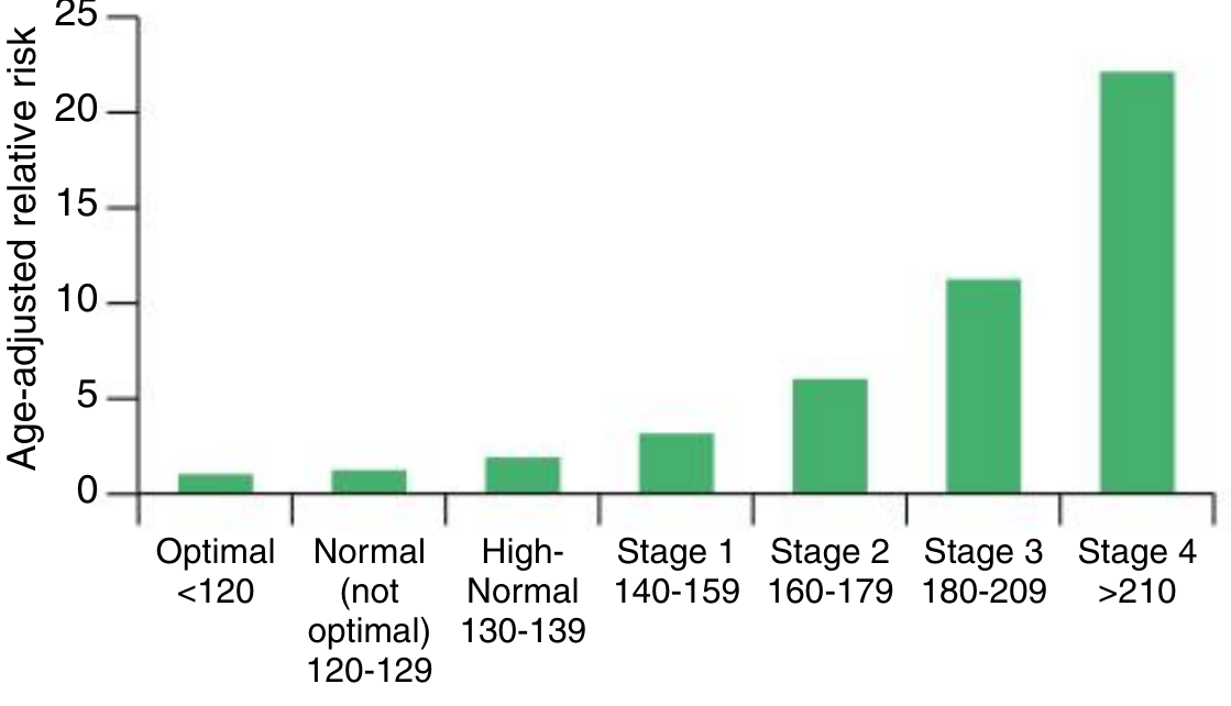
- Hypertension confers markedly increased risk of: stroke, coronary heart disease, peripheral vascular disease, vascular dementia, and heart failure in CKD patients
- CKD patients have a cardiovascular mortality risk 10-30x higher than the general population - much of this is BP-driven
Renal Outcomes
- Poorly controlled BP (SBP >160) → creatinine elevation in 10-20% of patients over time
- 2-5% progress to ESKD with poorly controlled SBP over 10-15 years
- Despite this being a relatively small fraction, hypertension is the second most common cause of ESKD after diabetes (USA and Europe) because it is so prevalent
APOL1 Polymorphism (Exam Trap)
- African Americans with hypertension have 2-6x higher ESKD incidence than white patients
- Driven partly by APOL1 gene polymorphisms - expressed in podocytes → lysosomal swelling, mitochondrial dysfunction, impaired autophagy → accelerated glomerulosclerosis
- Hypertensive African Americans show more severe arteriolosclerosis, more glomerulosclerosis
Part 3 - BP Targets in CKD (The Controversial Core)
The Key Trials You Must Know
| Trial | Population | Intensive Target | Standard Target | Key Finding |
|---|---|---|---|---|
| MDRD | Non-diabetic CKD + proteinuria | MAP 92 mmHg (≈125/75) | MAP 102 mmHg (≈140/90) | Benefit in subgroup with proteinuria >1g/day |
| AASK | African American, non-DM, CKD | MAP 92 mmHg | MAP 102-107 mmHg | No overall difference; but benefit with proteinuria >0.22 g/g |
| ACCORD | Type 2 DM, preserved GFR, high CV risk | SBP <120 mmHg | SBP <140 mmHg | No CV benefit; more hypotension, ↑creatinine, 10x more hyperkalemia |
| SPRINT | High CV risk, NO DM | SBP <120 mmHg | SBP <140 mmHg | CV benefit ✓ (reduced MI/stroke/HF/CV death); No CKD progression benefit; excluded >1g/day proteinuria |
| IDNT | Type 2 DM nephropathy | - | - | J-curve: lowest risk at SBP 120-130; ↑death below SBP 120 |
The J-Curve Problem
- A post-hoc IDNT analysis showed that in overt diabetic nephropathy:
- Lowest kidney event risk at SBP 120-130 mmHg
- Increased death risk below SBP 120 mmHg
- Increased events at very high and very low BP = J-curve
- The benefit of BP lowering has a plateau - going too low increases risk
Current Recommendations (2023-2024 Guidelines)
General CKD target: SBP <130 mmHg (ACC/AHA 2017, ESH 2023, KDIGO 2024)
| Guideline | CKD (no DM) | CKD + DM | CKD + Proteinuria >1g/day |
|---|---|---|---|
| ACC/AHA 2017 | <130/80 | <130/80 | <130/80 |
| ESH 2023 | <130/80 | <130/80 | <130/80 |
| KDIGO 2024 | SBP <120 (if tolerated) | <130/80 | <130/80 |
- The Cochrane 2024 meta-analysis [PMID 39403990] (6 RCTs, 7348 participants) found: lower targets vs standard showed little to no difference in mortality, cardiovascular events, or ESKD progression - moderate certainty evidence. However, lower targets did reduce stroke risk. This ongoing controversy means the target must be individualised.
Part 4 - Drug Treatment: What to Use and Why
Step 1 - RAAS Blockade (CORNERSTONE)
- CKD + proteinuria (albumin >30 mg/mmol or PCR >300 mg/g)
- CKD + diabetes (even microalbuminuria = ACR 3-30 mg/mmol)
- CKD + hypertension (with or without proteinuria)
- Systemic BP reduction
- Reduced intraglomerular pressure - blocks Ang II-mediated efferent arteriolar constriction → reduces glomerular filtration pressure → reduces proteinuria
- Anti-fibrotic effects - Ang II drives TGF-β production and interstitial fibrosis
- Reduced proteinuria - independent of BP - proteinuria itself is nephrotoxic
- Lewis 1993 (Captopril in Type 1 DM): 43% reduction in doubling of creatinine; significant reduction in death/dialysis/transplant - INDEPENDENT of BP lowering
- IDNT (Irbesartan, Type 2 DM): ARB reduced composite endpoint 20% vs placebo, 23% vs amlodipine (same BP achieved) - proves class-specific renal benefit beyond BP
- RENAAL (Losartan, Type 2 DM): Doubling of creatinine ↓25%, ESKD risk ↓28% vs placebo - same BP achieved
- REIN (Ramipril, non-DM proteinuric): 50% lower ESKD risk over 3 years with ramipril vs conventional Rx at same BP
- AASK: Ramipril superior to amlodipine AND metoprolol in slowing CKD in African Americans:
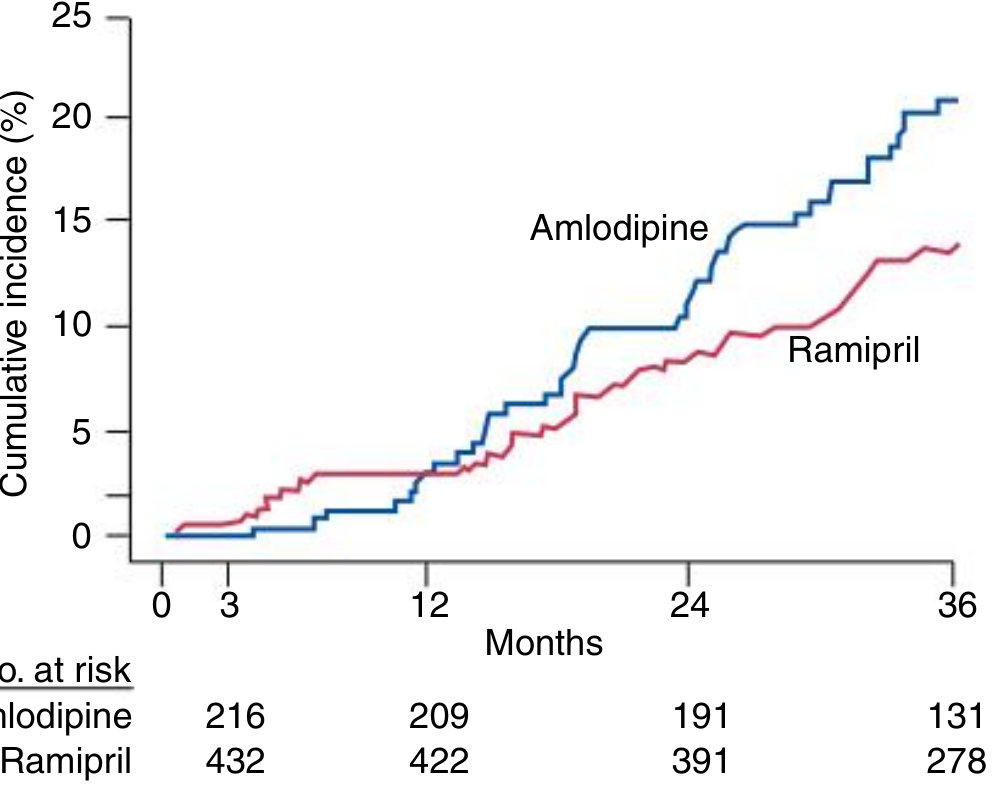
- Both are equivalent for renoprotection - choose based on tolerability
- ACEi → cough (bradykinin-mediated, ~10-15%): switch to ARB
- ARB → no cough, angioedema much rarer
- DO NOT combine ACEi + ARB (dual RAAS blockade) - ONTARGET trial showed no additional benefit but increased AKI, hyperkalaemia, hypotension
- Check creatinine and K⁺ at 1-2 weeks after initiation
- Creatinine rise ≤30% = acceptable (haemodynamic, reversible) - do NOT stop
- Creatinine rise >30% = investigate for bilateral RAS, volume depletion - withhold
- Hyperkalaemia (K⁺ >5.5-6.0 mmol/L) = reduce dose or use potassium binders
- Patiromer and Sodium Zirconium Cyclosilicate (SZC/Lokelma) - allow RAAS to be continued in patients who would otherwise require dose reduction due to hyperkalaemia
Step 2 - SGLT2 Inhibitors (THE GAME-CHANGER)
- Osmotic diuresis → volume depletion → BP reduction 4-6 mmHg systolic
- Glucosuria → caloric loss → weight reduction 1-4 kg
- Natriuresis
- Tubuloglomerular feedback (TGF) restoration - the key mechanism:
- Blocks SGLT2 in proximal tubule → ↑Na delivery to macula densa → restores TGF → afferent arteriolar constriction → ↓intraglomerular pressure
- This mirrors the mechanism of ACEi/ARB (efferent constriction) but acts on the afferent side
- Anti-inflammatory and anti-fibrotic effects
- Reduced hypoxia in renal medulla (reduces O₂ demand in proximal tubule)
| Trial | Drug | Population | Key CKD Result |
|---|---|---|---|
| CREDENCE | Canagliflozin | T2DM + overt diabetic nephropathy | 34% ↓ESKD risk; 34% ↓doubling of creatinine |
| DAPA-CKD | Dapagliflozin | CKD with or without DM, eGFR 25-75 | 44% ↓sustained ≥50% eGFR decline/ESKD/death; stopped early |
| EMPA-KIDNEY | Empagliflozin | CKD (low albumin subset included) | 28% ↓kidney progression/CV death |
Key exam point: DAPA-CKD included non-diabetic CKD patients - and still showed massive benefit. SGLT2i are no longer just for DM.
- Euglycaemic DKA: 13/1000 patient-years (withhold perioperatively/during fasting/illness)
- Genital mycotic infections (Candida)
- UTI risk (modest)
- NOT a major hyperkalaemia risk (unlike RAAS)
Step 3 - Finerenone (Nonsteroidal MRA - New Agent)
- Selective, nonsteroidal MRA - different from spironolactone/eplerenone (steroidal)
- More selective for mineralocorticoid receptor → less off-target sex hormone effects
- Higher tissue distribution in heart and kidney vs steroidal MRAs
- FIDELIO-DKD (T2DM + CKD stage III, UACR 852 mg/g median): 18% ↓ composite kidney failure/sustained 40% eGFR decline/renal death + reduced CV events
- FIGARO-DKD (T2DM + CKD, wider range): 13% ↓ composite major adverse CV events
Step 4 - Diuretics
| Diuretic | CKD Stage | Notes |
|---|---|---|
| Thiazides (e.g., chlorthalidone, indapamide) | eGFR ≥30 | Preferred for BP-lowering; chlorthalidone more potent than HCTZ; CLICK trial showed chlorthalidone effective in advanced CKD |
| Loop diuretics (furosemide, torasemide) | eGFR <30; or volume overloaded | Replace thiazides when GFR falls; higher doses needed; bid dosing often required |
| K⁺-sparing (spironolactone, amiloride) | With caution, eGFR >45 | Risk of hyperkalaemia; can be used with monitoring |
Step 5 - Calcium Channel Blockers (CCBs)
- Dihydropyridine CCBs (amlodipine, nifedipine): Effective BP reduction; vasodilatory; no direct renoprotection (inferior to RAAS in proteinuric disease as shown in AASK/IDNT); can worsen proteinuria by dilating afferent arteriole → ↑ intraglomerular pressure
- Non-dihydropyridine CCBs (diltiazem, verapamil): Also reduce proteinuria; preferred when CCB needed and proteinuria is present; additive with ACEi to reduce proteinuria
- CCBs are excellent add-on agents - combine well with RAAS blockers
Step 6 - Other Agents
| Drug | Role | Notes |
|---|---|---|
| Beta-blockers (carvedilol, bisoprolol, nebivolol) | CKD with heart failure/coronary disease | Carvedilol: alpha+beta; nebivolol: vasodilating (NO release); AVOID atenolol (renally cleared) |
| Alpha-1 blockers (doxazosin) | Resistant HTN or BPH coexistence | Less well studied; 4th/5th line |
| Hydralazine + nitrates | Alternative when RAAS not tolerated | Particularly in ESKD/dialysis |
| Minoxidil | Severe resistant HTN | Potent vasodilator; requires loop diuretic + beta-blocker; causes fluid retention and hirsutism |
| Clonidine | Short-term; dialysis patients | Central α2 agonist; useful in dialysis-related hypertension |
Part 5 - Treatment Algorithm
CKD + Hypertension
│
▼
Start ACEi or ARB (maximally tolerated dose)
+ Low-sodium diet (<2g Na/day), weight loss, exercise
│
├── DM present? → Add SGLT2 inhibitor early
│
├── eGFR still declining/proteinuria persistent?
│ → Optimise SGLT2 inhibitor + consider finerenone
│
├── BP still >130/80? → Add thiazide (if eGFR≥30)
│ or loop diuretic (eGFR<30)
│
├── Still uncontrolled? → Add CCB (dihydropyridine)
│
├── Still uncontrolled? → Add beta-blocker or alpha-blocker
│
└── Resistant HTN (≥4 drugs)?
→ Check adherence, ABPM (white coat?),
exclude secondary causes (OSA, primary aldosteronism, RAS)
Consider renal denervation (emerging)
- Sodium restriction: <2g/day (sodium, not NaCl) - critical in CKD
- DASH diet (high potassium foods - use with caution in advanced CKD due to hyperkalaemia risk)
- Weight reduction: each 1 kg loss → ~1 mmHg SBP fall
- Exercise: 30 minutes moderate intensity most days
- Alcohol restriction: ≤2 units/day
- Smoking cessation (accelerates CKD progression and CV risk)
Part 6 - Hypertension in Dialysis Patients (ESKD)
- As residual renal function approaches zero, volume overload becomes the near-sole driver
- Fluid removal with dialysis is the primary antihypertensive intervention in ESKD
- "Dry weight" optimisation - reducing post-dialysis target weight to eliminate excess fluid
- Intradialytic hypertension (BP rises during dialysis) - occurs in ~10-15% of haemodialysis patients - associated with endothelin release, RAAS activation, sympathetic activation
- Post-dialytic hypotension risk limits aggressive fluid removal
- BP measurement timing matters: pre-dialysis, post-dialysis, and ABPM give different readings
- Nocturnal/home dialysis achieves better BP control by more physiological fluid removal
- Loop diuretics have no role in anuric ESKD patients
- Hydralazine + beta-blocker + CCB combination common
- ACEi/ARB can be used in dialysis (though no longer filtering, CV benefit persists)
- Avoid renally-cleared drugs: atenolol, sotalol (accumulate)
Part 7 - Resistant Hypertension in CKD
- Volume overload (inadequate diuresis - under-dosed loop diuretics)
- Medication non-adherence (up to 50% in some studies)
- White coat hypertension (must exclude with ABPM/home BP)
- Primary aldosteronism (check renin-to-aldosterone ratio; common, underdiagnosed)
- Obstructive sleep apnoea (very common in CKD)
- Renovascular disease (ARAS - previous session)
- High dietary sodium intake
- ABPM to confirm true resistance
- Check renal sodium handling: 24h urine sodium
- Renin/aldosterone ratio (screen for primary aldosteronism)
- Renal artery duplex Doppler (screen for RAS)
- Sleep study (OSA)
- Add spironolactone 25-50mg (evidence from PATHWAY-2 trial: most effective 4th agent) - but watch K⁺ in CKD
- Renal denervation: SPYRAL HTN trials show benefit in resistant HTN; CKD-specific evidence emerging
Part 8 - Hypertensive Nephrosclerosis (Hypertensive CKD)
- Afferent arteriolar hyalinosis - PAS-positive homogeneous pink material replaces the arteriolar wall
- Interlobular artery fibromuscular intimal thickening (arteriosclerosis - "onion skinning" in malignant HTN)
- Ischaemic glomerulosclerosis - wrinkling/collapse of GBM, periglomerular fibrosis, capillary tuft retraction
- Tubular atrophy, interstitial fibrosis
- Long-standing uncontrolled hypertension (often >10-15 years)
- Bland urine sediment (minimal proteinuria, few cells/casts) - important distinction from glomerulonephritis
- Proteinuria usually <1g/day (non-nephrotic)
- Gradual slow decline in eGFR
- Fundoscopy: AV nipping, silver/copper wiring, cotton wool spots, flame haemorrhages (hypertensive retinopathy)
- BP typically >180/120 mmHg with end-organ damage
- Acute renal failure: fibrinoid necrosis of arterioles ("onion skinning")
- Haematuria + proteinuria (mimics GN) + red cell casts possible
- Associated with TMA (thrombotic microangiopathy): MAHA + thrombocytopenia
- Treatment: IV labetalol or nicardipine (reduce MAP by 20-25% in first hour; NOT more - risk of watershed infarcts)
- Renal recovery is possible with BP control even from AKI
Exam Summary Tables
Key Trials Mnemonics
- Captopril trial (Lewis 1993) - Type 1 DM
- AASK - African Americans
- Losartan = RENAAL - Type 2 DM
- MDRD - non-DM, proteinuria
- REIN - non-DM, ramipril, 50% ↓ESKD
- IDNT - Irbesartan, Type 2 DM
- DAPA-CKD (dapagliflozin, DM and non-DM)
- CREDENCE (canagliflozin, DM)
- EMPA-KIDNEY (empagliflozin)
Drug Selection at a Glance
| Situation | First Choice | Second | Avoid |
|---|---|---|---|
| CKD + proteinuria | ACEi or ARB | CCB (non-DHP) | ACEi+ARB combo |
| CKD + DM | ACEi/ARB + SGLT2i + finerenone | CCB, diuretic | - |
| CKD + HF | ACEi/ARB + beta-blocker | Loop diuretic | NSAIDs, CCB |
| CKD eGFR<30 | ACEi/ARB + loop diuretic | CCB | Thiazides (limited efficacy) |
| ESKD/Dialysis | Volume removal + CCB + hydralazine | Clonidine | Loop diuretics (if anuric) |
| Resistant HTN | Add spironolactone (if K⁺ allows) | Alpha-blocker | - |
Creatinine Rise Rules
| ACEi/ARB Creatinine Response | Action |
|---|---|
| Rise <30% - stabilises | CONTINUE - acceptable haemodynamic response |
| Rise >30% | INVESTIGATE: bilateral RAS? Hypovolaemia? |
| K⁺ 5.5-6.0 | Reduce dose + dietary K⁺ restriction + add binder |
| K⁺ >6.0 | Withhold; correct K⁺; consider binder before restarting |
Recent Evidence Update
Myeloma kidney
myeloma kidney cast nephropathy treatment bortezomib
Nephrotic syndrome
nephrotic syndrome podocyte glomerular disease treatment
nephrotic syndrome glomerulonephritis KDIGO guidelines
Top up done
Pulmonary Renal syndrome
pulmonary renal syndrome diffuse alveolar hemorrhage RPGN
anti-GBM disease ANCA vasculitis pulmonary hemorrhage treatment
Answer