
Femoral triangle
I'll look up the femoral triangle in the medical library for a thorough, textbook-quality answer.
Reading File
Now let me search the library for femoral triangle content in parallel:
Finding Sources
Finding Sources
Excellent. Now let me read the primary sections from Gray's Anatomy and THIEME Atlas in parallel:
Reading File
Reading File
Now let me read the femoral sheath/canal section and the clinical/surface anatomy section, plus fetch key images:
Reading File
Reading File
Loading Image
Loading Image
Now let me fetch the contents diagram and the surface anatomy image:
Loading Image
Loading Image
I now have comprehensive textbook content and four excellent diagrams. Here is a thorough, well-illustrated answer:
Femoral Triangle
The femoral triangle is a wedge-shaped (pyramidal) depression in the upper anterior thigh, at the junction between the anterior abdominal wall and the lower limb. It is a key anatomical region because major neurovascular structures pass through it from the abdomen into the lower extremity.
Boundaries
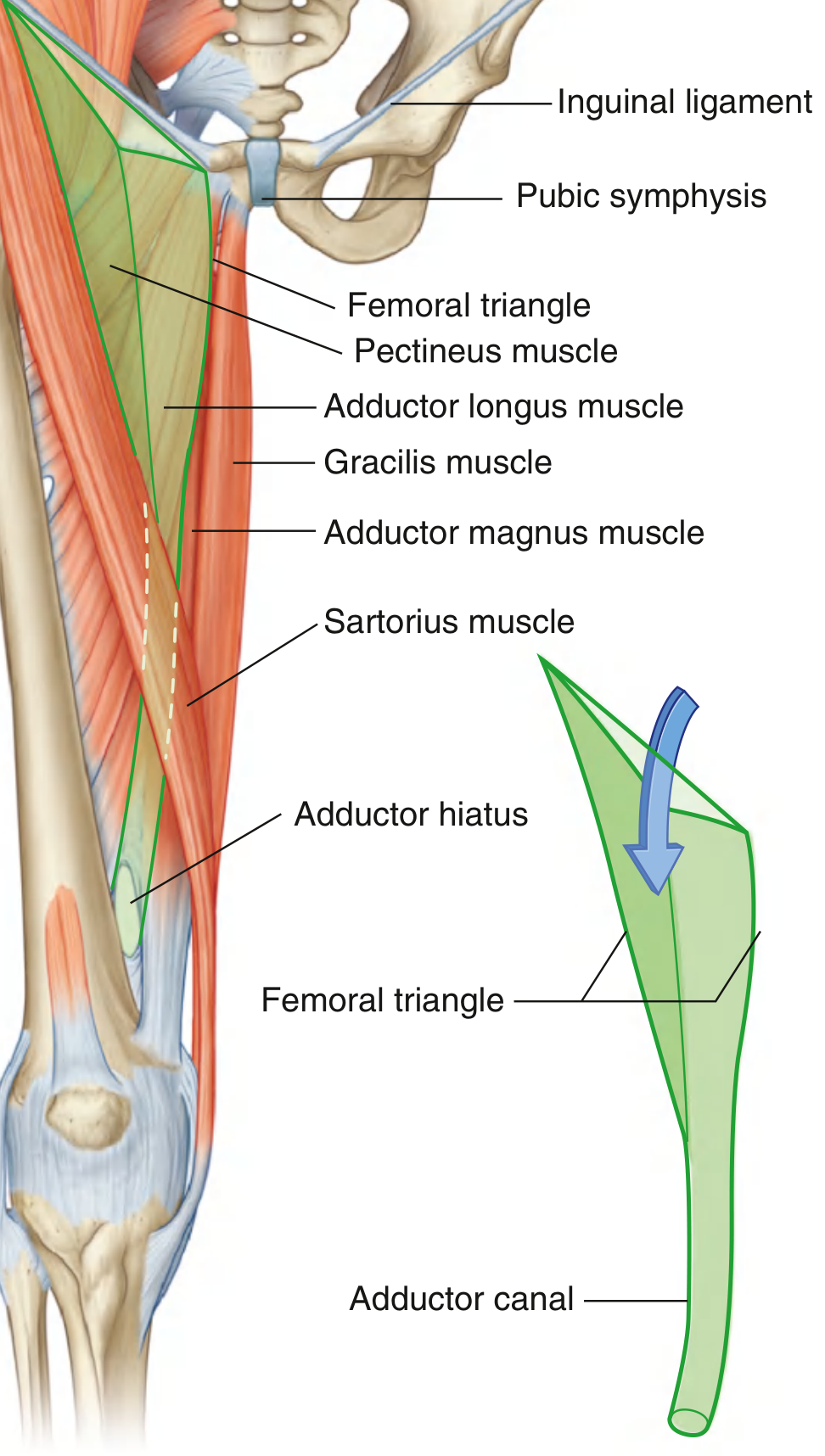
| Boundary | Structure |
|---|---|
| Base (superior) | Inguinal ligament |
| Lateral border | Medial margin of sartorius muscle |
| Medial border | Medial margin of adductor longus muscle |
| Apex (inferior) | Where the two borders meet; continuous with the adductor canal |
Floor (from lateral to medial): iliopsoas muscle (lateral half) + pectineus and adductor longus muscles (medial half).
Roof: Fascia lata (deep fascia of the thigh) + skin and superficial fascia. The great saphenous vein pierces the roof through the saphenous opening (fossa ovalis) to drain into the femoral vein.
Contents (Lateral to Medial)
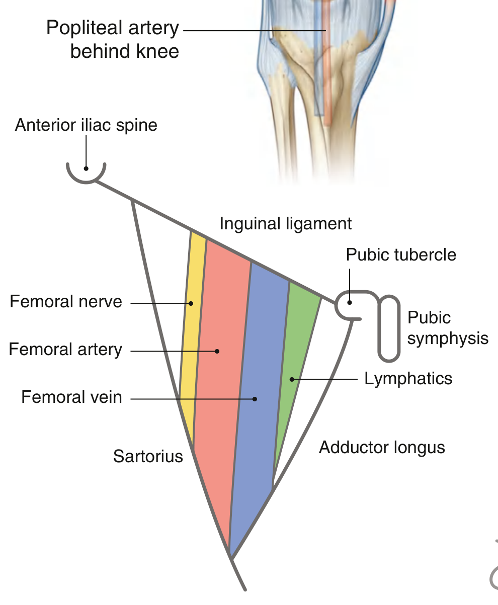
The classic mnemonic is NAVEL (Nerve, Artery, Vein, Empty space [femoral canal], Lymphatics):
| Structure | Notes |
|---|---|
| Femoral nerve | Most lateral; outside the femoral sheath |
| Femoral artery | Continuation of the external iliac artery |
| Femoral vein | Medial to the artery |
| Femoral canal | Most medial compartment; contains lymphatics (empty space = potential hernia site) |
Femoral Sheath
Within the femoral triangle, the femoral artery, vein, and lymphatics are enclosed in a funnel-shaped fascial sleeve called the femoral sheath:
- Continuous superiorly with transversalis fascia (anteriorly) and iliac fascia (posteriorly)
- Divided internally into three compartments:
- Lateral compartment - femoral artery
- Intermediate compartment - femoral vein
- Medial compartment (femoral canal) - lymphatics and areolar tissue
The femoral nerve is lateral to the sheath and is NOT enclosed within it.
Femoral Canal and Femoral Hernia
The femoral canal is the most medial, cone-shaped compartment of the femoral sheath. Its superior opening is the femoral ring, bounded by:
- Anteriorly: inguinal ligament
- Medially: lacunar ligament
- Posteriorly: pectineal (Cooper's) ligament and the pectineus muscle
- Laterally: femoral vein
The femoral ring is a potential weak point in the lower abdominal wall and is the site of femoral hernias. Femoral hernias are more common in women (wider pelvis) and emerge below and lateral to the pubic tubercle (distinguishing them from inguinal hernias, which emerge above and medial to the pubic tubercle).
The Femoral Artery
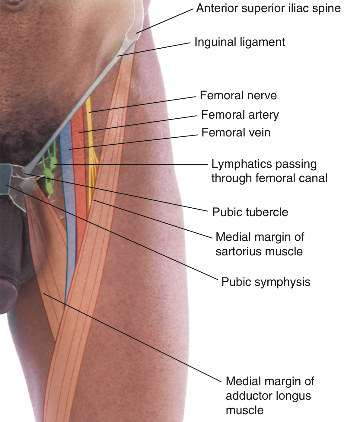
- The femoral artery is the continuation of the external iliac artery below the inguinal ligament.
- Its pulse is palpable just inferior to the inguinal ligament, midway between the anterior superior iliac spine (ASIS) and the pubic symphysis (the mid-inguinal point).
- In the femoral triangle it gives off the profunda femoris (deep femoral artery) - the principal artery supplying the thigh muscles.
- The femoral artery and vein exit the femoral triangle at its apex and pass into the adductor canal (subsartorial canal / Hunter's canal), emerging through the adductor hiatus to become the popliteal vessels behind the knee.
Vascular Compartment vs. Muscular Compartment (below the inguinal ligament)
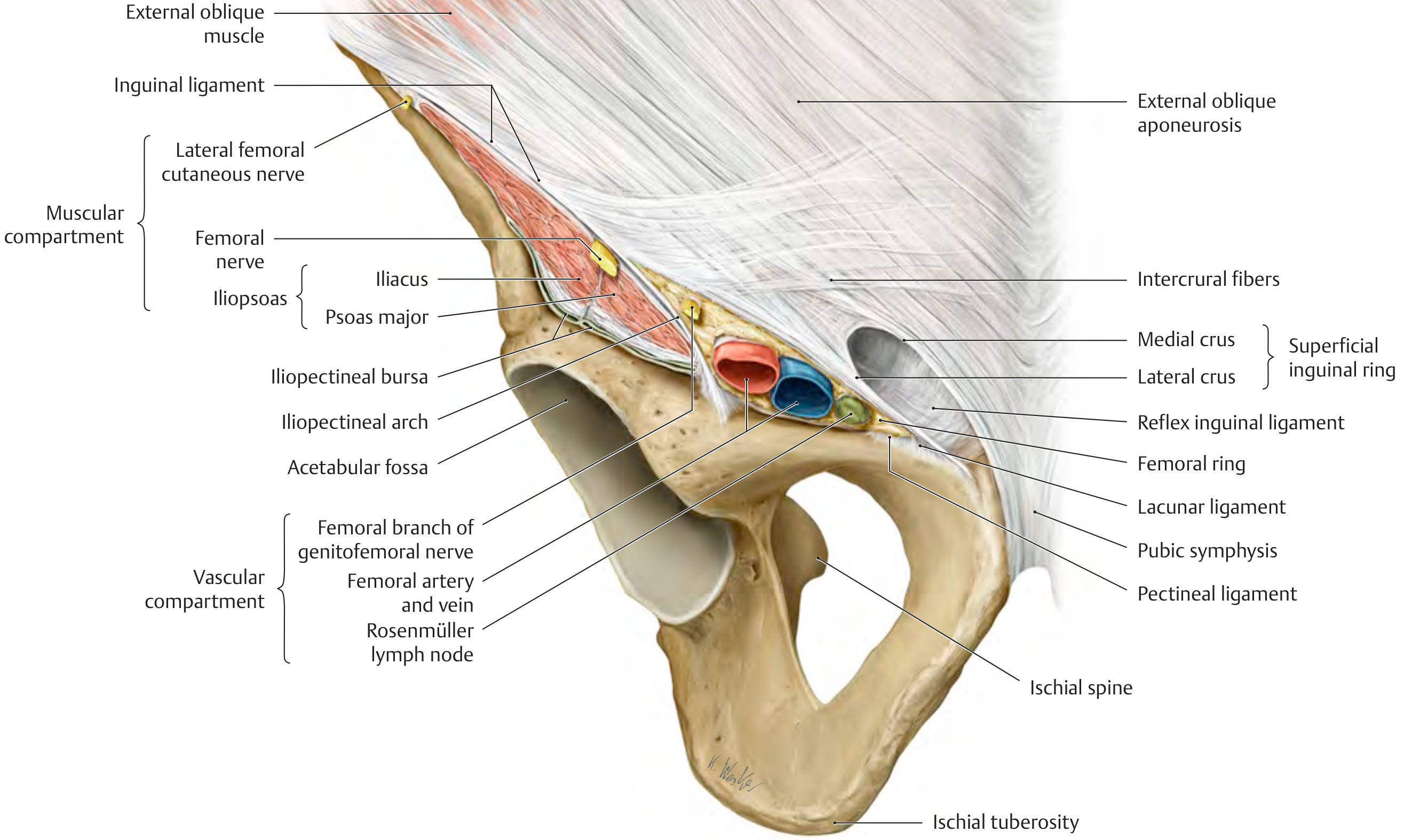
The space below the inguinal ligament is divided by the iliopectineal arch into:
- Muscular (lateral) compartment: iliopsoas, femoral nerve, lateral femoral cutaneous nerve
- Vascular (medial) compartment: femoral artery, femoral vein, femoral branch of the genitofemoral nerve, deep inguinal lymphatics (Rosenmüller node is contained within the femoral canal here)
Apex and Continuation
The apex of the femoral triangle points inferiorly and leads into the adductor canal, which runs medially down the thigh between the vastus medialis anterolaterally and the adductor longus/magnus posteromedially, roofed by the sartorius. This continuation carries the femoral artery, femoral vein, and saphenous nerve (branch of femoral nerve) into the popliteal fossa.
Clinical Relevance
-
Femoral pulse - First choice site for pulse palpation in cardiac arrest; used for femoral artery catheterization in angiography, coronary angioplasty, and cardiac catheterization.
-
Femoral vein access - Provides access to the right heart, pulmonary vessels, renal veins, and superior vena cava via catheter.
-
Femoral hernia - Abdominal contents prolapse through the femoral ring into the femoral canal. The rigid lacunar ligament medially makes strangulation a serious risk. Treatment is surgical.
-
Femoral nerve block - Anesthesia of the femoral nerve in the femoral triangle provides analgesia for the anterior thigh and knee (used in hip/femur fractures and knee surgery).
-
Lymph node enlargement - Inguinal/femoral lymph nodes in the triangle are often palpable in lower limb or perineal infections.
-
Deep vein thrombosis - The femoral vein is a common site for DVT and is accessed for thrombolysis or filter placement.
Sources: Gray's Anatomy for Students (9780323934237, pp. 675-676, 771); THIEME Atlas of Anatomy - General Anatomy and Musculoskeletal System (9781626237186, p. 573)
This is a shared conversation. Sign in to Orris to start your own chat.