
Dibetes mellitus Robbins only
Reading File
Searching sources...
Finding Sources
Finding Sources
Finding Sources
Reading File
Reading File
Reading File
Reading File
Reading File
Reading File
I now have enough information to provide a comprehensive, well-organized answer from Robbins. Let me also fetch the key images to include them.
Loading Image
Loading Image
Loading Image
I now have a thorough picture from Robbins. Let me compile the complete answer.
Diabetes Mellitus - Robbins, Cotran & Kumar Pathologic Basis of Disease
Definition
Diabetes mellitus is a group of metabolic disorders sharing the common feature of hyperglycemia caused by defects in insulin secretion, insulin action, or (most commonly) both. Chronic hyperglycemia leads to secondary damage in multiple organ systems - especially the kidneys, eyes, nerves, and blood vessels.
- Diabetes is the leading cause of end-stage renal disease, adult-onset blindness, and nontraumatic lower limb amputations in the United States.
Epidemiology
-
30 million Americans (~11% of the population) have diabetes; ~1.9 million have Type 1, the rest have Type 2.
- ~96 million US adults have "prediabetes" (impaired glucose tolerance not meeting T2D criteria).
- ~422 million people affected worldwide (WHO estimate).
- Native Americans, African Americans, and Hispanics are 1.5-2x more likely to develop diabetes than non-Hispanic Europeans.
- T2D rates in adolescents have increased dramatically in recent years ("diabetes epidemic").
Diagnosis (ADA/WHO Criteria)
- Fasting plasma glucose ≥ 126 mg/dL
- Random plasma glucose ≥ 200 mg/dL with symptoms of hyperglycemia (polyuria, polydipsia, unexplained weight loss)
- 2-hour plasma glucose ≥ 200 mg/dL during an oral glucose tolerance test (OGTT; 75 g glucose)
- HbA1c ≥ 6.5%
- Normal fasting blood glucose = 70-120 mg/dL
- Prediabetes / Impaired Glucose Tolerance: HbA1c 5.7-6.4%, or fasting glucose 100-125 mg/dL
Classification
Type 1 Diabetes (T1D)
- Autoimmune destruction of β cells → absolute insulin deficiency
Type 2 Diabetes (T2D)
- Peripheral insulin resistance + relative β-cell failure
Monogenic Forms (formerly MODY and others)
- Single-gene defects in β-cell function or insulin receptor signaling
Secondary / Other Specific Types
Exocrine pancreatic disease:
- Pancreatitis, pancreatectomy/trauma, pancreatic cancer, cystic fibrosis, hemochromatosis, fibrocalculous pancreatopathy
Endocrinopathies:
- Acromegaly, Cushing syndrome, hyperthyroidism, pheochromocytoma, glucagonoma
Infections:
- Cytomegalovirus, Coxsackie B virus, congenital rubella
Drugs:
- Glucocorticoids, thyroid hormone, interferon-α, protease inhibitors, β-adrenergic agonists, thiazides, nicotinic acid, phenytoin, Vacor
Genetic syndromes:
- Down syndrome, Klinefelter syndrome, Turner syndrome, Prader-Willi syndrome
Gestational Diabetes Mellitus (GDM)
Islet Cell Histology (Normal)
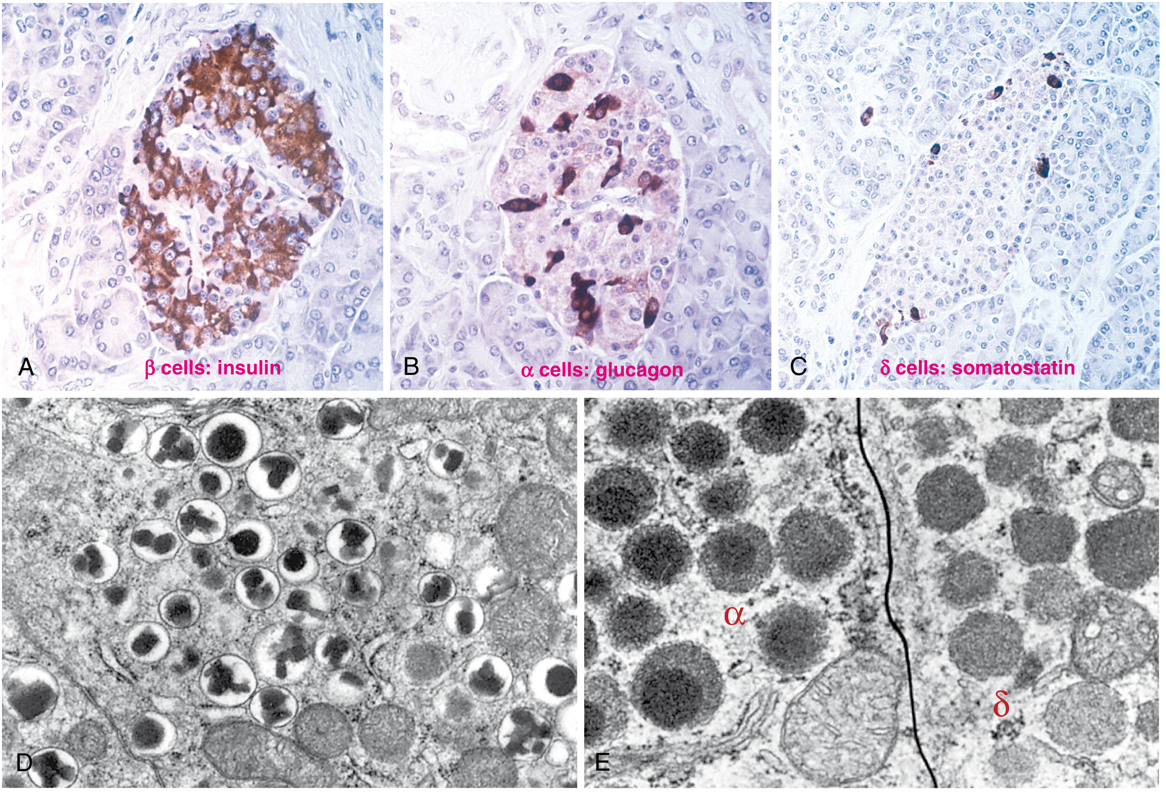
Fig. 24.27 - Hormone production in pancreatic islet cells. β cells secrete insulin (A), α cells secrete glucagon (B), δ cells secrete somatostatin (C).
Comparative Features: Type 1 vs Type 2 Diabetes
| Feature | Type 1 Diabetes | Type 2 Diabetes |
|---|---|---|
| Onset | Usually childhood/adolescence | Usually adult; increasing in adolescents |
| Body weight | Normal or weight loss | Majority obese (~80%) |
| Insulin levels | Progressive decrease | Elevated early; normal/moderate decrease late |
| Autoantibodies | Anti-GAD65, anti-IA2, anti-ZnT8 present | None |
| Ketoacidosis | Common if untreated | Nonketotic hyperosmolar coma more common |
| HLA linkage | MHC class II genes (HLA-DR3, DR4); also CTLA4, PTPN22, insulin gene VNTRs | No HLA linkage |
| Susceptibility genes | Immune-related | TCF7L2, PPARG, FTO (diabetogenic/obesity-related) |
| Pathogenesis | Dysfunction in T-cell selection and regulation; autoimmune β-cell destruction | Insulin resistance + β-cell dysfunction |
| Islet changes | Leukocytic infiltrate (insulitis); islet atrophy | Amyloid deposition; mild β-cell reduction |
Type 1 Diabetes - Pathogenesis
T1D results from autoimmune destruction of β cells in genetically susceptible individuals, triggered by environmental factors.
Genetic Susceptibility
- MHC class II (HLA-DR3, HLA-DR4, or both) accounts for ~50% of genetic susceptibility.
- HLA-DR3 or DR4: 3-5x increased risk
- HLA-DR3/DR4 heterozygotes: 10x increased risk
- Protective: HLA-DR2, HLA-DQ6
- Also: polymorphisms in CTLA4 (T-cell regulation), PTPN22 (T-cell signaling), insulin gene VNTRs (thymic insulin expression)
Environmental Triggers
- Viral infections (Coxsackie B, CMV, congenital rubella) may trigger autoimmunity via molecular mimicry or direct β-cell injury.
Immunological Mechanisms
- CD4+ T-helper cells and CD8+ cytotoxic T cells infiltrate the islets ("insulitis")
- Autoantibodies (anti-GAD65, anti-IA2, anti-ZnT8) appear years before clinical onset
- Progressive β-cell destruction → absolute insulin deficiency
Type 2 Diabetes - Pathogenesis
T2D results from the combination of insulin resistance and β-cell dysfunction.
1. Insulin Resistance
The primary defect is resistance to insulin action in target tissues (muscle, liver, adipose):
- Reduced uptake of glucose in muscle; reduced glycogen synthesis
- Failure of insulin to suppress hepatic gluconeogenesis
- Increased free fatty acid (FFA) release from adipocytes
Contributing factors:
- Obesity (especially central/visceral adiposity) - most important risk factor
- Physical inactivity
- Adipokines: TNF-α and IL-6 promote insulin resistance; adiponectin (protective) is reduced in obesity
- Excess FFAs lead to lipid accumulation in liver and muscle (lipotoxicity)
- Metabolic-associated steatotic liver disease (MASLD) is common in T2D/metabolic syndrome
2. β-Cell Dysfunction
β-cell function initially compensates (hyperinsulinism) but eventually "burns out":
- Lipotoxicity: excess FFAs impair β-cell function and attenuate insulin release
- Glucotoxicity: chronic hyperglycemia itself impairs β-cell function
- Abnormal incretin effect: reduced GIP and GLP-1 secretion → reduced insulin release
- Amyloid (IAPP) deposition in islets - present in >90% of long-standing T2D islets
- Genetic polymorphisms in insulin-secretion genes (TCF7L2, etc.)
The Incretin System
- GIP and GLP-1 are secreted by intestinal cells after oral food intake.
- They increase insulin secretion, suppress glucagon, delay gastric emptying, and promote satiety.
- DPP-4 degrades GLP-1/GIP → DPP-4 inhibitors slow this degradation.
- GLP-1 receptor agonists and tirzepatide (dual GIP/GLP-1 agonist) are now key treatments for T2D; also approved for obesity.
Insulin Action (Normal Physiology)
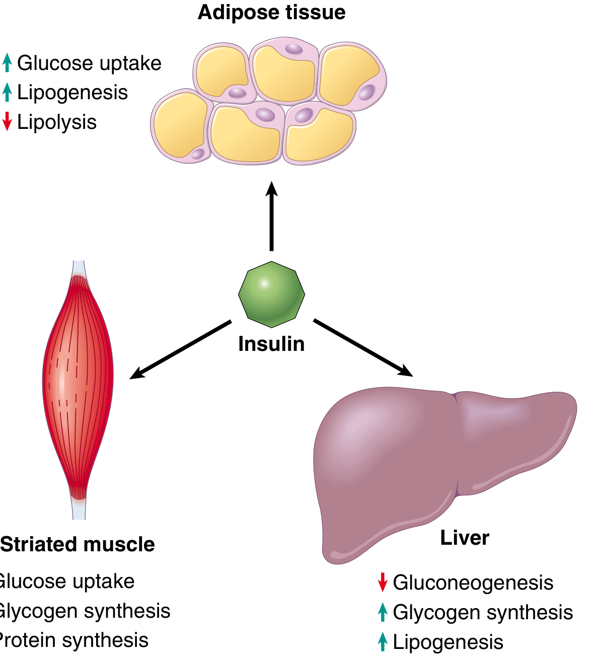
Fig. 24.29 - Insulin promotes glucose uptake and glycogen synthesis in muscle; lipogenesis and inhibits lipolysis in adipose tissue; glycogen synthesis and lipogenesis while suppressing gluconeogenesis in liver.
Insulin is the most potent anabolic hormone - principal targets are striated muscle (~2/3 of body weight) and adipocytes:
- Muscle: glucose → glycogen or ATP; amino acid uptake → protein synthesis
- Adipose: glucose → triglyceride synthesis (lipogenesis); inhibits lipolysis
- Liver: suppresses gluconeogenesis; promotes glycogen synthesis and lipogenesis
Monogenic Forms of Diabetes
MODY (Maturity-Onset Diabetes of the Young)
- Germline loss-of-function mutations; autosomal dominant, onset usually <25 years
- Glucokinase (GCK) mutations: altered "set point" for insulin secretion → hyperglycemia but usually no microvascular complications
- Other MODY forms (HNF-1α, HNF-4α, etc.) → frank diabetes with complications
- Insulin gene mutations also identified as a cause
Defects in Insulin Receptor Signaling
- Rare insulin receptor mutations → severe insulin resistance syndromes
Acute Metabolic Complications
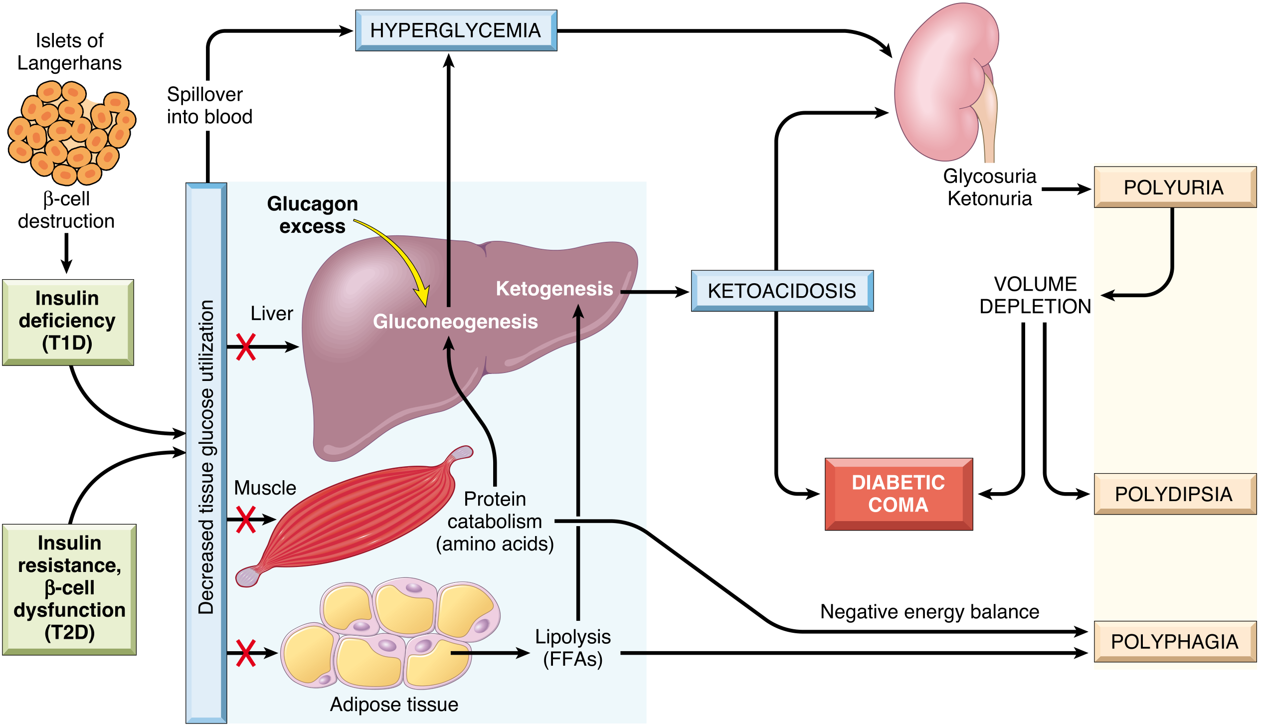
Fig. 24.33 - Metabolic cascade in insulin deficiency: decreased tissue glucose utilization → hyperglycemia → glycosuria → polyuria → polydipsia; lipolysis → FFAs → ketogenesis → ketoacidosis; protein catabolism → polyphagia; together culminating in diabetic coma.
Diabetic Ketoacidosis (DKA) - Primarily T1D
- Absolute insulin deficiency → lipolysis (FFAs) → hepatic ketogenesis
- Glucagon excess promotes gluconeogenesis
- Hyperglycemia → osmotic diuresis → glycosuria, ketonuria, polyuria
- Volume depletion → polydipsia
- Negative energy balance → polyphagia
- Kussmaul breathing, nausea, vomiting → coma if untreated
Hyperosmolar Nonketotic Coma - More common in T2D
- Extreme hyperglycemia (600-1200 mg/dL) without ketoacidosis (residual insulin prevents ketogenesis)
- Severe dehydration; no nausea/vomiting to prompt early medical attention
Hypoglycemia
- Common with insulin or sulfonylurea therapy
- Causes: missed meals, excess exercise, overdose
- Symptoms: dizziness, confusion, sweating, palpitations, tachycardia → loss of consciousness
- "Hypoglycemia unawareness" in long-standing disease
- Treat immediately with oral or IV glucose
Chronic Complications of Diabetes
Chronic complications first appear ~15-20 years after onset. Their severity correlates with both the degree and duration of hyperglycemia (evidenced by HbA1c control trials).
Glycemic Monitoring
- HbA1c = gold standard for long-term glycemic control (reflects ~120-day average)
- Target: HbA1c <7% in most patients
- Time-in-range (from continuous glucose monitors) is an emerging, potentially superior metric
Pathogenesis of Chronic Complications (4 Mechanisms)
-
Advanced Glycation End-Products (AGEs)
- Non-enzymatic glycosylation of proteins and lipids
- AGEs cross-link collagen in vessel walls → reduced compliance, thickening
- AGE receptors (RAGE) on macrophages/endothelium → cytokine release, ROS → vascular injury
- HbA1c is itself a product of this mechanism
-
Activation of Protein Kinase C (PKC)
- Intracellular hyperglycemia → diacylglycerol (DAG) → PKC activation
- PKC alters VEGF, TGF-β, NF-κB → vascular permeability, neovascularization, extracellular matrix synthesis, and inflammation
-
Polyol Pathway (Intracellular Glucose Metabolism)
- Aldose reductase converts excess glucose → sorbitol → fructose
- Intracellular sorbitol accumulation → osmotic stress, ROS
- Important in cells that do not require insulin for glucose uptake (lens, peripheral nerves, kidney)
-
Hexosamine Pathway
- Excess glucose → glucosamine-6-phosphate → alters O-GlcNAc modification of proteins → epigenetic changes, impaired signaling
Morphology of Chronic Diabetic Lesions
Diabetic Macrovascular Disease (Atherosclerosis)
- Accelerated atherosclerosis of aorta and large/medium muscular arteries
- 2-4x increased risk of coronary artery disease vs. nondiabetics; 4x higher risk of dying from cardiovascular complications
- Myocardial infarction equally common in diabetic females as in diabetic males (unlike non-diabetic women who are relatively protected in reproductive years)
- Hypertension in ~75% of T2D patients; dyslipidemia (↑TG, ↑LDL, ↓HDL) common
Hyaline Arteriolosclerosis
- Thickening and hyalinization of afferent (and sometimes efferent) arteriolar walls in the kidney
- Similar changes in other tissues; contributes to nephrosclerosis
Diabetic Nephropathy
One of the most important causes of chronic renal failure.
- Glomerular lesions (most characteristic):
- Glomerular basement membrane (GBM) thickening - earliest change
- Diffuse mesangial sclerosis: diffuse increase in mesangial matrix and cells; correlates with proteinuria and progression to renal failure
- Nodular glomerulosclerosis (Kimmelstiel-Wilson lesion): ovoid/spherical deposits of matrix in the mesangium periphery - pathognomonic of diabetic nephropathy; associated with microaneurysm formation
- Vascular lesions: hyaline arteriolosclerosis (efferent arteriole involvement is characteristic of diabetes)
- Tubular lesions: "Armanni-Ebstein lesion" - glycogen accumulation in tubular epithelial cells (now rarely seen with good glycemic control)
- Increased susceptibility to infection: pyelonephritis and papillary necrosis more common in diabetics
Diabetic Ocular Complications
- Cataract: glucose → sorbitol accumulation in lens → opacification
- Glaucoma: increased intraocular pressure → optic nerve damage
- Diabetic Retinopathy (most profound):
- Background (preproliferative) retinopathy: microaneurysms, dot/blot hemorrhages, hard exudates, cotton-wool spots, macular edema
- Proliferative retinopathy: neovascularization (VEGF-driven), vitreous hemorrhage, retinal detachment, blindness
Diabetic Neuropathy
- Affects up to 50% of diabetics overall; up to 80% of those with disease >15 years
- Peripheral symmetrical polyneuropathy most common (distal sensorimotor)
- Autonomic neuropathy: gastroparesis, bladder dysfunction, sexual dysfunction, orthostatic hypotension
- Mechanism: polyol pathway (sorbitol accumulation) + microvascular ischemia of nerves
Clinical Manifestations of Chronic Diabetes (Summary)
| Complication | Mechanism |
|---|---|
| Macrovascular disease (CAD, stroke, PVD) | Accelerated atherosclerosis |
| Diabetic nephropathy | Mesangial sclerosis, GBM thickening, nodular (K-W) lesions |
| Diabetic retinopathy | Background → proliferative; VEGF-driven neovascularization |
| Diabetic neuropathy | Polyol pathway + microvascular ischemia |
| Cataract | Lens sorbitol accumulation |
| Foot ulcers / amputations | Neuropathy + ischemia + infection |
| Susceptibility to infections | Impaired leukocyte function + vascular insufficiency |
Pancreatic Morphology in Diabetes
Type 1 Diabetes:
- Reduction in number and size of islets (β-cell depletion)
- Insulitis: lymphocytic infiltration (predominantly T cells) of islets
- Islet fibrosis in long-standing disease
Type 2 Diabetes:
- Subtle reduction in β-cell mass
- Amyloid (islet amyloid polypeptide / IAPP) deposition in >90% of islets - characteristic finding
- No insulitis
Source: Robbins, Cotran & Kumar - Pathologic Basis of Disease, Chapter 24 (The Endocrine Pancreas), pp. 1014-1030
This is a shared conversation. Sign in to Orris to start your own chat.