
Intravascular USG
intravascular ultrasound coronary artery IVUS image
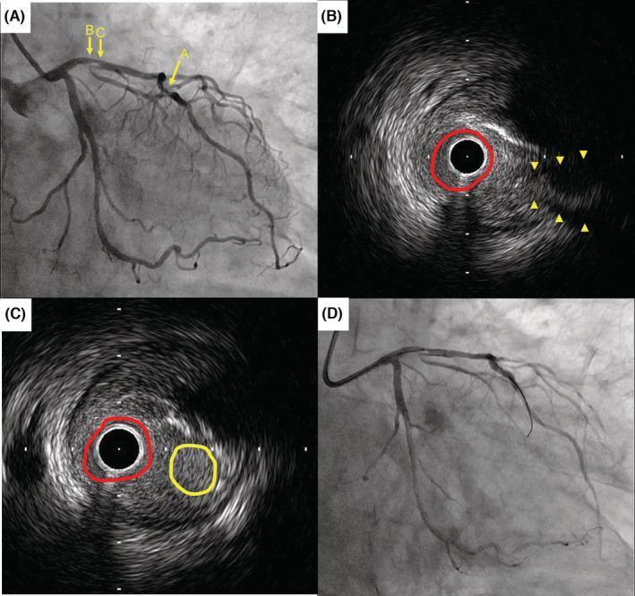
This composite diagnostic image illustrates coronary artery pathology and intravascular assessment in a patient with coronary artery disease. Panel A is a fluoroscopic coronary angiogram showing the left coronary system; arrow A highlights a 75% stenosis in the mid-left anterior descending (LAD) artery, while arrows B and C indicate the anatomical levels corresponding to the subsequent IVUS images. Panels B and C are Intravascular Ultrasound (IVUS) cross-sections. Panel B displays the vessel lumen (red circle) and the ostium of a side branch (yellow arrowheads). Panel C demonstrates a 'double lumen' appearance, where the red circle identifies the main LAD lumen and the yellow circle indicates the side branch lumen separated by an atherosclerotic plaque. Panel D is a fluoroscopic image documenting a failed reverse wire technique during a percutaneous coronary intervention (PCI) attempt. The content is designed for advanced medical education in interventional cardiology, focusing on the integration of angiography and IVUS for evaluating complex bifurcations and stenotic lesions prior to atherectomy and stenting.
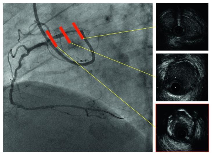
This composite diagnostic image features a coronary angiogram (left) alongside corresponding intravascular ultrasound (IVUS) cross-sections (right) of an anomalous right coronary artery (RCA). The angiogram displays a high-takeoff coronary artery with three red markers indicating the specific longitudinal segments analyzed via IVUS. The IVUS panels demonstrate diffuse, eccentric calcified plaque characterized by high-intensity echoic signals with posterior acoustic shadowing. Notably, the proximal intramural course of the ectopic artery exhibits an elliptical lumen shape, suggesting significant lateral compression from its position between the aorta and pulmonary artery. The cross-sectional views provide critical detail on vessel morphology, showing non-circular deformation and the burden of atherosclerotic plaque that is not fully captured by luminography alone. This material is designed for advanced cardiovascular training, illustrating the diagnostic utility of IVUS in assessing anatomical anomalies and plaque composition during percutaneous coronary intervention (PCI).
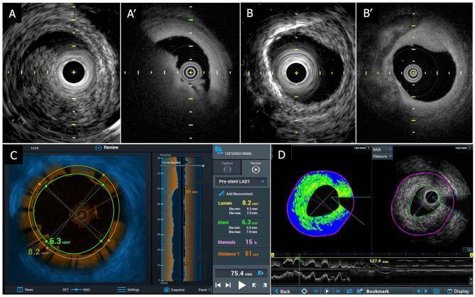
This composite diagnostic image showcases a TERUMO hybrid intravascular imaging system, comparing Intravascular Ultrasound (IVUS) and Optical Coherence Tomography (OCT) modalities in a cadaver coronary artery. Panels A and A' compare IVUS (grayscale, lower resolution) and OCT (higher resolution) for visualizing a thrombus, which appears as a distinct crescent-shaped mass in the OCT image. Panels B and B' demonstrate the visualization of vascular calcification; IVUS shows significant acoustic shadowing, while OCT provides higher superficial resolution of the calcified plaque. Panel C displays a fused IVUS-OCT interface with color mapping for tissue characterization and quantitative metrics, including lumen area (8.2 mm²), stent diameter (6.3 mm), and 15% stenosis. Panel D presents Integrated Backscatter IVUS (IB-IVUS) with color-coded plaque analysis (green and blue) and longitudinal vessel reconstruction. These images highlight the complementary nature of IVUS (greater penetration) and OCT (higher resolution) for intracoronary plaque assessment and percutaneous coronary intervention (PCI) guidance.
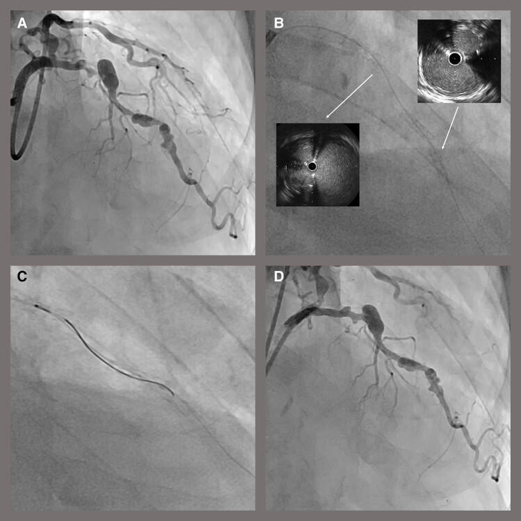
A composite of diagnostic images illustrating a percutaneous coronary intervention (PCI) procedure for coronary artery disease. Panel A is an initial coronary angiogram showing significant focal stenosis and irregular luminal diameter in a major coronary artery branch. Panel B displays an intra-procedural fluoroscopic image with two circular intravascular ultrasound (IVUS) insets; the IVUS cross-sections reveal a guidewire positioned outside the existing stent strut boundaries (extrastent tracking). Panel C demonstrates the corrective rewiring process using a double-lumen microcatheter to re-enter the true lumen. Panel D provides the final coronary angiogram following drug-coated balloon dilatation, showing a restored arterial lumen with smooth contours and resolved stenosis. This sequence serves as an educational tool for identifying stent-related complications, utilizing IVUS for diagnostic precision, and documenting successful coronary revascularization outcomes.
"intravascular ultrasound" AND "percutaneous coronary intervention"
intravascular ultrasound IVUS coronary intervention guidance current guidelines
Intravascular Ultrasound (IVUS)
What it is
How it works
- A rotating or electronic-array piezoelectric crystal at the catheter tip emits ultrasound and receives reflected signals, producing 360-degree cross-sectional images.
- Grayscale IVUS is derived from the amplitude of the radiofrequency signal.
- Based on echogenicity (not necessarily true histology), atheromas are classified into categories such as soft plaque (echolucent relative to adventitia), fibrous, calcified, and mixed plaque (Fuster and Hurst's The Heart, 15e, "Atheroma").
- Newer systems (e.g., hybrid IVUS-OCT catheters) combine IVUS's deeper tissue penetration with OCT's higher resolution for complementary plaque characterization (imaging shown below).
Clinical uses
- Sizing angioplasty balloons and stents and confirming post-stent apposition/expansion (Sabiston Textbook of Surgery, "Digital Subtraction Angiography").
- Assessing intermediate/ambiguous left main and bifurcation lesions where angiography underestimates disease severity.
- Detecting mechanisms of stent failure at follow-up: malapposition, underexpansion, neointimal hyperplasia, stent fracture, and positive/negative remodeling.
- Plaque characterization (soft/fibrous/calcified/mixed) and assessment of atherosclerotic burden (Fuster and Hurst's The Heart, 15e; Harrison's Principles of Internal Medicine, 22e).
- Surveillance for cardiac allograft vasculopathy after heart transplantation, where IVUS is considered a gold-standard early-detection tool since the disease is often angiographically and clinically silent (Braunwald's Heart Disease, 2-vol set).
- Adjunct to venography for planning endovascular or open treatment of venous disorders (Schwartz's Principles of Surgery, 11e).
- Bedside guidance for IVC filter placement, sometimes preferred over transabdominal ultrasound for visualization (Fischer's Mastery of Surgery, 8e).
IVUS vs. angiography vs. OCT
| Feature | Angiography | IVUS | OCT |
|---|---|---|---|
| View | Lumen silhouette only | Full cross-section of wall + lumen | Full cross-section, higher resolution |
| Tissue penetration | N/A | Greater (better for large plaque burden, calcification depth) | Lower (surface detail only) |
| Resolution | N/A | Lower | Higher |
| Best for | Overall vessel course, stenosis length | Vessel/plaque sizing, calcified/bulky lesions | Fine detail: thrombus, dissection, stent apposition |
Current guideline status (2024-2025 update)
- The 2024 ESC guidelines upgraded the recommendation for IVUS or OCT during complex PCI from Class IIa to Class I, reflecting robust trial evidence of improved outcomes.
- The 2021 AHA/ACC/SCAI revascularization guideline gives IVUS a Class 2a recommendation for procedural guidance, particularly for left main or complex stenting.
- A 2025 CVIT expert consensus document reinforces IVUS as standard of care for complex PCI guidance.
Recent evidence (PubMed, last ~2-3 years)
- Ahmed et al., 2025 (PMID: 39660907) - meta-analysis and meta-regression of RCTs comparing IVUS- vs angiography-guided PCI.
- Giacoppo et al., 2024, Circulation (PMID: 38344859) - network meta-analysis comparing angiography, IVUS, and OCT guidance.
- Kalsi et al., 2024 (PMID: 38350431) - focused on STEMI/stent thrombosis outcomes.
- Mrevlje et al., 2024 (PMID: 37657950) - unprotected left main disease specifically.