
Can you prepare a powerpoint presentation on Initial Assessment and Management in trauma only based on ATLS-11 guidelines, sabiston textbook of surgery and bailey and love . In 60-70 slides In simple way as a human prepares With pictures and flowcharts
ATLS primary survey ABCDE trauma flowchart
Glasgow Coma Scale chart
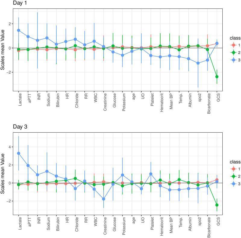
Two line graphs show the results of a latent profile analysis (LPA) comparing three patient subphenotypes (Classes 1, 2, and 3) based on clinical characteristics on Day 1 and Day 3 of ICU admission following cardiac arrest. The y-axis represents normalized Z-scores (scales mean value), while the x-axis lists clinical variables including metabolic markers (Lactate, Glucose, Bicarbonate), coagulation profiles (aPTT, INR, Platelet), organ function indicators (Bilirubin, Creatinine, UO), vital signs (HR, RR, Mean BP, Temp, spo2), and neurological status (Glasgow Coma Scale - GCS). Class 1 (red) represents a baseline phenotype with Z-scores near zero across most variables. Class 2 (green) is characterized primarily by neurological injury, demonstrated by a significantly negative Z-score for GCS by Day 3. Class 3 (blue) represents a multiple organ dysfunction phenotype, showing high positive Z-scores for Lactate, aPTT, INR, and Bilirubin on both days. This comparison chart serves as a tool for identifying clinical subphenotypes in post-cardiac arrest care, highlighting different trajectories of physiological recovery or failure.
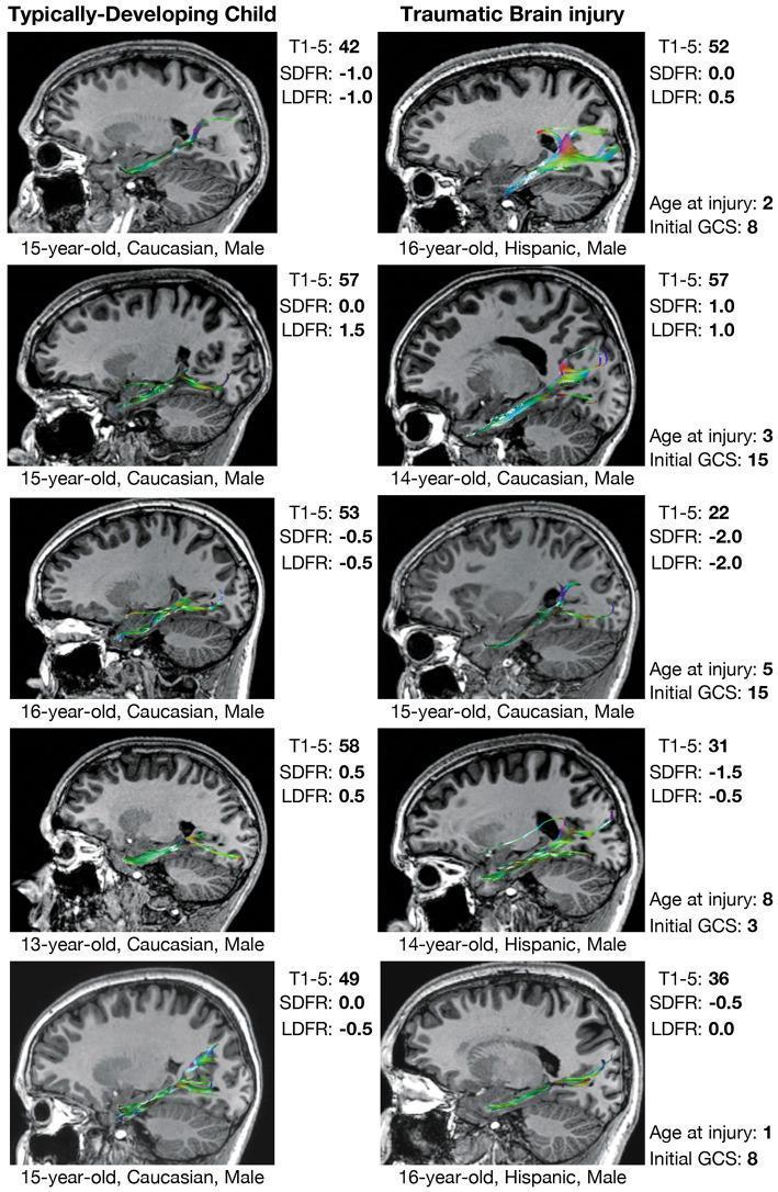
A comparison chart utilizing structural MRI and Diffusion Tensor Imaging (DTI) tractography to visualize the perforant pathway (PP) in pediatric subjects. The image is divided into two columns: Typically-Developing Children (TDC) on the left and children with a history of Traumatic Brain Injury (TBI) on the right. Each panel displays a sagittal T1-weighted brain section overlaid with color-coded white matter streamlines representing the PP, which projects toward the hippocampal formation. Data accompanying each image includes demographics (age, sex, race), injury metrics (age at injury, initial Glasgow Coma Scale score), and verbal memory performance metrics from the California Verbal Learning Test (CVLT), specifically T1-5 trials, short-delay free recall (SDFR), and long-delay free recall (LDFR). The visualization highlights qualitative differences in fiber tract morphology; TBI cases often demonstrate increased streamline density (hypertrophy) or diffused spatial distribution compared to the more compact and uniform tracts in TDC. The educational focus is on neuroplasticity and white matter developmental alterations following pediatric TBI and their correlation with long-term memory outcomes.
FAST focused assessment sonography trauma ultrasound
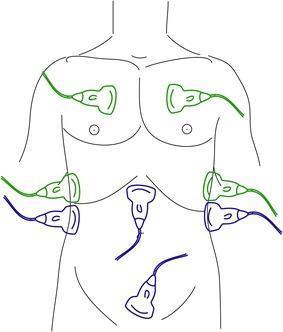
An anatomical diagram illustrating the transducer placement for the FAST (Focused Assessment with Sonography in Trauma) and eFAST (extended FAST) examinations on a human torso. The diagram uses color-coded ultrasound probes to distinguish between protocols. Blue probes indicate the standard FAST positions: the subxiphoid view (pericardial assessment), the right upper quadrant (Morison's pouch/hepatorenal space), the left upper quadrant (splenorenal space), and the suprapubic view (pelvic/rectovesical or recto-uterine pouch assessment) to detect free intraperitoneal fluid. Green probes represent the eFAST extensions: bilateral anterior thoracic views at the second to fourth intercostal spaces and mid-axillary views to assess for pneumothorax and hemothorax. This illustration is an educational tool for emergency medicine, trauma surgery, and radiology, demonstrating the systematic sonographic approach used to identify life-threatening hemorrhage or air in the thoracic and abdominal cavities during the primary survey of trauma patients.
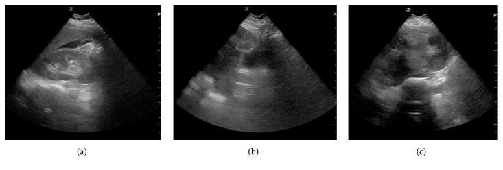
A composite of three grayscale ultrasound images (labeled a, b, and c) demonstrating clinical findings in a trauma setting. Image (a) shows a Focused Assessment with Sonography for Trauma (FAST) scan of the right upper quadrant. It reveals a wedge-shaped anechoic (black) area between the liver and the right kidney, representing free intraperitoneal fluid (hemoperitoneum) in Morison's pouch. Images (b) and (c) provide sagittal and transverse pelvic views, respectively, displaying the uterus. These images reveal incidental findings of leiomyomata uteri (uterine fibroids), appearing as multiple, well-circumscribed, hypoechoic to heterogeneously echogenic masses within the uterine myometrium. The presentation illustrates both acute traumatic pathology (positive FAST) and incidental gynecological findings that may complicate clinical management in emergency medicine and trauma surgery.
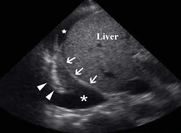
This diagnostic ultrasound image demonstrates a focused assessment of the right upper quadrant and thorax, commonly utilized in Focused Assessment with Sonography for Trauma (FAST) exams. The scan reveals a large anechoic to hypoechoic pleural collection (indicated by an asterisk) located superior to the diaphragm (delineated by white arrows). Within the pleural fluid, a collapsed right lower lung lobe is visible (marked by arrowheads), presenting with a tissue-like echogenicity relative to the surrounding effusion. The liver is visualized inferior to the diaphragm, showing typical parenchymal echotexture. Additionally, a smaller pocket of free anechoic intra-abdominal fluid (marked by a white star) is seen adjacent to the liver surface. This finding is clinically significant for the simultaneous detection of hemothorax and hemoperitoneum in a trauma setting, illustrating the utility of ultrasound in identifying pathological fluid across the diaphragmatic interface.
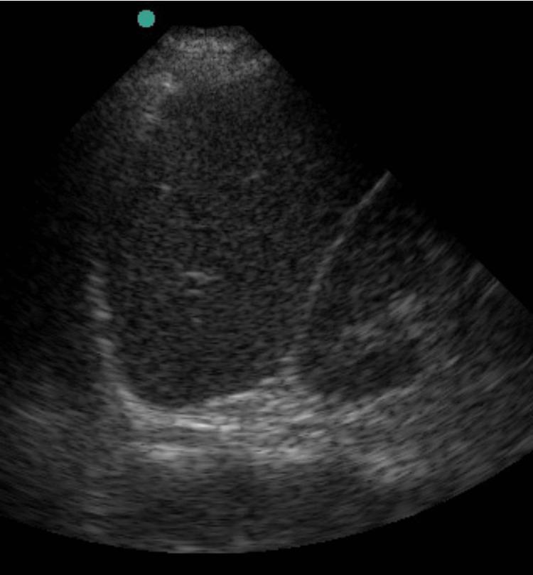
This grayscale diagnostic image is a B-mode ultrasound scan of the Left Upper Quadrant (LUQ), performed as part of a Focused Assessment with Sonography in Trauma (FAST) exam. The image displays a sector-shaped field of view focusing on the splenorenal interface. The primary organ visible is the spleen, which demonstrates a homogeneous, mid-level granular echotexture. Below the spleen, a hyperechoic (bright) linear interface is visible, representing the diaphragm and the superior pole of the left kidney (Koller's pouch). The clinical objective of this view in a trauma setting is to evaluate for the presence of anechoic (black) fluid, which would indicate hemoperitoneum or free intra-abdominal fluid in the potential space between the spleen and the kidney or in the subdiaphragmatic space. This specific scan does not show an obvious collection of free fluid, suggesting a negative LUQ FAST window at this cross-section. The image serves as a standard educational example of abdominal sonography for emergency medicine and surgical trauma protocols.
tension pneumothorax needle decompression chest tube
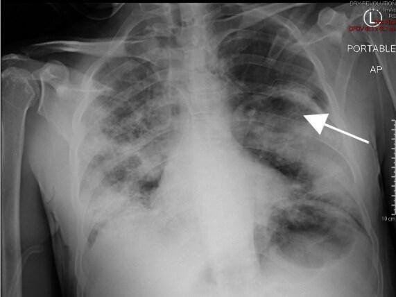
This diagnostic image is a portable anteroposterior (AP) chest radiograph illustrating the status of a patient following the treatment of a tension pneumothorax. A chest tube is visible in the left hemithorax, appropriately positioned within the pleural space. A prominent white arrow in the mid-to-upper left lung field points toward the visceral pleural line, indicating significant lung re-expansion towards the chest wall. Both lung fields exhibit diffuse, bilateral, patchy opacities and infiltrates, most prominent in the middle and lower zones, suggestive of underlying parenchymal disease or acute respiratory distress. The mediastinal contour appears stable post-decompression. This radiograph serves as a clinical example of the immediate radiological improvement and lung re-inflation achieved after emergent needle decompression and subsequent tube thoracostomy for tension pneumothorax. The image is highly relevant for medical education concerning emergency medicine, pulmonology, and critical care imaging.
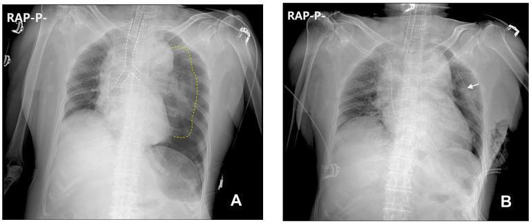
This figure presents two comparison chest radiographs (Anteroposterior view) demonstrating the presentation and resolution of a tension pneumothorax. Image A displays a massive left-sided tension pneumothorax. Key diagnostic features include a profound visceral pleural edge of the left lung (outlined with yellow dotted lines) showing total collapse toward the hilum, a significant mediastinal shift to the right (trachea highlighted by white dotted lines), and caudal displacement (flattening/depression) of the left diaphragm. These findings indicate high intrapleural pressure compromising venous return. Image B shows the clinical status following emergency intervention. A chest tube (indicated by a white arrow) is correctly positioned in the left hemithorax. The previously collapsed left lung has re-expanded, filling the hemithorax. The mediastinum and trachea have returned toward the midline, and the diaphragmatic contours show improved symmetry. This comparison illustrates the life-saving decompression of a tension pneumothorax and the restoration of normal thoracic anatomy through needle thoracostomy and tube drainage.
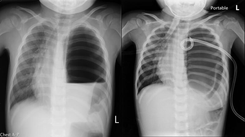
Anteroposterior (AP) chest radiographs of a pediatric patient illustrating a case of Congenital Diaphragmatic Hernia (CDH) mimicking a tension pneumothorax. The left panel shows a massive radiolucent area in the left hemithorax with a complete absence of normal lung markings and a prominent horizontal air-fluid level. A significant mediastinal shift to the right is visible, evidenced by the displacement of the heart and trachea. The right panel demonstrates the same patient after an attempted decompression with a pigtail catheter. The catheter tip is coiled within the left thoracic cavity, but the large air-filled space persists without significant lung re-expansion, a finding highly suggestive of a herniated hollow viscus (such as the stomach) rather than a simple pneumothorax. This comparison highlights the importance of distinguishing CDH from tension pneumothorax in pediatric emergencies to avoid unnecessary or harmful invasive procedures like needle decompression or tube thoracostomy.
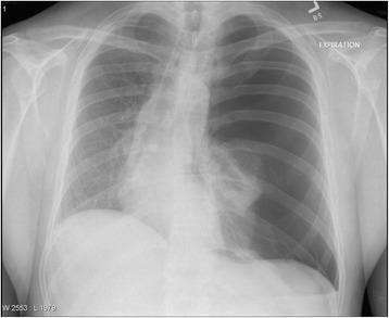
This diagnostic image is a posterior-anterior (PA) chest radiograph demonstrating a large left-sided tension pneumothorax. The left hemithorax shows marked hyperlucency with a complete absence of pulmonary vascular markings, characteristic of air in the pleural space. The left lung is fully collapsed (total atelectasis), visible as a visceral pleural edge retracted toward the hilum. There is significant mass effect evidenced by a rightward mediastinal shift, displacing the heart and trachea toward the contralateral side. Additional findings include widening of the left intercostal spaces and flattening of the left hemidiaphragm, indicating increased intrapleural pressure. This represents a medical emergency requiring immediate needle decompression or chest tube thoracostomy. The image serves as a classic educational example of tension pneumothorax for medical students and clinicians, illustrating the radiological hallmarks of pleural air accumulation and subsequent pressure-induced shift of mediastinal structures.
cricothyroidotomy surgical airway trauma
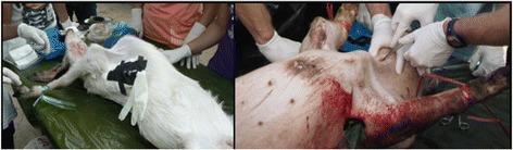
This composite of two clinical photographs illustrates the use of live animal models for advanced trauma life support training. The left image shows a goat model in the supine position being used for a cricothyroidotomy simulation; medical personnel are visible performing an incision on the anterior neck, where a black stabilization device is placed to represent the surgical airway site. The right image shows a pig model used for a chest tube thoracostomy procedure. Visible elements include an incision in the lateral thoracic wall with significant blood present, and a chest tube being manipulated by gloved personnel. These models are used in medical education to teach emergency interventions for upper airway obstruction, hemothorax, and tension pneumothorax when human cadaveric or high-fidelity synthetic simulators are unavailable. The educational focus is on identifying anatomical landmarks, surgical technique, and clinical management of life-threatening respiratory injuries in a trauma context.
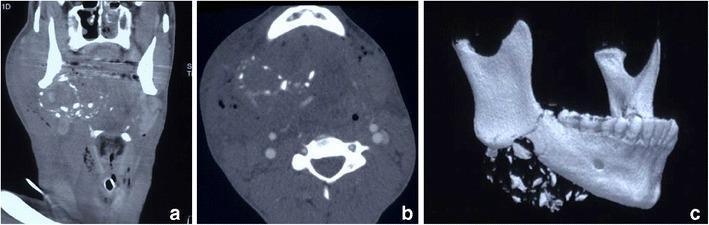
This composite diagnostic image presents a Multi-Detector Computed Tomography Angiography (MDCTA) study of a 16-year-old male with a high-velocity projectile injury to the right neck (Zone 2). Panel (a) is a coronal reformatted image showing a massive hyperdense haematoma in the right submandibular and sublingual spaces, extending laterally into the buccal soft tissues. Panel (b), an axial slice, demonstrates significant soft tissue oedema, haematoma, and bilateral surgical emphysema. Numerous in-driven bone fragments are dispersed within the right-sided soft tissue swelling. Due to severe airway distortion by haemorrhagic debris, a cricothyroidotomy cannula is visible in situ as a life-saving airway intervention. Panel (c) utilizes a 3D Volume Rendering Technique (VRT) to highlight a focal, highly comminuted fracture of the posterior right hemimandible. The 3D reconstruction clearly depicts the fragmentation and inferior dispersal of bone shards resulting from the impact. This case illustrates the utility of MDCTA in assessing complex maxillofacial trauma, secondary projectile damage from bone fragments, and the critical need for emergent surgical airway management in penetrating neck injuries.
pelvic binder hemorrhage control trauma
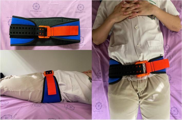
Clinical photographs demonstrating the design and application of a SAM Sling, a circumferential pelvic binder used in emergency medicine for suspected pelvic fractures. The top-left image shows the device laid flat, highlighting its components: a wide, blue padded fabric base with a black central section, a black perforated strap with precisely spaced holes for adjustable sizing, and a high-visibility orange buckle and pull-strap mechanism designed for controlled compression. The right-hand and bottom-left images show the binder applied to a patient in a supine position. The device is positioned around the greater trochanters to provide stabilization and compression to the pelvic ring. The buckle is centered anteriorly, and the strap is secured to achieve an optimal 'click' closure, indicating appropriate compressive force. This visual illustrates the correct anatomical placement (inferior to the iliac crests) and the snug, circumferential fit required for the pre-hospital or early hospital management of pelvic trauma to reduce internal pelvic volume and control hemorrhage.
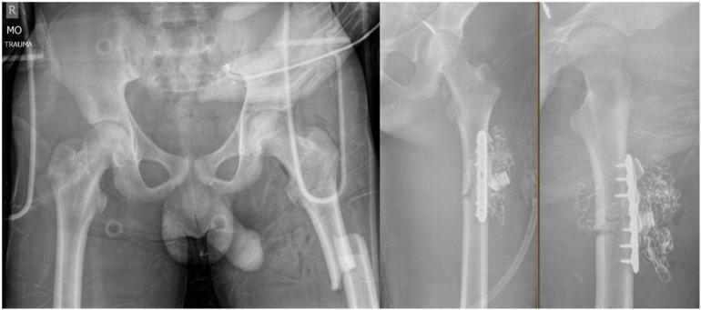
This composite diagnostic image consists of three x-ray radiographs of the pelvis and left femur in a trauma setting. The leftmost panel is an AP radiograph of the pelvis showing the bilateral hip joints, pubic symphysis, and sacroiliac regions. The middle and right panels show AP and lateral views, respectively, of the left femur. The femur radiographs reveal a subtrochanteric fracture that has undergone provisional stabilization. A radiopaque 3.5 mm locking compression plate (LCP) is affixed across the fracture site with multiple screws. Distinctive dense, coiled, radiopaque material is visible in the anterior thigh soft tissues adjacent to the plate, indicating surgical packing for damage control hemostasis. The images illustrate damage control orthopaedic management of high-energy musculoskeletal injuries, focusing on temporary internal fixation and hemorrhage control. External tubes and a pelvic binder/monitoring equipment are also visible, consistent with acute trauma management.
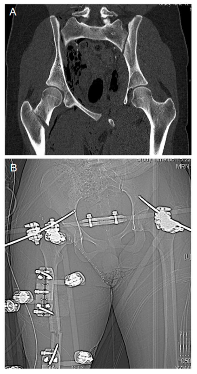
This composite figure illustrates damage control orthopedics in a severe trauma case. Figure A is a coronal CT scan image showing a displaced 'open book' pelvic ring fracture characterized by significant widening (diastasis) of the symphysis pubis and disruption of the sacroiliac joints. Additionally, a comminuted fracture of the right proximal femur with lateral displacement of the distal fragment is visible. Figure B is an anteroposterior (AP) x-ray radiograph (scout view) showing the post-operative state after emergency stabilization. An external fixation frame is in situ, utilizing percutaneous Schanz pins and connecting rods to bridge and stabilize the pelvic ring and the right femur fracture. A pelvic binder or similar radiopaque compression device is also visible across the mid-pelvis. The image demonstrates the transition from acute traumatic instability to mechanical stabilization as part of a life-saving orthopedic intervention to manage hemorrhage and prevent further soft tissue injury.
mkdir -p /tmp/workspace/trauma-atls-ppt/assets && cd /tmp/workspace/trauma-atls-ppt && python /tmp/skills/shared/scripts/fetch_images.py "https://cdn.orris.care/cdss_images/pmc_clinical_VQA_5060df12afa1185b81b7aa008a4dd32715d375d3aab5722b28d7f496591185a8.jpg" "https://cdn.orris.care/cdss_images/pmc_clinical_VQA_52fe35e51cd072c87b82ba77922fbe1f427af265117f7865127336faa7adce71.jpg" "https://cdn.orris.care/cdss_images/pmc_clinical_VQA_641460ddd79e45910461f2e1725e9909fa67e0457e22ce5d5fe7e2f735d47e9d.jpg" "https://cdn.orris.care/cdss_images/pmc_clinical_VQA_617de16245647da8c84c70b38ff161d3e3fadce705df59ed23fd53a42bf0d417.jpg" > fetch_out.json 2>fetch_err.log; python3 -c " import json d = json.load(open('fetch_out.json')) import base64 names = ['fast_diagram.jpg','tension_pneumo.jpg','cricothyroidotomy.jpg','pelvic_binder.jpg'] for item, name in zip(d, names): if item.get('base64'): b64 = item['base64'].split(',',1)[1] if ',' in item['base64'] else item['base64'] with open(f'assets/{name}','wb') as f: f.write(base64.b64decode(b64)) print('saved', name) else: print('FAILED', item.get('url'), item.get('error')) " ls -la assets/
~/trauma-atls-ppt/build_ppt.py
#!/usr/bin/env python3
"""
Initial Assessment and Management of the Trauma Patient
Based on ATLS 11th Edition, Sabiston Textbook of Surgery, Bailey & Love's Short Practice of Surgery
"""
from pptx import Presentation
from pptx.util import Inches, Pt, Emu
from pptx.dml.color import RGBColor
from pptx.enum.text import PP_ALIGN, MSO_ANCHOR
from pptx.enum.shapes import MSO_SHAPE, MSO_CONNECTOR
from pptx.oxml.ns import qn
import copy

# ---------- THEME ----------
NAVY = RGBColor(0x0B, 0x2545, 0x69)
BLUE = RGBColor(0x13, 0x67, 0xA3)
RED = RGBColor(0xC1, 0x29, 0x1D)
LIGHT_BLUE = RGBColor(0xDD, 0xEE, 0xF7)
LIGHT_GREY = RGBColor(0xF2, 0xF4, 0xF6)
DARK = RGBColor(0x22, 0x2A, 0x33)
WHITE = RGBColor(0xFF, 0xFF, 0xFF)
GREEN = RGBColor(0x1E, 0x7A, 0x3C)
ORANGE = RGBColor(0xD9, 0x7B, 0x0B)
GOLD = RGBColor(0xB8, 0x86, 0x0B)

FONT = "Calibri"

prs = Presentation()
prs.slide_width = Inches(13.333)
prs.slide_height = Inches(7.5)
BLANK = prs.slide_layouts[6]
SW, SH = prs.slide_width, prs.slide_height


def add_slide():
    return prs.slides.add_slide(BLANK)


def set_bg(slide, color=WHITE):
    bg = slide.background
    bg.fill.solid()
    bg.fill.fore_color.rgb = color


def add_rect(slide, x, y, w, h, color, line=False):
    shp = slide.shapes.add_shape(MSO_SHAPE.RECTANGLE, x, y, w, h)
    shp.fill.solid()
    shp.fill.fore_color.rgb = color
    if line:
        shp.line.color.rgb = color
    else:
        shp.line.fill.background()
    shp.shadow.inherit = False
    return shp


def add_text(slide, x, y, w, h, text, size=18, bold=False, color=DARK, align=PP_ALIGN.LEFT,
             font=FONT, anchor=MSO_ANCHOR.TOP, italic=False, line_spacing=1.0, wrap=True):
    tb = slide.shapes.add_textbox(x, y, w, h)
    tf = tb.text_frame
    tf.word_wrap = wrap
    tf.vertical_anchor = anchor
    tf.margin_left = 0
    tf.margin_right = 0
    tf.margin_top = 0
    tf.margin_bottom = 0
    lines = text.split("\n") if isinstance(text, str) else text
    for i, line in enumerate(lines):
        p = tf.paragraphs[0] if i == 0 else tf.add_paragraph()
        p.text = line
        p.alignment = align
        p.line_spacing = line_spacing
        for r in p.runs:
            r.font.size = Pt(size)
            r.font.bold = bold
            r.font.italic = italic
            r.font.name = font
            r.font.color.rgb = color
    return tb


def header(slide, kicker, title, color=NAVY):
    add_rect(slide, 0, 0, SW, Inches(1.15), color)
    add_rect(slide, 0, Inches(1.15), SW, Pt(3), RED)
    add_text(slide, Inches(0.55), Inches(0.12), Inches(11), Inches(0.35), kicker.upper(),
              size=13, bold=True, color=RGBColor(0xBF, 0xD7, 0xEA))
    add_text(slide, Inches(0.55), Inches(0.42), Inches(12), Inches(0.7), title,
              size=28, bold=True, color=WHITE)


def footer(slide, num):
    add_text(slide, Inches(0.55), Inches(7.18), Inches(8), Inches(0.3),
              "Initial Assessment & Management of Trauma  |  ATLS-11 \u2022 Sabiston \u2022 Bailey & Love",
              size=9, color=RGBColor(0x8A, 0x8A, 0x8A))
    add_text(slide, Inches(12.4), Inches(7.18), Inches(0.5), Inches(0.3), str(num),
              size=10, color=RGBColor(0x8A, 0x8A, 0x8A), align=PP_ALIGN.RIGHT)


def bullets_box(slide, x, y, w, h, items, size=17, color=DARK, gap=6, bold_lead=True):
    tb = slide.shapes.add_textbox(x, y, w, h)
    tf = tb.text_frame
    tf.word_wrap = True
    first = True
    for item in items:
        if isinstance(item, tuple):
            level, txt = item
        else:
            level, txt = 0, item
        p = tf.paragraphs[0] if first else tf.add_paragraph()
        first = False
        p.space_after = Pt(gap)
        p.line_spacing = 1.05
        marker = "\u25B8  " if level == 0 else "\u2013  "
        p.text = marker + txt
        p.level = 0
        r = p.runs[0]
        r.font.size = Pt(size if level == 0 else size - 2)
        r.font.name = FONT
        r.font.color.rgb = color if level == 0 else RGBColor(0x50, 0x58, 0x60)
        r.font.bold = False
        if level == 1:
            p.text = "     " + p.text
    return tb


def title_content_slide(kicker, title, bullets, num, note=None, color=NAVY, size=18):
    s = add_slide()
    set_bg(s)
    header(s, kicker, title, color)
    bullets_box(s, Inches(0.7), Inches(1.55), Inches(11.9), Inches(5.0), bullets, size=size)
    if note:
        add_rect(s, Inches(0.7), Inches(6.55), Inches(11.9), Inches(0.5), LIGHT_BLUE)
        add_text(s, Inches(0.85), Inches(6.6), Inches(11.6), Inches(0.4), note, size=12,
                  italic=True, color=NAVY, anchor=MSO_ANCHOR.MIDDLE)
    footer(s, num)
    return s


def two_col_slide(kicker, title, left_head, left_items, right_head, right_items, num, color=NAVY):
    s = add_slide()
    set_bg(s)
    header(s, kicker, title, color)
    colw = Inches(5.75)
    add_rect(s, Inches(0.7), Inches(1.55), colw, Inches(0.55), LIGHT_BLUE)
    add_text(s, Inches(0.9), Inches(1.55), colw, Inches(0.55), left_head, size=17, bold=True, color=NAVY, anchor=MSO_ANCHOR.MIDDLE)
    bullets_box(s, Inches(0.7), Inches(2.3), colw, Inches(4.6), left_items, size=15)

    add_rect(s, Inches(6.85), Inches(1.55), colw, Inches(0.55), RGBColor(0xFB, 0xE4, 0xE1))
    add_text(s, Inches(7.05), Inches(1.55), colw, Inches(0.55), right_head, size=17, bold=True, color=RED, anchor=MSO_ANCHOR.MIDDLE)
    bullets_box(s, Inches(6.85), Inches(2.3), colw, Inches(4.6), right_items, size=15)
    footer(s, num)
    return s


def section_slide(section_no, title, subtitle, num):
    s = add_slide()
    set_bg(s, NAVY)
    add_rect(s, 0, Inches(3.1), SW, Inches(0.05), RED)
    add_text(s, Inches(1), Inches(2.0), Inches(4), Inches(1), f"SECTION {section_no}", size=20, bold=True, color=RGBColor(0x8FBFE3, ) if False else RGBColor(0x8F,0xBF,0xE3))
    add_text(s, Inches(1), Inches(2.6), Inches(11.3), Inches(1.2), title, size=40, bold=True, color=WHITE)
    add_text(s, Inches(1), Inches(3.7), Inches(11), Inches(1), subtitle, size=18, italic=True, color=RGBColor(0xCF,0xE0,0xEF))
    footer_light(s, num)
    return s


def footer_light(slide, num):
    add_text(slide, Inches(0.55), Inches(7.18), Inches(8), Inches(0.3),
              "Initial Assessment & Management of Trauma  |  ATLS-11 \u2022 Sabiston \u2022 Bailey & Love",
              size=9, color=RGBColor(0x9F, 0xB6, 0xCC))
    add_text(slide, Inches(12.4), Inches(7.18), Inches(0.5), Inches(0.3), str(num),
              size=10, color=RGBColor(0x9F, 0xB6, 0xCC), align=PP_ALIGN.RIGHT)


def title_slide():
    s = add_slide()
    set_bg(s, NAVY)
    add_rect(s, 0, Inches(5.6), SW, Inches(0.08), RED)
    add_text(s, Inches(1), Inches(1.6), Inches(11.3), Inches(0.5), "TRAUMA CARE \u2022 TEACHING DECK", size=16, bold=True, color=RGBColor(0x8F,0xBF,0xE3))
    add_text(s, Inches(1), Inches(2.15), Inches(11.3), Inches(1.9), "Initial Assessment and\nManagement of the Trauma Patient", size=42, bold=True, color=WHITE, line_spacing=1.05)
    add_text(s, Inches(1), Inches(4.35), Inches(11), Inches(0.9), "The Primary Survey, Resuscitation, and Secondary Survey \u2013 A Practical, Step-by-Step Approach", size=19, italic=True, color=RGBColor(0xCF,0xE0,0xEF))
    add_text(s, Inches(1), Inches(6.0), Inches(11), Inches(0.6), "Based on ATLS 11th Edition \u2022 Sabiston Textbook of Surgery \u2022 Bailey & Love's Short Practice of Surgery", size=14, color=RGBColor(0xCF,0xE0,0xEF))
    add_text(s, Inches(1), Inches(6.6), Inches(11), Inches(0.4), "For undergraduate / postgraduate surgical teaching", size=12, color=RGBColor(0x9F,0xB6,0xCC))
    return s


def flow_box(slide, x, y, w, h, text, color, txt_color=WHITE, size=14, bold=True):
    shp = slide.shapes.add_shape(MSO_SHAPE.ROUNDED_RECTANGLE, x, y, w, h)
    shp.fill.solid(); shp.fill.fore_color.rgb = color
    shp.line.color.rgb = WHITE; shp.line.width = Pt(1.5)
    shp.shadow.inherit = False
    tf = shp.text_frame
    tf.word_wrap = True
    tf.margin_left = Pt(4); tf.margin_right = Pt(4); tf.margin_top = Pt(2); tf.margin_bottom = Pt(2)
    p = tf.paragraphs[0]
    p.text = text
    p.alignment = PP_ALIGN.CENTER
    for r in p.runs:
        r.font.size = Pt(size); r.font.bold = bold; r.font.color.rgb = txt_color; r.font.name = FONT
    tf.vertical_anchor = MSO_ANCHOR.MIDDLE
    return shp


def arrow_down(slide, x, y, w, h, color=NAVY):
    shp = slide.shapes.add_shape(MSO_SHAPE.DOWN_ARROW, x, y, w, h)
    shp.fill.solid(); shp.fill.fore_color.rgb = color
    shp.line.fill.background()
    shp.shadow.inherit = False
    return shp


def arrow_right(slide, x, y, w, h, color=NAVY):
    shp = slide.shapes.add_shape(MSO_SHAPE.RIGHT_ARROW, x, y, w, h)
    shp.fill.solid(); shp.fill.fore_color.rgb = color
    shp.line.fill.background()
    shp.shadow.inherit = False
    return shp


def abcde_flow_slide(num):
    s = add_slide(); set_bg(s)
    header(s, "Primary Survey", "The ABCDE Approach \u2013 Overview")
    items = [
        ("A", "Airway maintenance with\nC-spine protection", RED),
        ("B", "Breathing and\nventilation", ORANGE),
        ("C", "Circulation with\nhaemorrhage control", GOLD if False else RGBColor(0xB8,0x86,0x0B)),
        ("D", "Disability \u2013\nneurological status", GREEN),
        ("E", "Exposure /\nEnvironmental control", BLUE),
    ]
    n = len(items)
    total_w = Inches(11.9)
    gap = Inches(0.25)
    box_w = Inches((11.9 - 0.25*4) / 5)
    x = Inches(0.7)
    y = Inches(2.3)
    box_h = Inches(2.6)
    for i, (letter, label, color) in enumerate(items):
        bx = Inches(0.7) + i * (box_w + gap)
        box = slide_box_letter(s, bx, y, box_w, box_h, letter, label, color)
        if i < n - 1:
            arrow_right(s, bx + box_w + Inches(0.02), y + box_h/2 - Inches(0.15), gap - Inches(0.02), Inches(0.3), NAVY)
    add_text(s, Inches(0.7), Inches(5.2), Inches(11.9), Inches(0.5),
              "Perform in strict sequence \u2013 treat life-threatening problems as soon as they are found, then move on.",
              size=15, italic=True, color=NAVY, align=PP_ALIGN.CENTER)
    bullets_box(s, Inches(0.7), Inches(5.8), Inches(11.9), Inches(1.2), [
        "Simultaneous team approach in practice, but always assessed and re-assessed in this order",
        "Any deterioration at any point \u2192 go back to Airway and start again"], size=14)
    footer(s, num)
    return s


def slide_box_letter(slide, x, y, w, h, letter, label, color):
    box = slide.shapes.add_shape(MSO_SHAPE.ROUNDED_RECTANGLE, x, y, w, h)
    box.fill.solid(); box.fill.fore_color.rgb = color
    box.line.color.rgb = WHITE; box.line.width = Pt(2)
    box.shadow.inherit = False
    tf = box.text_frame
    tf.word_wrap = True
    tf.margin_top = Pt(6)
    p0 = tf.paragraphs[0]
    p0.text = letter
    p0.alignment = PP_ALIGN.CENTER
    p0.runs[0].font.size = Pt(40); p0.runs[0].font.bold = True; p0.runs[0].font.color.rgb = WHITE; p0.runs[0].font.name = FONT
    p1 = tf.add_paragraph()
    p1.text = label
    p1.alignment = PP_ALIGN.CENTER
    for r in p1.runs:
        r.font.size = Pt(13); r.font.bold = True; r.font.color.rgb = WHITE; r.font.name = FONT
    tf.vertical_anchor = MSO_ANCHOR.MIDDLE
    return box


def table_slide(kicker, title, headers, rows, num, col_widths=None, note=None, header_color=NAVY):
    s = add_slide(); set_bg(s)
    header(s, kicker, title)
    rowsn = len(rows) + 1
    colsn = len(headers)
    tbl_w = Inches(11.9); tbl_h = Inches(0.55 * rowsn)
    tbl_h = min(tbl_h, Inches(5.0))
    x, y = Inches(0.7), Inches(1.55)
    graphic_frame = s.shapes.add_table(rowsn, colsn, x, y, tbl_w, tbl_h)
    table = graphic_frame.table
    if col_widths:
        for i, cw in enumerate(col_widths):
            table.columns[i].width = Inches(cw)
    for j, htext in enumerate(headers):
        cell = table.cell(0, j)
        cell.text = htext
        cell.fill.solid(); cell.fill.fore_color.rgb = header_color
        for p in cell.text_frame.paragraphs:
            p.alignment = PP_ALIGN.CENTER
            for r in p.runs:
                r.font.bold = True; r.font.size = Pt(14); r.font.color.rgb = WHITE; r.font.name = FONT
    for i, row in enumerate(rows, start=1):
        for j, val in enumerate(row):
            cell = table.cell(i, j)
            cell.text = str(val)
            cell.fill.solid()
            cell.fill.fore_color.rgb = LIGHT_GREY if i % 2 == 0 else WHITE
            for p in cell.text_frame.paragraphs:
                p.alignment = PP_ALIGN.CENTER if j > 0 else PP_ALIGN.LEFT
                for r in p.runs:
                    r.font.size = Pt(12.5); r.font.color.rgb = DARK; r.font.name = FONT
    if note:
        add_text(s, Inches(0.7), y + tbl_h + Inches(0.15), Inches(11.9), Inches(0.6), note, size=13, italic=True, color=NAVY)
    footer(s, num)
    return s


def image_slide(kicker, title, img_path, bullets, num, caption=None, img_w=5.6):
    s = add_slide(); set_bg(s)
    header(s, kicker, title)
    pic_left = Inches(7.3)
    pic_top = Inches(1.7)
    pic = s.shapes.add_picture(img_path, pic_left, pic_top, width=Inches(img_w))
    if pic.height > Inches(4.6):
        ratio = Inches(4.6) / pic.height
        pic.height = Inches(4.6)
        pic.width = int(pic.width * ratio)
        pic.left = Inches(13.333 - 0.5) - pic.width
    if caption:
        add_text(s, pic_left, pic_top + pic.height + Inches(0.08), Inches(img_w), Inches(0.6), caption, size=11, italic=True, color=RGBColor(0x60,0x60,0x60))
    bullets_box(s, Inches(0.65), Inches(1.7), Inches(6.3), Inches(5.0), bullets, size=16)
    footer(s, num)
    return s


# =========================================================
# BUILD SLIDES
# =========================================================
n = 0
def nx():
    global n
    n += 1
    return n

# 1. Title
title_slide(); nx()

# 2. Objectives
title_content_slide("Introduction", "Learning Objectives", [
    "Understand the concept of the \u201cGolden Hour\u201d and time-critical trauma care",
    "Describe the sequence of the ATLS primary survey (ABCDE)",
    "Recognise and manage immediately life-threatening injuries",
    "Perform a systematic secondary survey (head-to-toe)",
    "Understand adjuncts to primary and secondary survey",
    "Appreciate the need for continuous re-evaluation and safe transfer",
], nx(), note="Approach is identical whether the mechanism is blunt, penetrating, or burn injury.")

# 3. Burden of trauma
title_content_slide("Introduction", "Why Trauma Care Matters", [
    "Trauma is one of the leading causes of death worldwide across all age groups",
    "In the USA, injury is the leading cause of death from age 1 to 44 years, and remains in the top 10 causes beyond age 65 (Sabiston)",
    "Overall trauma is the 4th leading cause of death, behind cardiac disease, malignancy and COVID-19",
    "Firearm injuries have now overtaken road traffic injuries as the leading cause of death in the 1\u201325 year age group in the USA",
    "Trauma deaths follow a trimodal distribution: immediate (seconds\u2013minutes), early (minutes\u2013hours \u2013 the window ATLS targets), and late (days\u2013weeks, from sepsis/organ failure)",
], nx())

# 4. Golden hour
title_content_slide("Introduction", "The \u201cGolden Hour\u201d Concept", [
    "Coined by R. Adams Cowley \u2013 the first hour after major injury is the most critical window to prevent death",
    "Rapid primary survey + resuscitation + transfer to definitive care saves lives",
    "\u201cGolden hour\u201d is not a strict 60 minutes \u2013 it means: treat fast, do not delay",
    "Goal of initial assessment: identify and treat life-threatening problems in order of priority, not necessarily reach a complete diagnosis immediately",
], nx(), note="Philosophy: \u2018Treat first what kills first.\u2019")

# 5. ATLS overview flow
s = add_slide(); set_bg(s); header(s, "Introduction", "The ATLS Sequence \u2013 Overview")
steps = ["Preparation", "Triage", "Primary\nSurvey (ABCDE)", "Resuscitation", "Adjuncts to\nPrimary Survey",
         "Secondary\nSurvey", "Adjuncts to\nSecondary Survey", "Re-evaluation", "Definitive Care /\nTransfer"]
colors = [NAVY, BLUE, RED, ORANGE, RGBColor(0xB8,0x86,0x0B), GREEN, BLUE, NAVY, RED]
box_w = Inches(1.28); gap = Inches(0.10)
xstart = Inches(0.55); y = Inches(2.6); box_h = Inches(1.7)
for i, (t, c) in enumerate(zip(steps, colors)):
    bx = xstart + i*(box_w+gap)
    flow_box(s, bx, y, box_w, box_h, t, c, size=12)
    if i < len(steps)-1:
        arrow_right(s, bx+box_w, y+box_h/2-Inches(0.12), gap, Inches(0.24), DARK)
add_text(s, Inches(0.55), Inches(4.7), Inches(12.2), Inches(1.6),
         "This is a repeatable cycle: at every stage the patient is continuously re-assessed, and the team returns to\n\u2018A\u2019 if the patient deteriorates at any point.", size=16, italic=True, color=NAVY, align=PP_ALIGN.CENTER)
footer(s, nx())

# ---- SECTION 1: Preparation & Triage ----
section_slide(1, "Preparation and Triage", "Getting ready before the patient arrives", nx())

title_content_slide("Preparation", "Pre-Hospital Phase", [
    "Coordination with receiving hospital \u2013 early notification of an incoming trauma call",
    "Airway maintenance, control of external haemorrhage and shock, immobilisation of the patient",
    "Rapid extrication and transport to the closest appropriate facility (trauma system / triage protocols)",
    "Time on scene should be minimised \u2013 \u201cscoop and run\u201d for unstable patients",
    "Gather AMPLE history and mechanism of injury information to hand over to the trauma team",
], nx())

title_content_slide("Preparation", "In-Hospital Phase", [
    "Resuscitation area prepared in advance: airway equipment, warmed IV fluids, monitors ready and checked",
    "Personal protective equipment donned by all staff before patient arrival",
    "Trauma team assembled with clearly assigned roles (see next slide)",
    "Laboratory and blood bank alerted; massive transfusion protocol placed on stand-by if indicated",
    "Relevant specialists (neurosurgery, orthopaedics, anaesthesia) informed early if mechanism suggests need",
], nx(), note="Sabiston: \u2018Early hospital notification allows the trauma team to assemble with roles assigned before arrival.\u2019")

# Trauma team roles flow
s = add_slide(); set_bg(s); header(s, "Preparation", "The Trauma Team \u2013 Roles")
roles = [("Team Leader", "Directs resuscitation,\nATLS-trained", NAVY),
         ("Airway Doctor", "Airway + C-spine\ncontrol", RED),
         ("Primary/Secondary\nSurvey Doctor", "Systematic\nexamination", ORANGE),
         ("Circulation Nurses (x2)", "IV access, fluids,\nblood, drugs", GREEN),
         ("Scribe", "Documents times,\nfindings, drugs", BLUE),
         ("Radiographer /\nOthers", "Imaging, specialty\nsupport on call", RGBColor(0xB8,0x86,0x0B))]
cols = 3
bw, bh = Inches(3.75), Inches(1.9)
gx, gy = Inches(0.25), Inches(0.35)
x0, y0 = Inches(0.7), Inches(1.7)
for i, (t, d, c) in enumerate(roles):
    r, cidx = divmod(i, cols)
    bx = x0 + cidx*(bw+gx)
    by = y0 + r*(bh+gy)
    box = flow_box(s, bx, by, bw, bh, t + "\n" + d, c, size=14)
add_text(s, Inches(0.7), Inches(6.35), Inches(11.9), Inches(0.6),
          "Clear role allocation + closed-loop communication = fewer errors, faster resuscitation.", size=14, italic=True, color=NAVY)
footer(s, nx())

title_content_slide("Triage", "Triage \u2013 Sorting Patients by Priority", [
    "Triage = sorting patients based on need for treatment and available resources",
    "Multiple casualties (number manageable): treat life-threatening + multi-system injuries first",
    "Mass casualty (number exceeds capacity): treat patients with the greatest chance of survival, using least time/resources/personnel first",
    "Triage categories commonly used: Immediate, Urgent, Delayed, Expectant",
    "Triage is re-assessed continuously as patients' conditions change",
], nx())

# ---- SECTION 2: Primary Survey Overview ----
section_slide(2, "The Primary Survey", "ABCDE \u2013 identify and treat what kills first", nx())

abcde_flow_slide(nx())

title_content_slide("Primary Survey", "General Principles", [
    "Primary survey = rapid, simultaneous assessment and treatment of life-threatening injuries",
    "Performed in the fixed order A-B-C-D-E, but in a real team many steps happen at the same time",
    "Vital functions are assessed quickly; if a problem is found, treat it immediately before moving to the next letter",
    "Continuously re-assess \u2013 if the patient deteriorates, always return to \u2018A\u2019",
    "History and detailed examination are deliberately deferred to the secondary survey",
], nx())

# ---- SECTION 3: Airway ----
section_slide(3, "A \u2013 Airway", "Airway maintenance with cervical spine protection", nx())

title_content_slide("A \u2013 Airway", "Assessing the Airway", [
    "Look, listen and feel: ask the patient a question \u2013 a clear verbal reply confirms a patent airway",
    "Look for agitation (may indicate hypoxia) or a depressed conscious level (may indicate hypercarbia)",
    "Look for foreign bodies, facial/mandibular/laryngeal/tracheal fractures that can cause obstruction",
    "Listen for abnormal sounds \u2013 snoring, gurgling, stridor (partial obstruction) or hoarseness",
    "Assume a cervical spine injury in any patient with multi-system trauma, especially with head/neck injury and altered consciousness",
], nx(), note="Bailey & Love / ATLS: manage airway and C-spine together \u2013 do not extend the neck.")

two_col_slide("A \u2013 Airway", "Airway Management Techniques",
    "Basic Manoeuvres", [
        "Chin lift / jaw thrust (avoid head tilt if spinal injury suspected)",
        "Suction to clear blood, vomit, secretions",
        "Remove visible foreign bodies",
        "Oropharyngeal airway (if no gag reflex)",
        "Nasopharyngeal airway (caution in suspected base-of-skull fracture)",
    ],
    "Definitive Airway (Tube in Trachea, Cuff Inflated)", [
        "Indications: apnoea, inability to protect airway, risk of aspiration, impending obstruction, GCS \u2264 8, severe facial trauma",
        "Orotracheal intubation with in-line C-spine immobilisation \u2013 preferred first choice",
        "Rapid sequence induction as needed",
        "Surgical airway if intubation fails or is not possible",
    ], nx())

image_slide("A \u2013 Airway", "Surgical Airway \u2013 Cricothyroidotomy",
    "/tmp/workspace/trauma-atls-ppt/assets/cricothyroidotomy.jpg",
    [
        "Indicated when the airway cannot be secured by intubation (\u201ccan't intubate, can't ventilate\u201d) or in severe maxillofacial/laryngeal trauma",
        "Needle cricothyroidotomy: temporary, provides oxygenation only (not ventilation) \u2013 used in children under ~12 years",
        "Surgical cricothyroidotomy: through the cricothyroid membrane, definitive surgical airway in older children/adults",
        "Tracheostomy is not a primary survey procedure \u2013 too slow in an emergency",
    ], nx(), caption="Simulation training for cricothyroidotomy and chest tube thoracostomy")

title_content_slide("A \u2013 Airway", "Cervical Spine Protection", [
    "Assume spinal injury until excluded, in any blunt trauma above the clavicles or with altered consciousness",
    "Maintain in-line manual immobilisation during airway manoeuvres, then apply a rigid collar + blocks/tape or a spine board",
    "Do NOT apply traction; maintain a neutral, neutral-aligned position",
    "Log-roll technique used to move the patient while keeping the spine straight",
    "Clinical + radiological clearance of the C-spine happens later, once life-threats are controlled",
], nx())

# ---- SECTION 4: Breathing ----
section_slide(4, "B \u2013 Breathing", "Breathing and ventilation", nx())

title_content_slide("B \u2013 Breathing", "Assessing Breathing", [
    "Expose the chest fully; look for rate, symmetry and effort of chest wall movement",
    "Look for tracheal deviation, distended neck veins, use of accessory muscles, cyanosis",
    "Feel for tenderness, crepitus (surgical emphysema/rib fracture), a flail segment",
    "Percuss for dullness (blood) or hyper-resonance (air)",
    "Auscultate for breath sounds bilaterally and heart sounds",
    "Give high-flow oxygen to every trauma patient with a reservoir mask",
], nx())

table_slide("B \u2013 Breathing", "Six Immediately Life-Threatening Chest Injuries",
    ["Injury", "Key Clinical Clue", "Immediate Action"],
    [
        ["Airway obstruction", "Stridor, gurgling", "Airway manoeuvres / definitive airway"],
        ["Tension pneumothorax", "Absent breath sounds, tracheal shift, distended neck veins, hypotension", "Immediate needle/finger decompression, then chest drain"],
        ["Open pneumothorax", "\u201cSucking\u201d chest wound", "Three-sided occlusive dressing + chest drain"],
        ["Massive haemothorax", "Absent breath sounds + shock, dull to percussion", "Chest drain, fluid/blood resuscitation, consider thoracotomy"],
        ["Flail chest with pulmonary contusion", "Paradoxical chest wall movement, hypoxia", "Oxygen, analgesia, may need ventilatory support"],
        ["Cardiac tamponade", "Beck's triad: hypotension, muffled heart sounds, distended neck veins", "Pericardiocentesis / thoracotomy"],
    ], nx(), col_widths=[3.3,4.9,3.7], note="These six must be actively excluded during every primary survey \u2013 they kill within minutes if missed.")

image_slide("B \u2013 Breathing", "Tension Pneumothorax",
    "/tmp/workspace/trauma-atls-ppt/assets/tension_pneumo.jpg",
    [
        "A clinical diagnosis \u2013 do NOT wait for a chest X-ray",
        "Signs: severe respiratory distress, absent breath sounds on affected side, tracheal deviation away from the injury, distended neck veins, hypotension",
        "Immediate treatment: needle decompression (2nd intercostal space, mid-clavicular line) or finger thoracostomy, followed promptly by a formal chest (tube) drain",
        "Reduces venous return \u2192 obstructive shock if untreated",
    ], nx(), caption="Left tension pneumothorax before and after chest tube decompression")

title_content_slide("B \u2013 Breathing", "Open Pneumothorax & Massive Haemothorax", [
    "Open pneumothorax: large chest wall defect equalises intrathoracic and atmospheric pressure \u2013 air preferentially enters the wound",
    "Management: cover with a sterile dressing taped on three sides (acts as a one-way flutter valve), followed by a chest drain sited away from the wound",
    "Massive haemothorax: > 1500 mL blood loss into the chest, or ongoing loss > 200 mL/hour",
    "Management: fluid/blood resuscitation + chest drain; persistent bleeding is an indication for thoracotomy",
], nx())

# ---- SECTION 5: Circulation ----
section_slide(5, "C \u2013 Circulation", "Circulation with haemorrhage control", nx())

title_content_slide("C \u2013 Circulation", "Recognising Shock", [
    "Haemorrhage is the leading cause of preventable death after injury",
    "Assess: level of consciousness, skin colour/temperature, and pulse rate/character",
    "Early compensated shock can have a near-normal blood pressure \u2013 do not wait for hypotension",
    "Tachycardia is often the earliest reliable sign; a narrowing pulse pressure suggests ongoing blood loss",
    "Look actively for the source \u2013 external bleeding is obvious, internal bleeding may not be",
], nx())

table_slide("C \u2013 Circulation", "Classes of Haemorrhagic Shock (ATLS)",
    ["Parameter", "Class I", "Class II", "Class III", "Class IV"],
    [
        ["Blood loss (%)", "< 15%", "15\u201330%", "31\u201340%", "> 40%"],
        ["Heart rate", "Normal", "Mildly \u2191", "\u2191\u2191 (>120)", "\u2191\u2191\u2191 (>140)"],
        ["Blood pressure", "Normal", "Normal", "\u2193", "\u2193\u2193"],
        ["Mental status", "Slightly anxious", "Mildly anxious", "Anxious/confused", "Confused/lethargic"],
        ["Management", "Crystalloid", "Crystalloid \u00b1 blood", "Crystalloid + blood", "Massive transfusion"],
    ], nx(), col_widths=[3.0,2.2,2.2,2.2,2.3], note="Compensatory mechanisms can mask early blood loss \u2013 always treat the trend, not one reading.")

title_content_slide("C \u2013 Circulation", "Controlling External Haemorrhage", [
    "Direct firm manual pressure on the bleeding point \u2013 first-line for almost all external bleeding",
    "Elevation and pressure dressings",
    "Tourniquet for severe, life-threatening limb haemorrhage not controlled by pressure",
    "Haemostatic dressings for junctional or difficult-to-compress wounds",
    "Do not waste time clamping vessels blindly in a bleeding wound \u2013 risks nerve injury",
], nx())

title_content_slide("C \u2013 Circulation", "Occult (Hidden) Sources of Blood Loss", [
    "\u201cBlood on the floor and four more\u201d \u2013 remember 5 places significant blood can be lost:",
    (1, "1. External (visible) haemorrhage"),
    (1, "2. Chest (haemothorax)"),
    (1, "3. Abdomen (solid organ / mesenteric injury)"),
    (1, "4. Pelvis / retroperitoneum (pelvic fracture)"),
    (1, "5. Long bones (femur, tibia \u2013 closed fractures)"),
    "FAST scan, chest & pelvis X-ray, and clinical exam of limbs help localise the source quickly",
], nx())

title_content_slide("C \u2013 Circulation", "Fluid Resuscitation & Blood Products", [
    "Two large-bore peripheral IV cannulae (or intraosseous access if difficult)",
    "Send blood for group & save/crossmatch, full blood count, and other baseline tests",
    "Warmed isotonic crystalloid as an initial bridge, but move early to blood products in significant haemorrhage",
    "Balanced (1:1:1) transfusion of packed red cells, fresh frozen plasma and platelets \u2013 avoid crystalloid-only resuscitation in major bleeding",
    "Activate the Massive Transfusion Protocol early if shock is severe or ongoing",
    "Permissive hypotension and damage-control resuscitation may be used until bleeding is surgically controlled",
], nx())

image_slide("C \u2013 Circulation", "Pelvic Fracture \u2013 External Compression",
    "/tmp/workspace/trauma-atls-ppt/assets/pelvic_binder.jpg",
    [
        "Suspect pelvic fracture with high-energy trauma, pelvic instability, or perineal bruising",
        "A pelvic binder (or improvised sheet) reduces pelvic volume and helps tamponade venous/bony bleeding",
        "Apply at the level of the greater trochanters, not the iliac crests",
        "Avoid \u201cspringing\u201d the pelvis repeatedly \u2013 this can dislodge a forming clot",
        "Definitive control may need angio-embolisation or surgical fixation",
    ], nx(), caption="SAM pelvic binder \u2013 correct positioning over the greater trochanters")

title_content_slide("C \u2013 Circulation", "Adjuncts in Severe Haemorrhage", [
    "Resuscitative thoracotomy: reserved for patients arresting from trauma with signs of life en route, particularly penetrating chest trauma",
    "REBOA (Resuscitative Endovascular Balloon Occlusion of the Aorta): temporary aortic occlusion to control non-compressible torso haemorrhage",
    "Recent UK trial data showed higher early mortality with REBOA versus standard care in broad-inclusion patients \u2013 use is now more selective and protocol-driven",
    "Both are bridges to definitive haemorrhage control in a hybrid theatre/operating room, never a substitute for surgery",
], nx(), note="Sabiston: REBOA \u2018should only take place within a trauma system capable of managing definitive surgical haemostasis.\u2019")

# ---- SECTION 6: Disability ----
section_slide(6, "D \u2013 Disability", "Rapid neurological assessment", nx())

title_content_slide("D \u2013 Disability", "Quick Neurological Assessment", [
    "Establish the level of consciousness using the Glasgow Coma Scale (GCS)",
    "Examine pupil size, symmetry and reaction to light",
    "Look for lateralising signs (limb weakness) suggesting a focal brain lesion",
    "Check level of spinal cord injury if suspected",
    "Always exclude and correct hypoxia and hypotension first \u2013 they alone can depress conscious level",
    "Exclude hypoglycaemia as a reversible cause of reduced GCS",
], nx())

table_slide("D \u2013 Disability", "Glasgow Coma Scale (GCS)",
    ["Response", "Finding", "Score"],
    [
        ["Eye opening", "Spontaneous / To voice / To pain / None", "4 / 3 / 2 / 1"],
        ["Verbal", "Orientated / Confused / Words / Sounds / None", "5 / 4 / 3 / 2 / 1"],
        ["Motor", "Obeys / Localises / Withdraws / Abnormal flexion / Extension / None", "6 / 5 / 4 / 3 / 2 / 1"],
    ], nx(), col_widths=[2.8,6.6,2.5],
    note="Best possible score = 15, lowest = 3. GCS \u2264 8 = severe injury, generally needs a definitive (protected) airway. (Bailey & Love, Table 28.4)")

title_content_slide("D \u2013 Disability", "AVPU \u2013 A Quick Alternative", [
    "A \u2013 Alert",
    "V \u2013 responds to Voice",
    "P \u2013 responds to Pain",
    "U \u2013 Unresponsive",
    "Useful for a very rapid first impression, but GCS should always follow for accurate documentation and trending",
], nx())

# ---- SECTION 7: Exposure ----
section_slide(7, "E \u2013 Exposure", "Exposure and environmental control", nx())

title_content_slide("E \u2013 Exposure", "Full Exposure & Temperature Control", [
    "Completely undress the patient to look for all injuries \u2013 \u201cyou can't treat what you can't see\u201d",
    "Log-roll to inspect the back, buttocks and perineum while protecting the spine",
    "Rectal examination if indicated by mechanism/findings",
    "Prevent hypothermia: warmed IV fluids/blood, warming blankets, cover the patient promptly once examined",
    "Hypothermia worsens coagulopathy and bleeding \u2013 part of the lethal triad with acidosis and coagulopathy",
], nx(), note="Preserve dignity and modesty while ensuring complete examination.")

# ---- SECTION 8: Adjuncts to Primary Survey ----
section_slide(8, "Adjuncts to the Primary Survey", "Monitoring, lines, imaging and reassessment", nx())

title_content_slide("Adjuncts", "Monitoring During Resuscitation", [
    "Continuous ECG monitoring \u2013 arrhythmias may indicate hypoxia, hypoperfusion or cardiac injury",
    "Pulse oximetry \u2013 continuous oxygen saturation",
    "Frequent blood pressure measurement (automated cuff or arterial line in unstable patients)",
    "End-tidal CO2 (capnography) in intubated patients to confirm tube placement and adequacy of ventilation",
    "Arterial blood gas \u2013 assesses oxygenation, ventilation and base deficit/lactate (markers of shock severity)",
    "Urinary catheter to monitor hourly urine output as a guide to resuscitation (unless urethral injury suspected)",
], nx())

title_content_slide("Adjuncts", "Gastric and Urinary Catheters \u2013 Caution", [
    "Nasogastric/orogastric tube decompresses the stomach, reduces aspiration risk",
    "Avoid the nasal route if base-of-skull or severe midface fracture is suspected \u2013 use the oral route instead",
    "Urinary catheter helps monitor output, but should NOT be inserted if urethral injury is suspected",
    "Signs of urethral injury: blood at the meatus, scrotal/perineal haematoma, high-riding prostate on rectal exam",
], nx())

image_slide("Adjuncts", "FAST \u2013 Focused Assessment with Sonography in Trauma",
    "/tmp/workspace/trauma-atls-ppt/assets/fast_diagram.jpg",
    [
        "Rapid bedside ultrasound performed during/immediately after the primary survey",
        "Standard FAST windows: subxiphoid (pericardium), right upper quadrant (Morison's pouch), left upper quadrant (splenorenal), suprapubic (pelvis)",
        "eFAST adds bilateral anterior chest views to detect pneumothorax",
        "Detects free fluid (blood) rapidly \u2013 helps decide need for urgent laparotomy in the unstable patient",
        "Operator-dependent; a negative FAST does not fully exclude injury",
    ], nx(), caption="Standard probe positions for FAST (blue) and extended eFAST (green) views")

title_content_slide("Adjuncts", "Primary Survey Imaging", [
    "Portable chest X-ray \u2013 identifies pneumothorax, haemothorax, widened mediastinum, rib fractures",
    "Pelvic X-ray \u2013 identifies pelvic ring fractures needing binder/early control",
    "These are done in the resuscitation room without moving an unstable patient",
    "CT scanning is deferred until the patient is haemodynamically stable enough to leave the resuscitation area",
], nx())

title_content_slide("Adjuncts", "Consider the Need for Transfer", [
    "At the end of the primary survey, decide: can this patient be definitively managed here, or do they need transfer?",
    "Do not delay transfer for detailed secondary survey or non-essential investigations if a higher level of care is needed",
    "Communicate clearly with the receiving centre \u2013 mechanism, findings, treatment given, response",
    "Stabilise what can be stabilised, but transfer promptly if resources (e.g. neurosurgery, interventional radiology) are unavailable locally",
], nx())

# ---- SECTION 9: Secondary Survey ----
section_slide(9, "The Secondary Survey", "Head-to-toe examination once primary survey is complete", nx())

title_content_slide("Secondary Survey", "When and Why", [
    "Begins only after the primary survey is complete, life-threats addressed, and the patient is responding to resuscitation",
    "A comprehensive head-to-toe examination plus a full history",
    "Aim: find all injuries, including subtle ones that do not threaten life immediately but matter for outcome",
    "If the patient deteriorates at any point during the secondary survey \u2013 STOP and go back to the primary survey (re-check A, B, C, D, E)",
], nx(), note="Sabiston: \u2018Many significant injuries may remain undetected after the primary survey \u2013 the secondary survey uncovers these.\u2019")

table_slide("Secondary Survey", "AMPLE History",
    ["Letter", "Stands For", "Why It Matters"],
    [
        ["A", "Allergies", "Avoid triggering reactions with drugs/dressings"],
        ["M", "Medications currently used", "Anticoagulants, antihypertensives affect management"],
        ["P", "Past illnesses / Pregnancy", "Comorbidities change risk and treatment priorities"],
        ["L", "Last meal", "Aspiration risk if anaesthesia/intubation needed"],
        ["E", "Events / Environment related to injury", "Mechanism clarifies likely injury pattern"],
    ], nx(), col_widths=[1.5,4.5,5.9])

title_content_slide("Secondary Survey", "Head & Maxillofacial Examination", [
    "Inspect and palpate scalp for lacerations and haematoma (may overlie a skull fracture)",
    "Look for Battle's sign (mastoid bruising) and \u2018raccoon eyes\u2019 (periorbital bruising) \u2013 suggest base-of-skull fracture",
    "Check for CSF rhinorrhoea/otorrhoea and haemotympanum",
    "Examine pupils, visual acuity, eye movements; remove contact lenses",
    "Palpate facial bones for tenderness, step-offs, crepitus; check for septal haematoma and dental/mouth injuries",
    "A full cranial nerve examination is performed where possible",
], nx())

title_content_slide("Secondary Survey", "Neck & Cervical Spine", [
    "Maintain immobilisation until the C-spine is formally cleared clinically and/or radiologically",
    "Inspect and palpate posterior neck for tenderness, deformity, step-off",
    "Check for surgical emphysema, expanding haematoma, tracheal deviation",
    "Palpate carotid pulses and auscultate for bruits (may suggest vascular injury)",
    "Voice change may indicate laryngeal injury or airway swelling",
], nx())

title_content_slide("Secondary Survey", "Chest, Abdomen and Pelvis", [
    "Chest: re-inspect for bruising/wounds, palpate ribs/sternum/clavicles, re-auscultate for evolving pneumo/haemothorax",
    "Abdomen: inspect for bruising (seatbelt sign), palpate for tenderness/guarding, note any distension \u2013 a clinically difficult abdomen may need repeated FAST/CT or diagnostic laparoscopy/laparotomy",
    "Pelvis: assess stability once only (repeated manipulation can dislodge clots); look for perineal/scrotal bruising, blood at the meatus",
    "Rectal examination if indicated: tone, blood, high-riding prostate",
    "Vaginal examination if indicated in female patients with pelvic trauma",
], nx())

title_content_slide("Secondary Survey", "Musculoskeletal & Neurological Examination", [
    "Inspect and palpate all four limbs for deformity, wounds, tenderness, swelling",
    "Check distal pulses, capillary refill, and peripheral neurology in every injured limb",
    "Look for compartment syndrome signs \u2013 pain out of proportion, pain on passive stretch",
    "Perform a full neurological examination: GCS, cranial nerves, motor/sensory levels, reflexes",
    "Document a spinal cord injury level clearly if present",
    "Log-roll (with C-spine control) to examine the entire back and spine",
], nx())

title_content_slide("Secondary Survey", "Adjuncts to the Secondary Survey", [
    "Further, more detailed imaging once the patient is stable: CT head/chest/abdomen/pelvis, CT angiography, specific limb X-rays",
    "Contrast studies if hollow viscus/urological injury suspected",
    "Specialist review \u2013 orthopaedics, neurosurgery, maxillofacial, plastics as indicated",
    "Tetanus prophylaxis and antibiotics for contaminated/open wounds",
    "Splint fractures and dress wounds properly once life-threats are controlled",
], nx())

# ---- SECTION 10: Reevaluation & Definitive care ----
section_slide(10, "Re-evaluation and Definitive Care", "Never stop reassessing", nx())

title_content_slide("Re-evaluation", "Continuous Monitoring & Reassessment", [
    "Trauma patients are re-evaluated constantly \u2013 vital signs, urine output, GCS trend, pain",
    "New findings can appear as swelling develops or the patient becomes more cooperative",
    "Any deterioration = immediately repeat the primary survey from \u2018A\u2019",
    "Adequate pain relief is part of good trauma care and does not mask a deteriorating exam if titrated carefully",
    "Tetanus status, analgesia, and antibiotics (if indicated) should not be forgotten",
], nx())

title_content_slide("Definitive Care", "Damage Control Principles", [
    "In the physiologically exhausted patient (acidotic, coagulopathic, hypothermic \u2013 the \u2018lethal triad\u2019), prioritise survival over complete anatomical repair",
    "Damage-control surgery: rapid haemorrhage/contamination control, temporary abdominal closure, transfer to ICU to correct physiology",
    "Damage-control resuscitation: permissive hypotension, early balanced blood products, minimising crystalloid",
    "Definitive reconstructive surgery is staged once the patient is physiologically stable",
], nx())

title_content_slide("Definitive Care", "Documentation and Communication", [
    "Accurate, timed documentation of findings, treatments and the patient's response is essential (medico-legal and clinical value)",
    "A dedicated scribe during resuscitation improves accuracy",
    "Structured handover (e.g. ATMIST/SBAR) when transferring care between teams or to another hospital",
    "Keep the patient and family informed as appropriate throughout care",
], nx())

title_content_slide("Definitive Care", "Triage for Transfer to Higher-Level Care", [
    "Transfer if local resources cannot provide definitive care \u2013 e.g. no neurosurgery, no interventional radiology, no paediatric trauma service",
    "Do the primary survey, resuscitate, and stabilise as much as possible before transfer \u2013 do not delay transfer for non-essential tests",
    "Clear communication with the receiving centre in advance",
    "A trained team should accompany an unstable patient during transfer, with monitoring continued throughout",
], nx())

# ---- SECTION 11: Special populations ----
section_slide(11, "Special Situations", "Children, pregnancy and the elderly", nx())

two_col_slide("Special Situations", "Paediatric Trauma",
    "Key Anatomical/Physiological Differences", [
        "Larger head, shorter neck, more flexible airway structures",
        "Large occiput causes neck flexion \u2013 use a shoulder roll to keep neutral position",
        "Greater physiological reserve masks shock until sudden decompensation",
        "Higher risk of hypothermia due to larger surface area to body-mass ratio",
    ],
    "Practical Points", [
        "Same ABCDE approach as adults, adapted to size",
        "Weight-based drug and fluid dosing",
        "Hypoxia is the most common cause of paediatric cardiac arrest \u2013 secure airway/oxygenation early",
        "Involve child life/family support where possible",
    ], nx())

two_col_slide("Special Situations", "Pregnancy & The Elderly",
    "Pregnant Trauma Patient", [
        "Two patients \u2013 mother and fetus; mother's stabilisation is the priority",
        "After ~20 weeks, tilt the patient/uterus leftward (or manually displace uterus) to relieve caval compression and improve venous return",
        "Rh status and fetal monitoring should follow once mother is stable",
    ],
    "Elderly Trauma Patient", [
        "Reduced physiological reserve \u2013 normal vital signs can hide significant shock",
        "Beta-blockers may blunt the tachycardic response to blood loss",
        "Comorbidities and anticoagulant use increase bleeding risk and complicate management",
        "Have a lower threshold for early, aggressive resuscitation and monitoring",
    ], nx())

# ---- Summary ----
s = add_slide(); set_bg(s); header(s, "Summary", "Putting It All Together")
steps2 = ["Preparation\n& Triage", "A \u2013 Airway\n+ C-spine", "B \u2013 Breathing", "C \u2013 Circulation\n+ Haemorrhage",
          "D \u2013 Disability", "E \u2013 Exposure", "Adjuncts &\nResuscitation", "Secondary\nSurvey", "Definitive Care\n/ Transfer"]
colors2 = [NAVY, RED, ORANGE, RGBColor(0xB8,0x86,0x0B), GREEN, BLUE, NAVY, RED, GREEN]
box_w2 = Inches(1.28); gap2 = Inches(0.10)
xstart2 = Inches(0.55); y2 = Inches(1.9); box_h2 = Inches(1.7)
for i, (t, c) in enumerate(zip(steps2, colors2)):
    bx = xstart2 + i*(box_w2+gap2)
    flow_box(s, bx, y2, box_w2, box_h2, t, c, size=11.5)
    if i < len(steps2)-1:
        arrow_right(s, bx+box_w2, y2+box_h2/2-Inches(0.12), gap2, Inches(0.24), DARK)
arrow_down_box = s.shapes.add_shape(MSO_SHAPE.CURVED_DOWN_ARROW, Inches(10.5), Inches(3.8), Inches(2), Inches(1.2))
bullets_box(s, Inches(0.7), Inches(4.1), Inches(12), Inches(2.8), [
    "Golden rule: treat life-threats in order \u2013 A before B before C before D before E",
    "Re-evaluate constantly; if the patient worsens, restart from \u2018A\u2019",
    "Primary survey saves life; secondary survey saves function; definitive care completes recovery",
    "Team-based, protocol-driven care (ATLS) reduces preventable trauma deaths",
], size=16)
footer(s, nx())

title_content_slide("Summary", "Key Take-Home Points", [
    "The primary survey (ABCDE) identifies and treats immediately life-threatening problems first",
    "Haemorrhage remains the leading cause of preventable death \u2013 find it and stop it early",
    "Adjuncts (monitoring, FAST, X-rays) support but never replace clinical assessment",
    "The secondary survey is a systematic head-to-toe search for all remaining injuries, guided by AMPLE history",
    "Continuous re-evaluation throughout care is what prevents missed, deteriorating injuries",
    "Good documentation, communication and timely transfer complete safe trauma care",
], nx())

# References
s = add_slide(); set_bg(s, NAVY)
add_rect(s, 0, Inches(1.15), SW, Pt(3), RED)
add_text(s, Inches(0.55), Inches(0.42), Inches(12), Inches(0.7), "References", size=28, bold=True, color=WHITE)
add_rect(s, 0,0, SW, Inches(1.15), NAVY)
bullets_box(s, Inches(0.8), Inches(1.8), Inches(11.5), Inches(4.5), [
    "American College of Surgeons Committee on Trauma. Advanced Trauma Life Support (ATLS) Student Course Manual, 11th Edition.",
    "Sabiston Textbook of Surgery: The Biological Basis of Modern Surgical Practice \u2013 Chapter on Initial Management of the Trauma Patient.",
    "Bailey & Love's Short Practice of Surgery, 28th Edition \u2013 Chapters on Trauma / Principles of Trauma Management, Head Injury.",
], size=17, color=WHITE)
footer_light(s, nx())

prs.save("/tmp/workspace/trauma-atls-ppt/Initial_Assessment_and_Management_of_Trauma.pptx")
print("Saved. Total slides:", n)
cd /tmp/workspace/trauma-atls-ppt && python3 build_ppt.py
~/trauma-atls-ppt/build_ppt.py
Successfully replaced 1 instance(s) of the string in '/tmp/workspace/trauma-atls-ppt/build_ppt.py'
cd /tmp/workspace/trauma-atls-ppt && python3 build_ppt.py
cd /tmp/workspace/trauma-atls-ppt && ls -la *.pptx && python3 -c " from pptx import Presentation p = Presentation('Initial_Assessment_and_Management_of_Trauma.pptx') print('Slides:', len(p.slides.__iter__.__self__._sldIdLst)) "
Initial Assessment and Management of Trauma
Presentation · PPTX
- Title + Introduction (slides 1-5) – objectives, burden of trauma, the Golden Hour, ATLS sequence overview (flowchart)
- Preparation & Triage (6-10) – pre-hospital/in-hospital prep, trauma team roles diagram, triage principles
- Primary Survey Overview (11-12) – ABCDE flowchart with colored boxes and arrows
- A – Airway (13-16) – assessment, techniques, cricothyroidotomy (real clinical image), C-spine protection
- B – Breathing (17-20) – assessment, table of the 6 immediately life-threatening chest injuries, tension pneumothorax (real chest X-ray image), open pneumothorax/haemothorax
- C – Circulation (21-27) – shock recognition, Class I-IV shock table, haemorrhage control, "blood on the floor and four more," fluid/blood resuscitation, pelvic binder (real clinical photo), REBOA/thoracotomy
- D – Disability (28-30) – neuro assessment, GCS table, AVPU
- E – Exposure (31) – full exposure, hypothermia prevention
- Adjuncts to Primary Survey (32-36) – monitoring, catheters, FAST exam (real anatomical diagram), imaging, transfer decision
- Secondary Survey (37-43) – AMPLE history table, head-to-toe exam (head/face, neck/spine, chest/abdomen/pelvis, MSK/neuro), adjunct imaging
- Re-evaluation & Definitive Care (44-47) – continuous reassessment, damage control, documentation, transfer criteria
- Special Situations (48-50) – paediatric, pregnancy, elderly
- Summary (51-52) – full recap flowchart + key takeaways
- References (last slide) – ATLS-11, Sabiston, Bailey & Love