
uterine fibroid intramural management perimenopausal
uterine leiomyoma treatment management
uterine fibroid intramural ultrasound pelvis bulky uterus
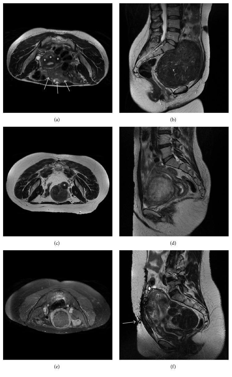
This composite of magnetic resonance imaging (MRI) scans illustrates various clinical presentations of uterine fibroids (leiomyomas) that impact suitability for magnetic resonance-guided focused ultrasound (MRgFUS). (a) Axial T2-weighted MRI shows small, hypointense intramural fibroids (asterisks) with multiple bowel loops (arrows) interposed between the abdominal wall and the uterus, presenting a risk of thermal injury to the bowel. (b) Sagittal T2-weighted MRI displays a massive fibroid occupying the pelvis, noted for its proximity to the sacrum and nerves. (c) Axial T2-weighted MRI identifies a small, pedunculated subserosal fibroid (asterisk). (d) Sagittal T2-weighted MRI depicts a 'bright' hyperintense cellular fibroid, which typically responds poorly to thermal ablation. (e) Axial T1-weighted fat-saturated post-contrast image reveals a non-enhancing fibroid, indicating pre-existing avascularity. (f) Sagittal T2-weighted MRI highlights a bulky abdominal wall scar (arrow), which can distort or absorb ultrasound energy during treatment. The collection serves to teach screening criteria, focusing on size, location, signal intensity, enhancement patterns, and acoustic window accessibility.
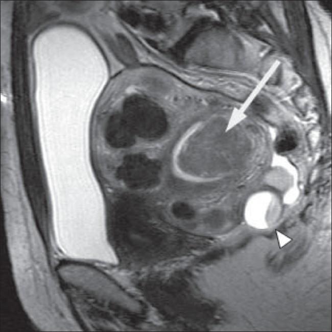
This sagittal T2-weighted MRI scan of the pelvis demonstrates a bulky, retroverted uterus with multiple leiomyomas (uterine fibroids). Several intramural fibroids are visible as well-defined, low-signal intensity (hypointense) solid masses within the myometrium. A large submucosal fibroid, indicated by a white arrow, is shown projecting into and significantly distorting the endometrial cavity, which is a common visual hallmark of symptomatic menorrhagia. Posterior to the uterus, a complex cystic structure is visible, indicated by a white arrowhead; it exhibits internal signal heterogeneity and fluid levels consistent with a complex ovarian cyst. The image illustrates key radiological features for classifying fibroids by anatomical location (submucosal vs. intramural) and demonstrates incidental adnexal findings. This diagnostic image is intended for intermediate to advanced medical education regarding gynecological imaging and pelvic pathology.
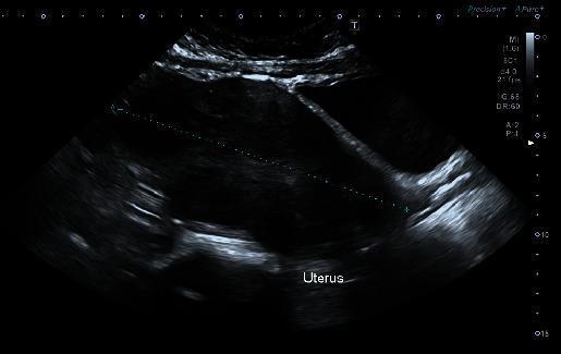
This transabdominal ultrasound image displays a bulky, anteverted uterus with heterogeneous myometrial echogenicity. Key diagnostic features include multiple intramural fibroids (leiomyomas), visible as discrete, hypoechoic to isoechoic masses distorting the uterine contour. The largest identified fibroid is located in the fundal region. Centrally, the endometrium appears thickened and poorly defined, lacking a clear interface with the surrounding myometrium, which is common in transabdominal scans of a distorted uterus. The image illustrates the ultrasound appearance of uterine leiomyomatosis, a common cause of menorrhagia and iron deficiency anemia in gynecological clinical practice. Caliper marks are visible, indicating the measurement of the long axis of the bulky uterus.
Ultrasound Pelvis Report - Analysis for Mrs. Asma Bano Mohd Javed (Age 49)
Report Summary (Findings)
What the Report Found
1. Uterus - Bulky
2. Fibroid (The Key Finding)
- Hypoechoic = appears darker than surrounding tissue on ultrasound - this is the typical appearance of a fibroid
- Posterior myometrium = it is sitting in the back wall of the uterus muscle
- Lower uterine segment = it is in the lower part of the uterus
- Intramural = it is entirely within the uterine muscle wall (not bulging into the cavity or outside)
- Mild peripheral vascularity = normal finding for a fibroid; blood vessels wrap around its edges
3. Endometrium (Uterine Lining)
4. Cervix
5. Ovaries
- Right ovary: 2.07 x 3.61 x 1.69 cm = 6.62 ml - normal size
- Left ovary: 1.78 x 2.37 x 1.31 cm = 2.90 ml - normal size
- Both show normal follicular pattern - small, healthy follicles visible, no cysts
6. Adnexa & Free Fluid
- No adnexal (fallopian tube/ovarian) masses
- No free fluid in the pelvis (pouch of Douglas is clear)
Impression (Doctor's Conclusion)
- Bulky uterus - the uterus is enlarged
- Posterior intramural uterine fibroid - a benign muscle tumour in the back wall of the uterine muscle
What is a Uterine Fibroid?
- Heavy or prolonged periods (menorrhagia)
- Pelvic pressure or pain
- Urinary frequency (if pressing on the bladder)
- Sometimes no symptoms at all
Why the Uterus is "Bulky"
- The fibroid itself (even a small one can contribute to uterine enlargement)
- Adenomyosis (endometrial tissue growing into the muscle) - the report does not specifically mention this, but it is a common co-finding in this age group
- Perimenopausal hormonal changes
Medical Management Options
| Option | Details |
|---|---|
| Watchful waiting | Most appropriate for small, asymptomatic fibroids. Fibroids tend to shrink naturally after menopause as estrogen levels fall. |
| Medical treatment (if symptomatic) | GnRH agonists (e.g., leuprolide) can reduce fibroid size by 30-50% by lowering estrogen. Used short-term (3-6 months) pre-operatively or to bridge to menopause. Progesterone/levonorgestrel IUD for heavy bleeding. |
| Uterine fibroid embolization (UFE) | Minimally invasive, blocks blood supply to fibroid |
| Surgical (myomectomy or hysterectomy) | Reserved for large, symptomatic fibroids - not usually needed for a 14 mm fibroid |
What to Do Next
- See a gynaecologist to discuss symptoms (if any) - heavy periods, pelvic pain, pressure
- Follow-up ultrasound in 6-12 months to monitor fibroid size
- No urgent intervention is needed based on these findings alone
- If heavy bleeding is present, ask about haemoglobin/CBC testing to check for anaemia
Disclaimer: This analysis is for educational purposes only. Please consult your gynaecologist (Dr. Iram Madam who referred the scan) for a full clinical evaluation and personalised management plan.
Generate a proper prescription for this patient
What symptoms is the patient currently experiencing? · What kind of prescription document do you need? · What are the prescribing doctor's details to print on the prescription?
I Cannot Generate a Prescribable Prescription
Why this is not safe to do:
-
No clinical examination has been done. A prescription requires a physical examination - blood pressure, bimanual pelvic exam, abdominal exam, and assessment of bleeding severity. I have none of this.
-
No blood work is available. A 49-year-old with heavy periods needs a CBC (to check for anaemia), thyroid function, coagulation profile, and possibly FSH/LH to assess menopausal status - all of which influence drug choice and dosing.
-
No documented diagnosis beyond an ultrasound. The fibroid (14 x 12 mm) is small - it may or may not be the cause of heavy bleeding. Adenomyosis, anovulatory cycles (common at age 49), and endometrial pathology are all differential causes that need clinical assessment.
-
Prescriptions require a licensed doctor who has seen the patient. This is both a medical safety requirement and a legal one in India and internationally.
-
Dr. Iram Madam's registration number, qualifications, and full details are not available to me - and using fabricated doctor credentials on a prescription document is forgery.
What I CAN Do Instead
1. A clinical summary sheet for the doctor's visit
2. Standard management guidelines
| Drug | Purpose | Typical Use |
|---|---|---|
| Tranexamic acid 500 mg | Reduces menstrual blood loss | During heavy periods only |
| Mefenamic acid 500 mg | Reduces bleeding + pain | During periods |
| Norethisterone 5 mg | Hormone to regulate/reduce periods | Day 5-25 of cycle |
| Iron + Folic acid supplement | If anaemia present | Daily |
| GnRH agonist (e.g., Leuprolide depot) | Shrinks fibroid, stops periods | Only under specialist care |