
i am going to study surgery for my block examination of 4th year mbbs i will send you my topic of studies can you send me he content i will earn you can use bailey surgey dograr surgery and others if you like Primary references I'll use: Bailey & Love's Short Practice of Surgery (gold standard) Dogar's Surgery (high-yield for Pakistani MBBS exams) Schwartz's Principles of Surgery (for concepts and pathophysiology) Sabiston Textbook of Surgery (where helpful) Current surgical guidelines (ASCRS, WSES, ACS, NICE, etc.) when relevant How I'll explain each topic For every disease or condition, I'll cover: Definition Classification Etiology / Risk factors Pathophysiology (step-by-step, easy to understand) Pathology / Morphology (if important) Clinical features Investigations Laboratory Imaging Gold standard investigation Differential diagnosis Complications Management Initial stabilization Conservative treatment Surgical treatment Operative principles Important viva questions Frequently asked MBBS examination points Clinical pearls and mnemonics Flowcharts and tables for quick revision If the topic is a surgical procedure, I'll explain: Definition Indications Contraindications Relevant anatomy Preoperative preparation Instruments Step-by-step procedure Postoperative care Complications Viva questions If the topic is trauma, I'll include: ATLS approach (ABCDE) Assessment Investigations Definitive management Damage control surgery Complications If the topic is a malignancy, I'll include: Epidemiology Risk factors Molecular pathogenesis (when appropriate) Clinical presentation Staging (TNM) Investigations Treatment Prognosis I can also provide: High-yield exam notes Clinical scenarios similar to ward tests Past-paper style long questions Short notes Viva questions MCQs with explanations Flowcharts Summary tables Memory tricks (mnemonics) Diagrams (ASCII or illustrated explanations where useful) Since you're preparing for a 4th year MBBS block examination, I'll focus on the depth expected in Pakistani medical universities—detailed enough to understand the topic, but also emphasizing high-yield points commonly tested in written papers, vivas, and ward exams.
-
General surgery and perioperative care
Fluid/electrolyte balance, shock, sepsis, wounds, burns, nutrition, surgical infections, preoperative and postoperative care. -
Trauma and emergency surgery
ATLS assessment, head/chest/abdominal trauma, fractures, hemorrhage control, damage-control surgery. -
Gastrointestinal and hepatopancreatobiliary surgery
Esophagus, stomach, small bowel, appendix, colorectal disease, hernias, liver, biliary tract, pancreas, spleen. -
Breast, endocrine, vascular, and urology
Breast lumps/cancer, thyroid/parathyroid, arterial and venous disorders, urinary tract and prostate conditions. -
Procedures, exam practice, and revision tools
Operations, instruments, clinical cases, long and short questions, viva, MCQs, flowcharts, and mnemonics.
Choose the area for your first detailed study note.
-
General surgery and perioperative care
Fluid/electrolyte balance, shock, sepsis, wounds, burns, nutrition, surgical infections, preoperative and postoperative care. -
Trauma and emergency surgery
ATLS assessment, head/chest/abdominal trauma, fractures, hemorrhage control, damage-control surgery. -
Gastrointestinal and hepatopancreatobiliary surgery
Esophagus, stomach, small bowel, appendix, colorectal disease, hernias, liver, biliary tract, pancreas, spleen. -
Breast, endocrine, vascular, and urology
Breast lumps/cancer, thyroid/parathyroid, arterial and venous disorders, urinary tract and prostate conditions. -
Procedures, exam practice, and revision tools
Operations, instruments, clinical cases, long and short questions, viva, MCQs, flowcharts, and mnemonics.
- Acute appendicitis
- Intestinal obstruction
- Inguinal hernia
- Acute cholecystitis
- Shock
- Burns
- Breast carcinoma
- Thyroid swelling
- Head injury
- Perforated peptic ulcer
Enlarged Prostate – IPSS scoring, watchful waiting, medical management, TURP and open prostatectomy
"benign prostatic hyperplasia" AND treatment
EAU Guidelines 2026 male lower urinary tract symptoms benign prostatic obstruction recommendations IPSS TURP open prostatectomy
http://uroweb.org/guidelines/management-of-non-neurogenic-ma…
benign prostatic hyperplasia prostate transition zone anatomy diagram
PMID: 42095468
site:uroweb.org guidelines non-neurogenic male LUTS alpha blockers 5 alpha reductase inhibitors watchful waiting TURP open prostatectomy recommendations
https://uroweb.org/guidelines/management-of-non-neurogenic-m…
Enlarged Prostate: BPH, IPSS, Watchful Waiting, Medical Therapy, TURP and Open Prostatectomy
1. Core definition and terminology
- Benign prostatic enlargement (BPE): anatomical enlargement
- Benign prostatic obstruction (BPO): bladder outlet obstruction due to the enlarged prostate
- LUTS: lower urinary tract symptoms
Not every enlarged prostate causes LUTS, and not every man with LUTS has BPH or obstruction.
Pathology and mechanism
- Ageing and androgenic stimulation, especially dihydrotestosterone (DHT), promote stromal and glandular hyperplasia.
- Nodules arise in the transition/periurethral zone.
- Enlarging lateral lobes and sometimes a median lobe compress or distort the prostatic urethra.
- This causes increased bladder outlet resistance.
- The detrusor initially hypertrophies, giving a trabeculated bladder.
- Later, the bladder decompensates, producing residual urine, recurrent UTI, stones, chronic retention, hydroureteronephrosis, and renal impairment.
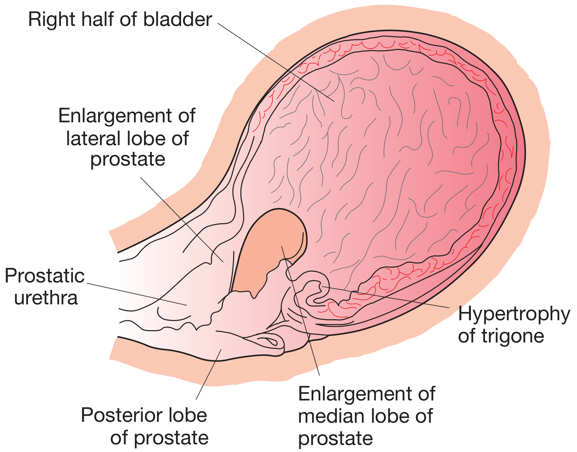
Bailey & Love notes that the relation among prostate size, LUTS, and true urodynamic obstruction is complex. - Bailey & Love's Short Practice of Surgery, pp. 1543-1545
2. Clinical features: LUTS
A. Voiding or obstructive symptoms
- Hesitancy
- Poor urinary stream
- Intermittency
- Straining to void
- Terminal dribbling
- Sensation of incomplete emptying
- Acute or chronic urinary retention
B. Storage or irritative symptoms
- Frequency
- Urgency
- Nocturia
- Urge incontinence
3. IPSS: International Prostate Symptom Score
- Quantify symptom severity
- Assess the degree of bother
- Monitor response to watchful waiting, drug treatment, or surgery
Seven IPSS questions
-
Incomplete emptying
“How often have you had the sensation of not emptying the bladder completely?” -
Frequency
“How often have you had to pass urine again within 2 hours?” -
Intermittency
“How often have you found that you stopped and started again while passing urine?” -
Urgency
“How often have you found it difficult to postpone urination?” -
Weak stream
“How often have you had a weak urinary stream?” -
Straining
“How often have you had to push or strain to begin urination?” -
Nocturia
“How many times do you usually get up at night to pass urine?”
Scoring
| Score | Meaning |
|---|---|
| 0 | Not at all |
| 1 | Less than 1 in 5 times |
| 2 | Less than half the time |
| 3 | About half the time |
| 4 | More than half the time |
| 5 | Almost always |
Interpretation
| Total IPSS | Severity |
|---|---|
| 0-7 | Mild |
| 8-19 | Moderate |
| 20-35 | Severe |
- 0: Delighted
- 6: Terrible
Viva pearl
IPSS measures symptom severity, but it does not prove bladder outlet obstruction. Definitive assessment of obstruction, when necessary, is by pressure-flow urodynamic studies.
4. Basic assessment before treatment
History
- LUTS and IPSS
- Duration and impact on quality of life
- Hematuria, dysuria, fever, UTI
- Retention episodes
- Drug history: anticholinergics, sympathomimetics, opioids, diuretics
- Diabetes, Parkinson disease, stroke, spinal disease
- Fluid, caffeine, alcohol intake
- Sexual function and erectile dysfunction
Examination
- Abdomen: palpable distended bladder
- External genitalia and meatus
- Neurological examination where indicated
- Digital rectal examination (DRE):
- BPH: smooth, enlarged, firm-rubbery, non-tender prostate with obliterated median sulcus
- Carcinoma: hard, irregular, nodular, asymmetrical prostate
Investigations
| Investigation | Purpose |
|---|---|
| Urinalysis and urine culture | Exclude UTI, hematuria, glycosuria |
| Serum creatinine, electrolytes | Detect renal impairment, especially in retention |
| PSA | If life expectancy is adequate and result would alter management; also predicts prostate volume/progression |
| Uroflowmetry | Measures Qmax |
| Post-void residual, PVR | Detects incomplete emptying/retention |
| Ultrasound KUB/prostate | Prostate volume, residual urine, hydronephrosis, stones |
| Cystoscopy | Hematuria, stricture, bladder stone/tumor, preoperative assessment |
| Urodynamics | Diagnostic uncertainty, neurological disease, failed surgery, near-normal flow with severe symptoms |
Uroflowmetry values
| Qmax | Interpretation |
|---|---|
| >15 mL/s | Usually normal |
| 10-15 mL/s | Equivocal |
| <10 mL/s | Suggestive of obstruction or weak detrusor |
5. Watchful waiting
Definition
Suitable patient
- IPSS usually 0-7, or selected men with moderate symptoms
- Symptoms do not significantly impair quality of life
- No retention, recurrent UTI, bladder stones, renal impairment, or recurrent significant hematuria
- Low residual urine and no high-pressure chronic retention
- Patient understands when to return urgently
Measures
- Explain the benign nature after appropriate assessment
- Avoid excessive fluids and “fluid binges”
- Reduce evening fluid intake, especially if nocturia
- Reduce caffeine and alcohol
- Timed voiding and bladder training
- Review drugs that worsen voiding
- Treat constipation
- Manage diabetes, heart failure, sleep apnea, and other causes of nocturia
- Repeat IPSS, DRE, PVR, renal function, and PSA when indicated
Do not choose watchful waiting if there is:
- Refractory or recurrent retention
- Renal impairment due to obstruction
- Hydroureteronephrosis
- Recurrent UTI
- Recurrent gross hematuria due to BPH
- Bladder stones or diverticulum
- Severe bothersome symptoms despite conservative measures
6. Medical management
A. Alpha-1 blockers
Examples
- Tamsulosin
- Alfuzosin
- Silodosin
- Doxazosin
- Terazosin
Mechanism
- Prostate
- Prostatic urethra
- Bladder neck
Clinical use
- First-line drug for moderate-to-severe bothersome LUTS
- Particularly useful when rapid improvement is desired
- Benefit begins within days to weeks
- They improve symptoms but do not shrink the prostate or substantially prevent long-term progression.
Adverse effects
- Postural hypotension
- Dizziness and fatigue
- Headache
- Nasal congestion
- Retrograde ejaculation or reduced ejaculation
- Intraoperative floppy iris syndrome, especially with tamsulosin, relevant before cataract surgery
B. 5-alpha reductase inhibitors, 5-ARIs
Examples
- Finasteride
- Dutasteride
Mechanism
Effects
- Prostate shrinks by roughly 20-25%
- Reduces risk of acute urinary retention
- Reduces future need for surgery
- Onset is slow: meaningful benefit requires 4-6 months, often longer
- Serum PSA falls by approximately 50% after adequate treatment. Interpret PSA accordingly.
Indications
- Moderate-to-severe LUTS
- Enlarged prostate, generally >40 mL
- PSA suggestive of increased prostate volume/progression
- Concern about disease progression, retention, or surgery
Adverse effects
- Reduced libido
- Erectile dysfunction
- Ejaculatory dysfunction
- Reduced semen volume
- Gynecomastia or breast tenderness, less common
C. Combination therapy: alpha-blocker + 5-ARI
Indication
Rationale
- Alpha-blocker gives rapid symptom relief.
- 5-ARI reduces prostate size and long-term risk of retention/surgery.
D. Other useful drugs
| Drug group | Example | When helpful | Key caution |
|---|---|---|---|
| PDE-5 inhibitor | Tadalafil daily | LUTS with erectile dysfunction | Nitrates contraindicated |
| Antimuscarinic | Solifenacin, tolterodine | Persistent storage symptoms: urgency/frequency | Avoid or use carefully if high PVR/retention risk |
| Beta-3 agonist | Mirabegron | Storage symptoms/overactive bladder features | Monitor BP |
| Desmopressin | Selected nocturnal polyuria | Significant nocturia due to nocturnal polyuria | Hyponatremia risk, especially elderly |
7. Indications for surgery in BPH
Absolute or strong indications
- R: Refractory or recurrent urinary retention
- U: Upper tract dilatation, hydroureter/hydronephrosis
- S: Stones in the bladder
- H: Hematuria, recurrent and attributable to BPH after exclusion of other causes
- S: Sepsis or recurrent UTI due to obstruction
- Renal impairment due to obstruction
- Large bladder diverticulum with obstruction
- Severe symptoms affecting quality of life despite conservative/medical therapy
8. TURP: Transurethral resection of prostate
Definition
TURP removes the obstructing transition-zone adenoma. It is not a radical prostatectomy and does not remove the entire prostate.
Indications
- Bothersome moderate-to-severe LUTS refractory to medical therapy
- Recurrent/refractory urinary retention
- Recurrent UTI, bladder stones, hematuria, diverticulum, renal impairment from BPO
- Usually preferred for prostate size approximately 30-80 mL, depending on surgeon skill, anatomy, and available technology
Contraindications or situations requiring caution
- Untreated UTI
- Uncorrected coagulopathy
- Unfit for anesthesia
- Very large prostate, where open simple prostatectomy or enucleation may be preferable
- Suspected untreated prostate cancer requires appropriate evaluation first
Preoperative preparation
- History, IPSS, DRE
- Urine culture and treat infection
- CBC, renal function, electrolytes, coagulation screen
- Ultrasound for prostate size, PVR, upper tract if indicated
- PSA and prostate cancer assessment when appropriate
- Stop/adjust anticoagulants where safe
- Informed consent: bleeding, infection, TUR syndrome, retrograde ejaculation, erectile dysfunction, incontinence, stricture, bladder-neck contracture, reoperation
Basic operative principle
- Spinal or general anesthesia.
- Resectoscope introduced per urethra.
- Cystoscopy is performed to assess urethra, prostate, bladder, stones, and tumors.
- Resection begins with obstructing tissue, commonly median lobe if present.
- Prostatic tissue is resected in chips while preserving:
- Ureteric orifices
- External urethral sphincter, distal to verumontanum
- Prostatic capsule
- Hemostasis is achieved.
- Chips are evacuated and sent for histopathology.
- A three-way Foley catheter is inserted for continuous bladder irrigation.
Postoperative care
- Continuous bladder irrigation until urine clears
- Monitor catheter patency, urine color, clots, input-output
- Check hemoglobin and electrolytes if significant bleeding or prolonged procedure
- Analgesia and antibiotics according to local protocol
- Catheter generally removed after 1-3 days when hematuria settles
- Trial of void and measurement of PVR
- Warn about temporary dysuria, frequency, urgency, and hematuria during healing
Complications of TURP
Early
- Hemorrhage and clot retention
- UTI/sepsis
- Bladder perforation
- Capsular perforation and extravasation
- Failure to void
- DVT/PE, anesthetic complications
- TUR syndrome
TUR syndrome
- Nausea/vomiting
- Restlessness, confusion, seizures
- Bradycardia
- Hypertension initially
- Pulmonary edema
- Hyponatremia
- Stop surgery and irrigation.
- ABC resuscitation and oxygen.
- Check electrolytes and ECG.
- Restrict fluid and give loop diuretic if fluid overloaded.
- Severe symptomatic hyponatremia requires carefully monitored hypertonic saline in a high-dependency/ICU setting.
- Bipolar TURP uses normal saline irrigation and markedly reduces this risk.
Late
- Retrograde ejaculation: common
- Erectile dysfunction
- Urethral stricture
- Bladder-neck contracture
- Persistent storage symptoms/urge incontinence
- Rare stress urinary incontinence due to sphincter injury
- Recurrent adenoma and need for re-treatment
9. Open simple prostatectomy
Definition
It is different from radical prostatectomy, which removes the whole prostate, seminal vesicles, and often pelvic lymph nodes for prostate cancer.
Indications
- Very large gland, commonly >80-100 mL
- Large median lobe/large intravesical protrusion
- Coexisting bladder stone requiring open cystolithotomy
- Large bladder diverticulum requiring diverticulectomy
- Inability to perform or lack of availability of endoscopic enucleation
- Failed prior endoscopic procedure in selected cases
Types
| Operation | Route | Important point |
|---|---|---|
| Freyer prostatectomy | Transvesical/suprapubic | Bladder opened, adenoma enucleated through bladder neck |
| Millin prostatectomy | Retropubic | Adenoma enucleated without opening bladder |
| Laparoscopic/robotic simple prostatectomy | Minimally invasive | Alternative where facilities and expertise exist |
A. Freyer's transvesical prostatectomy
Main steps
- Lower midline or Pfannenstiel incision.
- Open the bladder.
- Inspect bladder, ureteric orifices, median lobe, stones, and diverticula.
- Circumferential mucosal incision at the bladder neck around the adenoma.
- Finger enucleation of adenoma along the surgical capsule.
- Achieve hemostasis by suturing or packing.
- Insert urethral Foley catheter, usually with suprapubic drainage depending on local practice.
- Close bladder and provide drainage.
Advantages
- Direct access to bladder
- Easy removal of concomitant large bladder stones
- Useful for very large median lobe and huge adenoma
Disadvantages
- More invasive
- Greater blood loss
- Longer admission and catheter duration
- Wound-related complications
B. Millin's retropubic prostatectomy
Main principle
Advantages
- Avoids cystotomy
- Useful for large prostate when bladder surgery is not needed
Disadvantages
- Less suitable if large bladder stones/diverticulum need simultaneous treatment
- Risks include bleeding, urinary leak, and injury to surrounding structures
10. TURP versus open simple prostatectomy
| Feature | TURP | Open simple prostatectomy |
|---|---|---|
| Access | Transurethral, endoscopic | Open abdominal incision |
| Typical prostate size | About 30-80 mL | Usually >80-100 mL |
| Hospital stay | Shorter | Longer |
| Blood loss | Less, but can be significant | Usually greater |
| Recovery | Faster | Slower |
| TUR syndrome | Possible with monopolar TURP | Not applicable |
| Best for | Standard-sized obstructing prostate | Very large gland or need for concomitant open bladder surgery |
| Retrograde ejaculation | Common | Common |
| Histology | TUR chips | Enucleated adenoma |
11. High-yield management flowchart
Male with LUTS
       ↓
History + IPSS + DRE + urinalysis/culture
Creatinine, PVR, uroflowmetry ± PSA/ultrasound
       ↓
Exclude: UTI, cancer, stricture, stones, neurogenic bladder,
diabetes, overactive bladder, drugs
       ↓
Mild symptoms, not bothered, no complication?
       ↓
Yes → Watchful waiting + lifestyle modification + follow-up
       ↓
No / bothersome moderate-severe symptoms
       ↓
Alpha-1 blocker
       ↓
Large prostate (>40 mL) or high progression risk?
       ↓
Add 5-alpha reductase inhibitor
       ↓
Persistent symptoms or complication of BPO?
       ↓
Surgery
       ↓
30-80 mL → TURP / bipolar TURP / suitable endoscopic option
>80-100 mL → HoLEP if available, or open/robotic simple prostatectomy
12. Important viva questions
-
What is BPH?
Histological hyperplasia of stromal and epithelial elements in the transition zone of the prostate. -
Where does BPH occur?
Transition zone/periurethral region. -
Where does prostate carcinoma commonly arise?
Peripheral zone. -
What is the IPSS range?
0 to 35. -
Interpret IPSS.
Mild 0-7, moderate 8-19, severe 20-35. -
Does a high IPSS prove obstruction?
No. It quantifies symptoms; obstruction is confirmed by pressure-flow studies when required. -
How do alpha-blockers work?
Relax smooth muscle at prostate and bladder neck, reducing dynamic obstruction. -
How do 5-ARIs work?
Inhibit conversion of testosterone to DHT, shrinking prostate and reducing progression. -
Which drug class lowers PSA by about 50%?
5-alpha reductase inhibitors. -
What is the gold-standard operation for BPH?
TURP for appropriately sized prostates. -
What is TUR syndrome?
Dilutional hyponatremia and fluid overload due to absorption of hypotonic irrigating fluid during monopolar TURP. -
Name indications for surgery in BPH.
Retention, renal impairment/hydronephrosis, recurrent UTI, recurrent hematuria, stones, diverticulum, and failed medical therapy with severe bothersome symptoms. -
What is the difference between simple and radical prostatectomy?
Simple prostatectomy removes/enucleates adenoma for BPH. Radical prostatectomy removes the entire prostate for cancer.
One-minute revision
- BPH = transition-zone stromal + glandular hyperplasia.
- IPSS = 7 urinary symptoms, total 0-35; mild 0-7, moderate 8-19, severe 20-35.
- Watchful waiting = mild/non-bothersome uncomplicated LUTS.
- Alpha-blocker = rapid symptom relief; relaxes prostate/bladder-neck smooth muscle.
- 5-ARI = slow action, reduces prostate volume and progression; PSA falls about 50%.
- TURP = standard operation for moderate-sized glands.
- Open simple prostatectomy = large prostate, generally >80-100 mL, or associated large bladder stone/diverticulum.
- TUR syndrome = water intoxication/dilutional hyponatremia from hypotonic irrigation.
Carcinoma of prostate – clinical presentation and management
"prostatic neoplasms" AND guideline
EAU guidelines prostate cancer 2026 diagnosis staging treatment localized metastatic management
prostate cancer metastatic osteoblastic bone scan image prostate carcinoma
PMID: 42457454
https://uroweb.org/guidelines/prostate-cancer?e-page-2e4fdd4…
"prostatic neoplasms" AND treatment
PMID: 38182303
site:uroweb.org/guidelines/prostate-cancer chapter treatment metastatic hormone sensitive prostate cancer androgen deprivation radiotherapy recommendations
Carcinoma of Prostate: Clinical Presentation and Management
Definition
| Feature | BPH | Prostate carcinoma |
|---|---|---|
| Usual zone | Transition/periurethral zone | Peripheral, usually posterior zone |
| DRE | Smooth, firm-rubbery, symmetrical | Hard, irregular, nodular, asymmetrical |
| Early LUTS | Common | Usually absent |
| Metastases | No | Commonly bone, especially axial skeleton |
- Bailey & Love's Short Practice of Surgery, p. 1553
- Robbins & Kumar Basic Pathology, pp. 661-662
Clinical presentation
1. Early localized prostate cancer
- Raised PSA on risk-based testing
- Abnormal DRE: hard nodule, induration, asymmetry, or irregularity
- Incidental carcinoma in TURP tissue for presumed BPH:
- T1a: tumor in ≤5% of resected tissue
- T1b: tumor in >5% of resected tissue
- MRI followed by targeted/systematic biopsy
Early prostate cancer does not usually cause LUTS because it arises peripherally, away from the urethra.
2. Locally advanced disease
Symptoms
- Lower urinary tract obstruction:
- Poor stream, hesitancy, intermittency
- Incomplete emptying
- Acute or chronic retention
- Hematuria
- Pelvic or perineal pain
- Ureteric obstruction causing hydronephrosis and renal failure
- Rarely rectal symptoms: tenesmus or constipation due to rectal compression
3. Metastatic disease
Features of metastases
- Persistent lower back, pelvic, hip, rib, or bone pain
- Pathological fracture
- Spinal metastasis with cord compression:
- Severe back pain
- Limb weakness
- Sensory loss
- Bladder/bowel dysfunction
- Malaise, anorexia, weight loss
- Anemia or pancytopenia due to marrow infiltration
- Lower-limb edema from pelvic lymph-node obstruction
- Renal failure from bilateral ureteric obstruction
- Bailey & Love's Short Practice of Surgery, pp. 1553-1555
Examination
General examination
- Pallor and weight loss
- Bony tenderness
- Leg edema
- Neurological deficit or signs of spinal cord compression
- Palpable distended bladder in obstruction/retention
- Supraclavicular lymphadenopathy in advanced disease
DRE findings
- Hard or stony hard
- Irregular or nodular
- Asymmetrical
- May have obliteration of median sulcus
- May be fixed in locally advanced disease
Investigations
Initial tests
| Investigation | Role |
|---|---|
| PSA | Risk assessment, supports diagnosis, monitoring and follow-up. Not diagnostic alone |
| DRE | Detects suspicious palpable lesion |
| Urinalysis/culture | Exclude UTI and hematuria causes |
| CBC | Anemia or marrow involvement |
| Renal function/electrolytes | Detect obstructive renal failure |
| ALP | May rise with extensive osteoblastic bone metastases |
PSA: high-yield points
- PSA is prostate-specific, not cancer-specific.
- PSA rises in cancer, BPH, prostatitis, urinary retention, recent instrumentation, and after ejaculation.
- PSA must be interpreted with age, DRE, prostate volume, MRI, and clinical risk.
- It is particularly useful for post-treatment monitoring:
- After radical prostatectomy, PSA should become very low/undetectable.
- Rising PSA after treatment may indicate biochemical recurrence.
Confirming diagnosis
Multiparametric MRI, mpMRI
- Identify suspicious lesions
- Assign PI-RADS score
- Guide targeted biopsy
- Assess local extension
Biopsy: diagnostic gold standard
- Transperineal MRI-targeted biopsy plus systematic regional cores, or
- Transrectal ultrasound-guided biopsy where appropriate
Staging investigations
| Clinical situation | Preferred staging approach |
|---|---|
| Low-risk localized cancer | Often no metastatic imaging needed |
| Intermediate/high-risk disease | MRI for local staging plus PSMA PET/CT where available |
| If PSMA PET/CT unavailable | CT abdomen/pelvis plus bone scan |
| Bone pain, high PSA, high-grade disease | Bone scan or PSMA PET/CT |
| Suspected cord compression | Urgent MRI spine |
Histology and grading
Gleason score
Primary pattern + Secondary pattern = Gleason score
Example: 3 + 4 = 7
ISUP Grade Groups
| Gleason score | ISUP Grade Group | Usual interpretation |
|---|---|---|
| 3 + 3 = 6 | 1 | Low grade |
| 3 + 4 = 7 | 2 | Favorable intermediate grade |
| 4 + 3 = 7 | 3 | Unfavorable intermediate grade |
| 8 | 4 | High grade |
| 9-10 | 5 | Very high grade |
TNM staging: simplified MBBS version
| Stage | Meaning |
|---|---|
| T1 | Clinically inapparent, not palpable or visible on imaging |
| T2 | Tumor confined within prostate |
| T3a | Extracapsular extension |
| T3b | Seminal-vesicle invasion |
| T4 | Invades bladder, rectum, external sphincter, levator muscles, or pelvic wall |
| N1 | Regional pelvic lymph-node metastasis |
| M1 | Distant metastasis, commonly bone |
Management principles
- Stage: localized, locally advanced, nodal, metastatic
- PSA
- Gleason score/ISUP grade
- Life expectancy, age, comorbidities, and performance status
- Patient preference
- Urinary, erectile, and bowel function
Confirmed prostate cancer
        ↓
Stage + PSA + ISUP grade + life expectancy
        ↓
Localized low risk → Active surveillance or curative treatment
Localized intermediate/high risk → Radical treatment
Locally advanced/N1 → Radiotherapy + long-term hormonal therapy
Metastatic disease → Systemic hormonal treatment ± intensified therapy
1. Localized low-risk cancer
Active surveillance
Follow-up includes
- Serial PSA
- Periodic DRE
- Repeat MRI when indicated
- Repeat biopsy according to protocol
- Curative treatment if grade, volume, PSA, MRI, or biopsy indicates progression
Suitable patients
- Low-risk localized cancer
- Long enough life expectancy for monitoring to matter
- Reliable for follow-up
| Active surveillance | Watchful waiting |
|---|---|
| Curative treatment is planned if cancer progresses | Palliative treatment only if symptoms develop |
| Usually for low-risk localized cancer | Often for frail/elderly patients with limited life expectancy |
| Regular PSA, MRI, and repeat biopsy | Less intensive follow-up |
2. Localized cancer suitable for curative treatment
A. Radical prostatectomy
Definition
- Entire prostate
- Prostatic capsule
- Seminal vesicles
- Usually vas deferens segments
- Open retropubic surgery
- Laparoscopic surgery
- Robot-assisted laparoscopic surgery
Indications
- Localized disease in a fit patient
- Usually life expectancy >10 years
- Selected locally advanced disease as part of multimodal therapy
Major complications
- Erectile dysfunction
- Incontinence, especially stress urinary incontinence
- Retrograde ejaculation/infertility, although ejaculation is absent after removal of seminal vesicles
- Bleeding
- Lymphocele after lymph-node dissection
- Urethrovesical anastomotic stricture
- Rectal injury, rare
- DVT/PE
B. Radical radiotherapy
- External beam radiotherapy, EBRT
- Brachytherapy in selected localized cases
- Hypofractionated schedules are increasingly used
Often combined with androgen-deprivation therapy, ADT
- Intermediate-risk: short course ADT may be used with radiotherapy.
- High-risk or locally advanced disease: long-term ADT with radiotherapy.
Complications
- Urinary frequency, urgency, dysuria
- Radiation cystitis and hematuria
- Radiation proctitis, diarrhea, rectal bleeding
- Erectile dysfunction
- Urethral stricture, less common
3. Locally advanced disease: T3/T4, M0
Common treatment
Manage complications
- Urinary retention: catheterization, suprapubic catheter if needed, selected palliative TURP
- Ureteric obstruction: ureteric stent or percutaneous nephrostomy
- Hematuria: bladder irrigation, endoscopic control, radiotherapy/embolization in selected cases
- Spinal cord compression: emergency corticosteroids, urgent MRI, radiation oncology/neurosurgical referral
4. Node-positive disease: N1, M0
- Radiotherapy to the prostate and pelvic nodes
- Long-term ADT
- In selected high-risk cases, treatment intensification with an androgen receptor pathway inhibitor may be considered under oncology guidance.
5. Metastatic prostate cancer
A. Metastatic hormone-sensitive prostate cancer, mHSPC
Methods of ADT
| Method | Examples/notes |
|---|---|
| Surgical castration | Bilateral orchiectomy, immediate and permanent |
| Medical castration | LHRH/GnRH agonists or antagonists |
| Antiandrogen therapy | Used in selected regimens, often with other agents |
LHRH agonists
LHRH antagonists
Treatment intensification
- Androgen receptor pathway inhibitor:
- Abiraterone plus steroid
- Enzalutamide
- Apalutamide
- Darolutamide
- Docetaxel chemotherapy in appropriate patients, particularly with high-volume disease
B. Metastatic castration-resistant prostate cancer, mCRPC
- Continue ADT
- Androgen receptor pathway inhibitors
- Docetaxel or cabazitaxel
- PARP inhibitor therapy for eligible tumors with homologous-recombination repair mutations, such as BRCA alterations
- PSMA-targeted radioligand therapy in eligible settings
- Radium-223 for selected symptomatic bone-predominant disease without visceral metastases
- Palliative radiotherapy for painful bone metastases
Management of bone metastases
- Analgesia using the WHO pain ladder
- Palliative external beam radiotherapy for painful focal lesions
- Urgent management of impending fracture or spinal cord compression
- Orthopedic stabilization where needed
- Bone-protective agents in suitable patients:
- Denosumab
- Zoledronic acid
- Calcium/vitamin D assessment and dental review before antiresorptive therapy because of the risk of osteonecrosis of the jaw
High-yield viva questions
-
What is the commonest histological type?
Adenocarcinoma. -
Which zone is commonly affected?
Peripheral zone, usually posteriorly. -
Why does early carcinoma often not cause LUTS?
It arises in the peripheral zone and does not initially compress the prostatic urethra. -
What is the commonest site of distant metastasis?
Bone, especially axial skeleton. -
Are bone metastases lytic or blastic?
Typically osteoblastic/sclerotic. -
What is the gold standard for diagnosis?
Histopathology from prostate biopsy. -
Is PSA diagnostic of prostate cancer?
No. It is prostate-specific, not cancer-specific. -
What are the three major factors for risk stratification?
PSA, clinical TNM stage, and Gleason score/ISUP Grade Group. -
What is the treatment for low-risk localized disease?
Active surveillance is often preferred; radical prostatectomy or radiotherapy are alternatives in selected patients. -
What are definitive curative treatments?
Radical prostatectomy and radical radiotherapy. -
What is the first-line foundation for metastatic disease?
Androgen-deprivation therapy. -
What is the difference between simple and radical prostatectomy?
Simple prostatectomy enucleates adenoma for BPH. Radical prostatectomy removes the whole prostate and seminal vesicles for cancer.
One-minute exam summary
Prostate carcinoma:
- Usually adenocarcinoma in peripheral zone.
- Early disease: asymptomatic, raised PSA, abnormal DRE, or incidental TURP finding.
- Advanced disease: BOO, hematuria, pelvic pain, renal failure.
- Metastases: osteoblastic bone lesions → back/bone pain, fracture, cord compression, anemia.
- Diagnosis: PSA + DRE + mpMRI, confirmed by targeted/systematic biopsy.
- Staging: TNM, PSA, Gleason/ISUP, PSMA PET/CT or CT plus bone scan in higher-risk disease.
- Low-risk: active surveillance.
- Localized curable disease: radical prostatectomy or radiotherapy.
- Locally advanced/N1: radiotherapy + long-term ADT.
- Metastatic: ADT plus systemic intensification when appropriate.