
periapical radiopacity of jaw
Reading File
Reading File
Finding Sources
Finding Sources
Searching PubMed
"periapical" AND "radiopaque" AND "jaw"
Reading File
Reading File
Finding Sources
Searching Images
periapical radiograph radiopaque lesion at root apex cemento-osseous dysplasia or cementoblastoma
Searching Images
cementoblastoma periapical radiograph radiodense lesion lucent rim
Finding Sources
Periapical radiopacity of the jaw
A periapical radiopacity is a dense area at or around a tooth root apex. Most of the differential comes down to two questions: is the tooth vital, and is the lesion attached to the root?
Main causes
| Entity | Key features |
|---|---|
| Focal cemento-osseous dysplasia (periapical COD) | Non-neoplastic. Often several periapical lesions at mandibular incisors, usually in women over 30. The teeth are vital. |
| Benign cementoblastoma | True neoplasm fused to the root. Usually the mandibular first molar in young adults. Often painful. |
| Condensing osteitis / focal sclerosing osteomyelitis | Reactive sclerosis from low-grade pulpal inflammation. This comes from my own knowledge, not the library. |
| Idiopathic osteosclerosis (dense bone island) | Incidental, with no lesion at the root. This also comes from my own knowledge. |
| Hypercementosis | Thickened cementum around the root. Seen in Paget disease, where roots can fuse with alveolar bone and cause ankylosis. |
| Odontoma and other mixed lesions | An irregular radiopaque mass, often between tooth roots and associated with unerupted teeth. Adenomatoid odontogenic tumour can show radiopaque foci. |
What the textbooks say
Focal cemento-osseous dysplasia. Focal COD is the more common form. It appears as multiple periapical lesions of mandibular incisors, found on routine radiographs. Early lesions are rounded radiolucencies. They become increasingly radiopaque, keeping a narrow radiolucent rim, as bone and cementum-like tissue are deposited. The related teeth are vital, unlike with periapical granulomas or radicular cysts. COD is self-limiting and rarely needs treatment. - Scott-Brown's Otorhinolaryngology Head & Neck Surgery, p. 25.13 section "Cemento-osseous dysplasias". Florid COD affects all four quadrants and may expand the jaw. - K J Lee's Essential Otolaryngology lists the same features: radiopaque, well demarcated or poorly defined, with the periapical type in the anterior mandible around the tooth root.
Benign cementoblastoma. Radiographically it is a well-defined, rounded, radiopaque or mixed-density lesion replacing the apical third of the root, surrounded by a narrow radiolucent zone. The tooth is vital. Treatment is extraction of the tooth with complete enucleation, and recurrence is common. - Scott-Brown's Otorhinolaryngology Head & Neck Surgery, p. 459.
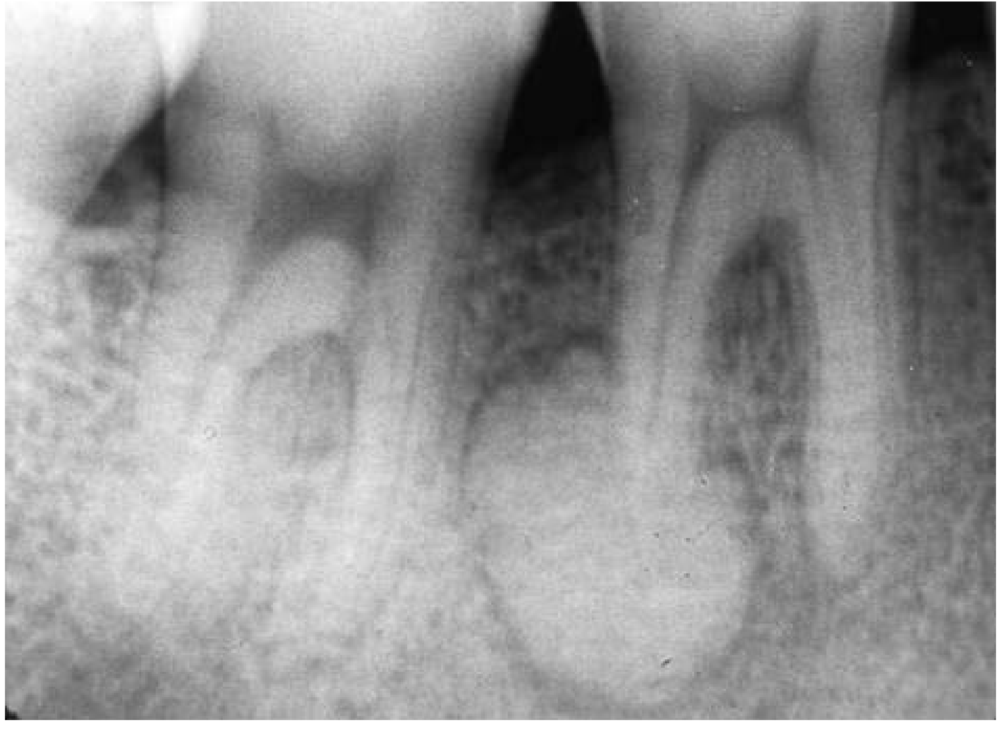
Figure 25.19, Scott-Brown's: periapical radiograph of benign cementoblastoma, radiodense with a lucent rim.
Odontoma. An irregular radiopaque mass, often between tooth roots and associated with unerupted teeth. - K J Lee's Essential Otolaryngology
Practical approach
- Test pulp vitality with ice or electric pulp testing.
- A vital tooth points to COD, cementoblastoma or idiopathic osteosclerosis.
- A non-vital or heavily restored tooth points to condensing osteitis. It can also mean a chronic apical lesion that is healing.
- Check the margins.
- A radiolucent rim with the lesion attached to the root suggests cementoblastoma.
- A rim that fades as the lesion matures suggests COD.
- Check size and number. Multiple lesions in the anterior mandible suggest COD. Diffuse lesions in all four quadrants suggest florid COD.
- Check symptoms. Pain, expansion or a growing lesion suggest cementoblastoma. They can also suggest a rare malignancy such as osteosarcoma. Asymptomatic, stable lesions suggest COD or osteosclerosis.
- Management depends on the diagnosis.
- Asymptomatic COD or osteosclerosis needs only periodic review.
- Biopsy of COD is best avoided, because it can cause infection and non-healing in this poorly vascularised bone. This is from my own knowledge.
- Condensing osteitis is treated by endodontic treatment or extraction of the cause.
- Cementoblastoma is treated surgically.
Recent evidence check
A PubMed search of the last 2 years found only systematic reviews on AI detection of periapical lesions (for example PMID 38851523). None of them changes the above.
I found no matching library images beyond the cementoblastoma figure. I can narrow this down if you have a specific case, such as the tooth, its vitality and the lesion's margins.
This is a shared conversation. Sign in to Orris to start your own chat.