Help me prepare for neet pg and inicet by making rapido revision notes from standard textbook of surgery only as if thought by a excellent mbbs professor which should also include pyqs and pyts on the topic breast which can also contain images, flowcharts and mnemonics
Reading File
Finding Sources
Finding Sources
Searching the Web
NEET PG INICET breast surgery previous year questions MCQs
Finding Sources
Finding Sources
Finding Sources
Reading File
Reading File
Reading File
Reading File
Reading File
Loading Image
Loading Image
I now have everything needed. Let me compile the complete, professor-quality rapid revision notes:
🏥 BREAST - RAPID REVISION NOTES
For NEET PG & INICET | Based on Bailey & Love 28e, Sabiston 21e, S Das 13e
Prepared as taught by an MBBS Professor | High-Yield | PYQ-Integrated
SECTION 1 - ANATOMY (HIGH-YIELD)
Breast Anatomy
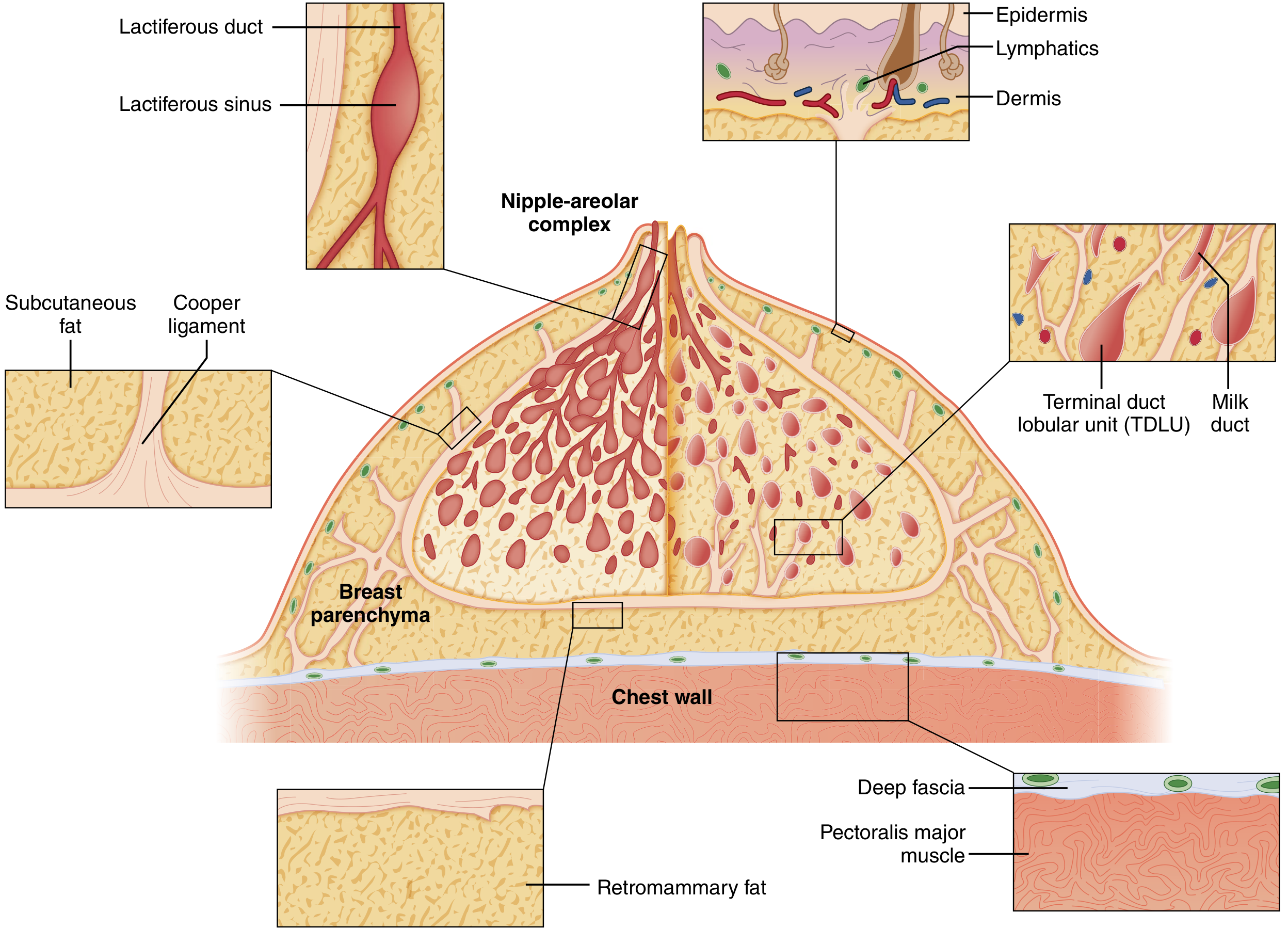
Layers (superficial to deep):
Skin → Subcutaneous fat → Superficial fascia → Breast parenchyma → Deep fascia → Pectoralis major
3 tissue types of mature breast:
- Glandular epithelium
- Fibrous stroma
- Adipose tissue
Ductal system:
- 15-20 lobes, each draining through a lactiferous duct at nipple
- Below nipple: lactiferous sinus (dilated segment)
- End unit: Terminal Duct Lobular Unit (TDLU) = acini + terminal ductules
- TDLU is the site of origin of most breast carcinomas
Cooper's (Suspensory) Ligaments:
- Run from chest wall fascia to dermis
- Provide shape and structure
- When infiltrated by cancer → skin dimpling / peau d'orange (orange peel appearance)
Professor's Pearl: "Peau d'orange = Cooper ligament infiltration = NEVER forget this association. It has appeared in NEET PG almost every cycle."
Blood Supply
- Medial: Internal mammary (internal thoracic) artery - main supply
- Lateral: Lateral thoracic artery + thoracoacromial artery
- Intercostals: 2nd-4th perforators
Lymphatic Drainage (MOST IMPORTANT for MCQs)
BREAST LYMPHATIC DRAINAGE

Upper outer quadrant (most common site of cancer)
         |
         v
   Axillary nodes (75% of drainage)
   [Level I → Level II → Level III]
         |
         v
   Subclavian vein → Right lymphatic/thoracic duct

   Medial quadrant → Internal mammary nodes
   Lower → Subdiaphragmatic → Liver (metastasis route)
Axillary Node Levels (BERRY's levels by relation to pectoralis minor):
| Level | Location | Key |
|---|---|---|
| I | Lateral to pec minor | Low axillary |
| II | Behind pec minor | Mid axillary |
| III | Medial to pec minor | Apical/infraclavicular |
Mnemonic - "I'm LBM" (Lateral, Behind, Medial)
Rotter's nodes = interpectoral nodes (between pec major and minor)
Sentinel node = first draining node = Level I (anterior group / anterior pectoral group)
SECTION 2 - ANDI (ABERRATIONS OF NORMAL DEVELOPMENT AND INVOLUTION)
The ANDI Concept (Bailey & Love - Chapter 58)
ANDI classifies benign breast conditions as aberrations of the normal physiological process:
ANDI FRAMEWORK

PHASE         NORMAL PROCESS          ABERRATION          DISEASE
----------------------------------------------------------------------
Development   Lobule formation        Fibroadenoma        Giant FA
(15-25 yr)    Duct development        Nipple inversion     --

Cyclical      Cyclical changes        Cyclical mastalgia   Incapacitating pain
(25-40 yr)    Lobular involution      Cyclical nodularity  Diffuse fibroadenosis

Involution    Lobular involution      Macrocysts           Tension cyst
(35-55 yr)    Duct involution         Sclerosing adenosis
              Fibrosis                Duct ectasia         Periductal mastitis
SECTION 3 - BENIGN BREAST DISEASES
3.1 Fibroadenoma
| Feature | Detail |
|---|---|
| Age | 15-25 years (most common lump in this age) |
| Origin | Hyperplasia of a lobule |
| Size | Usually 2-3 cm |
| Feel | Firm, well-defined capsule, SLIPS AWAY from fingers ("breast mouse") |
| Imaging | Confirmed on USG - may observe without biopsy if typical |
| Biopsy | Needed if age >25 or atypical USG features |
Giant fibroadenoma: >5 cm, occurs at puberty, excised via submammary incision
Risk of cancer:
- Simple FA: RR = 1.5-1.7
- FA + epithelial hyperplasia: RR = 3.4-3.7
- Complex FA + family history: RR = 3.0-4.0 (especially lobular carcinoma)
Indications for surgical excision of FA:
- Age >30 years
- Suspicious features on imaging (microlobulation)
- Atypia on histology
- Size >5 cm
- Family history of breast cancer
- Patient's preference
Mnemonic for FA excision: "SAFE-P" - Size >5cm, Age >30, Features atypical, Evidence of atypia, Preference of patient
3.2 Phyllodes Tumour
| Feature | Detail |
|---|---|
| Old name | Cystosarcoma phyllodes |
| Age | Usually >30 years |
| Appearance | Large, bosselated surface, mobile (Figure 58.15 Bailey) |
| Type | True mixed neoplasm - epithelial + mesenchymal elements |
| Spread | Hematogenous (NOT lymphatic) - to lungs |
Classification by histology:
| Type | Mitotic rate per 10 HPF |
|---|---|
| Benign | <4 |
| Borderline | 4-9 |
| Malignant | >10 |
Treatment: Wide local excision (WLE) with 2-cm margin + overlying skin + pectoralis major fascia
- High recurrence rate if inadequate margin
PYQ 2019 - NEET PG: "Treatment of choice for cystosarcoma phyllodes?" - Wide local excision with 2 cm clear margin. NOT simple enucleation.
3.3 Breast Cysts
- Common in 35-50 age group (involution phase)
- Macro-cysts: >3 mm, may be multiple, bilateral
- Management: Aspiration - if clear fluid and lump disappears, no further action needed
- Re-aspiration or excision if: blood-stained aspirate, mass doesn't disappear, recurrence after 2 aspirations
3.4 Breast Abscess
Lactational (puerperal) mastitis:
- Organism: Staph aureus (most common)
- Entry: via crack in nipple
- Treatment: Continue breastfeeding + antibiotics; if fluctuant → incision & drainage (radial incision, away from areola)
Non-lactational:
- Periductal mastitis (young women, smokers) → organism: mixed aerobic + anaerobic
- Subareolar abscess: recurrent, associated with duct ectasia
PYQ - AIIMS: "Incision for breast abscess?" - Radial incision (Langer's lines radiate from nipple). Circumareolar only for central/subareolar abscess.
3.5 Duct Ectasia (Plasma Cell Mastitis)
- Perimenopausal women
- Dilated major ducts filled with inspissated material
- Presents with: thick, greenish/creamy nipple discharge; nipple retraction; sub-areolar mass
- Discharge color: green/brown (NOT bloody unlike duct papilloma)
3.6 Duct Papilloma
- Most common cause of bloodstained nipple discharge
- Usually solitary, in major duct
- Located just behind the nipple
- Treatment: Microdochectomy (excision of affected duct)
- Multiple papillomas → slightly increased cancer risk
PYQ (NEET PG 2021): "Most common cause of bloodstained nipple discharge?" Answer: Duct papilloma (intraductal papilloma)
3.7 Galactocele
- Milk-filled cyst during/just after lactation
- Subareolar, solitary
- Treatment: Single aspiration (usually curative); encourage continued breastfeeding
- Surgery rarely needed
3.8 Mastalgia (Breast Pain)
Types:
| Type | Features | Treatment |
|---|---|---|
| Cyclical | 2 weeks premenstrual, bilateral upper outer quadrant, resolves post-menstruation | Evening primrose oil → bromocriptine → danazol |
| Non-cyclical | Unilateral, constant, burning, post-menopausal | NSAIDs, exclude other causes |
| Chest wall (Tietze syndrome) | Costochondral junction tenderness, not true breast pain | NSAIDs + steroid injection |
SECTION 4 - BREAST CANCER
4.1 Epidemiology
- Most common cancer in women worldwide
- Most common cancer causing cancer death in women (second after lung in West; first in India)
- Lifetime risk in general population: ~12% (1 in 8)
- Peak incidence: postmenopausal (55-65 years)
- Male breast cancer: rare, ~1% of all breast cancers; BRCA2 mutation most commonly implicated
4.2 Risk Factors
RISK FACTORS MNEMONIC: "FARM BEL HORMONES"

F - Family history (1st degree relative doubles risk; BRCA1/BRCA2)
A - Age (increases with age)
R - Radiation exposure (chest irradiation in childhood)
M - Menarche early / Menopause late (prolonged estrogen exposure)
B - BRCA mutations (BRCA1: 65-80% lifetime risk; BRCA2: 40-70%)
E - Estrogen use (HRT, OCP)
L - Lobular carcinoma in situ (LCIS) - RR 8-10x
H - History of breast cancer (contralateral risk)
O - Obesity (postmenopausal, BMI >30)
R - Race (White > Black; but Black women diagnosed at younger age)
M - Mammographic density (dense breast = ↑risk)
O - Oral contraceptives (slight increase)
N - Nulliparity / No breastfeeding
E - Early first live birth protective (before age 20)
S - Smoking, alcohol
BRCA1 vs BRCA2:
| Feature | BRCA1 | BRCA2 |
|---|---|---|
| Gene location | Chr 17q21 | Chr 13q12 |
| Lifetime breast cancer risk | 65-80% | 40-70% |
| Ovarian cancer risk | 40-60% | 15-20% |
| Male breast cancer | Low | High |
| Associated with | Triple-negative breast cancer | -- |
Protective factors: Early first pregnancy (<20 yrs), prolonged breastfeeding, oophorectomy, exercise, maintaining normal BMI
4.3 Pathology of Breast Cancer
Origin: TDLU (Terminal Duct Lobular Unit) is site of origin for most carcinomas
BREAST CARCINOMA

NON-INVASIVE (In situ)                 INVASIVE (Infiltrating)
        |                                        |
   _____|_____                        ___________|___________
   |          |                       |                     |
  DCIS       LCIS              IDC (NST)              ILC
(ductal)   (lobular)       (No Special Type)      (Lobular)
                               (Most common)

Special Types of IDC:
- Mucinous (colloid) - Good prognosis
- Tubular - Best prognosis
- Medullary - Better prognosis (despite high grade)
- Papillary
- Cribriform
DCIS (Ductal Carcinoma In Situ):
- Malignant cells confined to ducts, no basement membrane breach
- Subtypes: comedo (most aggressive), cribriform, micropapillary, solid
- Treatment: Wide excision + radiation; mastectomy for extensive disease
- Comedo DCIS: necrosis + calcification visible on mammogram
LCIS (Lobular Carcinoma In Situ):
- Not a true cancer - a marker of bilateral cancer risk
- Usually incidental finding on biopsy
- Risk: 8-10x increased risk of cancer in EITHER breast (not just ipsilateral)
- Treatment: Observation or chemoprevention (tamoxifen/raloxifene); prophylactic mastectomy in high-risk patients
- Does NOT need wide excision with clear margins
IDC (NST) - Most Common (70-80%):
- Hard, stellate, irregular mass
- "Schirrous" type (most common sub-type with dense fibrous stroma)
- Fixation to Cooper ligaments → dimpling
- Fixation to skin → peau d'orange
- Fixation to pec major → immobility on tensing pec
ILC (Invasive Lobular Carcinoma):
- Single file arrangement ("Indian file" pattern)
- Loss of E-cadherin expression
- Multicentric and bilateral more common than IDC
- Metastasizes to peritoneum, GI tract, meninges (unusual sites)
- Poorer detection on mammogram (no distinct mass)
Paget's Disease of Breast:
- Eczema-like eruption of nipple + areola with underlying DCIS or invasive cancer
- Paget cells: large cells with clear halo in epidermis
- Always investigate for underlying cancer
- Key differentiator from eczema: unilateral, starts at nipple (eczema spares nipple)
PYQ (NEET PG 2018): "A woman presents with unilateral eczematous lesion of nipple. What is the diagnosis?" Answer: Paget's disease of breast - must undergo biopsy.
Inflammatory Breast Cancer:
- Diffuse erythema + edema + warmth + peau d'orange (NO discrete lump)
- Due to tumor emboli blocking dermal lymphatics
- Clinically staged T4d
- Worst prognosis
- Treatment: Neoadjuvant chemotherapy FIRST (surgery not primary treatment)
4.4 Molecular/Receptor Classification
BREAST CANCER MOLECULAR SUBTYPES

LUMINAL A              LUMINAL B              HER2-enriched        TRIPLE NEGATIVE
ER+, PR+               ER+, PR+               ER-, PR-             ER-, PR-, HER2-
HER2-                  HER2+ or HER2-         HER2+++              BRCA1 mutations
Low Ki67               High Ki67              --                   Basal-like
Best prognosis         Intermediate           Intermediate         Worst prognosis
Hormone therapy        Hormone +              Anti-HER2            Chemo only
                       Chemo ± anti-HER2
Professor's Teaching Point: "Triple-negative breast cancer (TNBC) is the worst. No targeted therapy available. Only chemotherapy. Associated with BRCA1 mutations. Young, premenopausal women. Medullary carcinoma is often TNBC but paradoxically has better prognosis."
4.5 Staging - TNM (AJCC 8th Edition)
T Staging:
| T | Description |
|---|---|
| T1 | ≤2 cm |
| T1a | >0.1 but ≤0.5 cm |
| T1b | >0.5 but ≤1 cm |
| T1c | >1 but ≤2 cm |
| T2 | >2 but ≤5 cm |
| T3 | >5 cm |
| T4 | Any size with chest wall/skin involvement |
| T4a | Chest wall extension (NOT pec muscle alone) |
| T4b | Skin edema/ulceration/satellite nodules |
| T4c | Both 4a and 4b |
| T4d | Inflammatory carcinoma |
PYQ (NEET PG 2025): "Identify the stage of breast cancer depicted in image showing diffuse erythema, peau d'orange of entire breast." - Answer: T4d (Inflammatory carcinoma)
N Staging:
| N | Description |
|---|---|
| N0 | No regional LN involvement |
| N1 | Mobile ipsilateral axillary LN (Level I-II) |
| N2a | Fixed/matted ipsilateral axillary LN |
| N2b | Clinically detected internal mammary LN (no axillary) |
| N3a | Ipsilateral infraclavicular LN (Level III) |
| N3b | Internal mammary + axillary LN |
| N3c | Ipsilateral supraclavicular LN |
M Staging:
- M0: No distant metastasis
- M1: Distant metastasis (bone most common site of metastasis in breast cancer)
OVERALL STAGING:
- Stage I: T1N0M0
- Stage II: T1N1M0 or T2N0M0 or T2N1M0 or T3N0M0
- Stage III: T3N1M0 / Any T4 / N2/N3
- Stage IV: Any M1
Most common metastasis sites (Mnemonic: "BLiBs"):
- Bone (most common overall - osteolytic > osteosclerotic)
- Liver
- Lung
- Brain
SECTION 5 - TRIPLE ASSESSMENT
The Gold Standard Workup
TRIPLE ASSESSMENT

    Clinical examination
           +
    Imaging (Mammography / USG)
           +
    Tissue diagnosis (FNAC / Core biopsy)

ALL THREE must be concordant
If any ONE is suspicious → proceed to biopsy/surgery
Choice of imaging by age:
- <35 years: USG (dense breast, less radiation)
-
35 years: Mammography (better for microcalcifications)
- Equivocal findings / dense breast: MRI breast
PYQ (INICET 2022): "A 28-year-old female with a breast lump. Best initial investigation?" - USG breast (not mammography, due to dense breast in young women)
BI-RADS Classification
| Category | Assessment | Malignancy Risk | Action |
|---|---|---|---|
| 0 | Incomplete | N/A | Additional imaging needed |
| 1 | Negative | ~0% | Routine annual screening |
| 2 | Benign | ~0% | Routine annual screening |
| 3 | Probably benign | <2% | Short-term follow-up (6 months) |
| 4a | Low suspicion | 2-10% | Biopsy considered |
| 4b | Intermediate | 10-50% | Biopsy considered |
| 4c | Moderate-high | 50-95% | Biopsy considered |
| 5 | Highly suspicious | ≥95% | Biopsy required |
| 6 | Biopsy-proven malignancy | -- | Treatment |
USG features of MALIGNANCY:
- Solid mass (not cystic)
- Taller than wide (anti-parallel orientation) - most specific
- Angular/irregular/spiculated margins
- Posterior acoustic shadowing
- Hyperechoic rim
Mammographic features of MALIGNANCY:
- Irregular, spiculated opacity
- Microcalcifications (sand-grain, clustered) - hallmark of DCIS
- Skin thickening, nipple retraction
- Architectural distortion
Tissue Diagnosis Options
| Method | Advantage | Disadvantage |
|---|---|---|
| FNAC | Quick, cheap, outpatient | Cytology only, cannot assess invasion |
| Core needle biopsy (CNB) | Histology, receptor status | Needs local anesthesia |
| Excision biopsy | Definitive | Operative, wait for results |
Professor's Point: "FNAC tells you IF it's cancer (cytology). CNB tells you WHAT TYPE of cancer (histology + receptors). For treatment planning, always prefer CNB."
SECTION 6 - SCREENING
| Method | Age group | Frequency |
|---|---|---|
| BSE (Breast Self Examination) | From age 20 | Monthly (7-10 days after menstruation) |
| Clinical breast examination | 20-40 years | Every 3 years |
| Mammography | 40-70 years (India: ACS recommends starting 40) | Annual |
| MRI + Mammography | High-risk (BRCA+, >20% lifetime risk) | Annual |
PYQ (AIIMS 2019): "Gold standard screening modality for breast cancer?" - Mammography
SECTION 7 - TREATMENT OF BREAST CANCER
7.1 Surgical Options
SURGERY FOR BREAST CANCER

CONSERVATIVE (BCS)                    MASTECTOMY
      |                                     |
Wide Local Excision (WLE)         __________|__________
+ Radiation MANDATORY             |                   |
                                 Simple           Modified Radical
                                Mastectomy      Mastectomy (MRM)
                                (skin + breast    + axillary clearance
                                 NO axillary)     Level I, II, III

                              Radical Mastectomy (Halsted)
                              (MRM + pec major + pec minor)
                              Historical - rarely done now
Halsted's Radical Mastectomy (Historical):
- Removal: entire breast + pec major + pec minor + axillary LNs (Level I, II, III)
- Developed by William S Halsted in 1894
- Complication: lymphedema, restricted shoulder movement
Modified Radical Mastectomy (Patey's operation - MRM):
- Removal: entire breast + Level I, II, III axillary LNs + pec minor
- Pec major preserved
- Operation of choice for invasive breast cancer when BCS not suitable
Simple Mastectomy (Total mastectomy):
- Breast only, no axillary clearance
- Used for: DCIS, prophylactic, Paget's disease
Breast Conserving Surgery (BCS / Lumpectomy / WLE):
- Excision of tumor with clear margins (>2 mm)
- ALWAYS followed by radiation therapy
- Contraindications to BCS:
- Multicentric disease
- Previous chest radiation
- Diffuse malignant microcalcifications
- Inability to achieve clear margins
- Large tumor:breast ratio
- Pregnancy (1st/2nd trimester, radiation contraindicated)
- Connective tissue disease (scleroderma, lupus)
- Patient preference for mastectomy
PYQ (NEET PG 2020): "A 45-year-old woman has a 2 cm lump in breast. What is the preferred surgical option?" - Breast conserving surgery + radiotherapy (if eligible).
Sentinel Lymph Node Biopsy (SLNB):
- Used for clinically node-negative patients
- Technique: Technetium-99m labeled sulfur colloid + blue dye (patent blue / methylene blue) injected around tumor
- Identifies first draining node (sentinel node)
- If sentinel node negative → no axillary clearance needed
- If sentinel node positive → axillary lymph node dissection (ALND)
PYQ: "Which dye is used for SLNB?" Answer: Patent blue dye / Isosulfan blue (or Methylene blue) + Technetium-99m colloid
7.2 Systemic Therapy
Chemotherapy:
- Neoadjuvant: given BEFORE surgery (downstage tumor, assess response)
- Adjuvant: given AFTER surgery
- Regimens: AC-T (Adriamycin + Cyclophosphamide → Taxane); CMF (older)
Hormone Therapy:
- For ER+ / PR+ tumors
- Premenopausal: Tamoxifen (SERM - blocks ER) for 5-10 years; also consider ovarian suppression
- Postmenopausal: Aromatase inhibitors (Anastrozole, Letrozole, Exemestane) - superior to tamoxifen in postmenopausal
Mnemonic: "Pre-T, Post-AI" - Premenopausal = Tamoxifen; Postmenopausal = Aromatase Inhibitor
Tamoxifen side effects:
- Endometrial cancer risk (↑4x)
- Thromboembolism
- Hot flashes, vaginal dryness
- Bone protective in postmenopausal
Anti-HER2 Therapy:
- For HER2+++ tumors
- Trastuzumab (Herceptin): monoclonal antibody; given with chemotherapy
- Complication: Cardiotoxicity (cardiomyopathy) - monitor ECHO
- Pertuzumab: used with trastuzumab in metastatic HER2+ disease
CDK4/6 Inhibitors:
- Palbociclib, Ribociclib, Abemaciclib - for HR+/HER2- advanced disease
- Combined with aromatase inhibitor
PARP Inhibitors:
- Olaparib, Talazoparib - for BRCA1/2 mutant HER2-negative advanced breast cancer
7.3 Radiotherapy
- Mandatory after BCS (reduces local recurrence by ~75%)
- Also given after mastectomy when: ≥4 positive axillary nodes, tumor >5 cm, positive margins
- Targets: breast/chest wall ± regional nodes
- Side effects: radiation dermatitis, fatigue, pneumonitis, rib fractures, secondary malignancy
SECTION 8 - SPECIAL SITUATIONS
8.1 Breast Cancer in Pregnancy
- Most common cancer in pregnancy (after cervical cancer)
- Majority are IDC, usually ER-negative
- Mammography: safe in pregnancy (shielded); USG preferred
- FNAC/CNB: safe
- Surgery: safe in all trimesters; MRM preferred
- Chemotherapy: safe after 1st trimester (AC regimen)
- Radiation: CONTRAINDICATED in pregnancy
- Termination does NOT improve prognosis
- Prognosis: worse stage-for-stage than non-pregnant (delayed diagnosis)
PYQ (NEET PG): "Breast cancer diagnosed at 26 weeks. What is contraindicated?" Answer: Radiotherapy (contraindicated throughout pregnancy).
8.2 Male Breast Cancer
- Rare, ~1% of all breast cancers
- Associated with: BRCA2 mutations, Klinefelter syndrome, exogenous estrogen, cirrhosis
- Mostly IDC; ER/PR positive in most
- Presents late (no awareness, less breast tissue)
- Treatment: MRM + tamoxifen
8.3 Locally Advanced Breast Cancer (LABC)
- Stage IIIA, IIIB, IIIC
- T4 disease or N2/N3
- Approach: Neoadjuvant chemotherapy FIRST → surgery → radiation
- Inflammatory breast cancer is LABC by definition
SECTION 9 - NIPPLE DISCHARGE - DIFFERENTIAL DIAGNOSIS
NIPPLE DISCHARGE GUIDE

COLOR/TYPE              MOST LIKELY CAUSE
Milky (bilateral)    →  Galactorrhea (prolactinoma, drugs, hypothyroidism)
Clear / serous       →  Fibrocystic disease
Green / brown        →  Duct ectasia
BLOODY (unilateral)  →  DUCT PAPILLOMA (most common)
                     →  Carcinoma (if >40 years)
Purulent             →  Breast abscess/mastitis
SECTION 10 - COMPARISON TABLE (HIGH-YIELD)
Fibroadenoma vs Fibroadenosis vs Phyllodes
| Feature | Fibroadenoma | Fibroadenosis | Phyllodes |
|---|---|---|---|
| Age | 15-25 years | 25-45 years | >30 years |
| Consistency | Firm, rubbery | Tender nodularity | Large, bosselated |
| Mobility | Very mobile ("breast mouse") | Part of breast | Mobile initially |
| Pain | Painless | Cyclical pain | Painless |
| Number | Usually single | Multiple/bilateral | Usually single |
| Treatment | Observe / excise | Reassurance, hormones | WLE 2 cm margin |
| Cancer risk | 1.5-3.7x | Slight | Malignant type |
DCIS vs LCIS
| Feature | DCIS | LCIS |
|---|---|---|
| Location | Ducts | Lobules |
| Mammographic finding | Microcalcifications | Usually not seen |
| Malignant potential | True precursor (ipsilateral) | Bilateral risk marker |
| Treatment | WLE + radiation or mastectomy | Observation / chemoprevention |
| Margins important? | YES | NO |
SECTION 11 - MNEMONICS MASTER LIST
KEY MNEMONICS FOR BREAST

1. ANDI: Aberrations of Normal Development and Involution
   (all benign breast disease)

2. Risk factors: "FARM BEL HORMONES" (see Section 4.2)

3. Triple Assessment: Clinical + Imaging + Tissue = C.I.T.

4. Surgery types (historical order):
   "Halsted → Patey → Auchincloss → BCS"
   (More radical → Less radical → Conservation)

   Halsted = Radical (pec major + minor removed)
   Patey = MRM (pec minor removed, pec major kept)
   Auchincloss = MRM variant (pec minor kept)

5. Axillary node levels = "I Love III"
   (I = Lateral, II = Behind/posterior, III = Medial to pec minor)

6. Cooper's ligament infiltration → peau d'orange
   Mnemonic: "COPPER makes ORANGE juice"

7. Hormone therapy:
   "Pre-T, Post-AI"
   Premenopausal → Tamoxifen
   Postmenopausal → Aromatase Inhibitor

8. Most common breast lump by age:
   <25 yrs: Fibroadenoma
   25-45 yrs: Fibroadenosis (ANDI/fibrocystic)
   >45 yrs: Carcinoma

9. SLNB agents: "Tech + Blue makes True"
   Technetium-99m + Blue dye → Sentinel node identification

10. T4d = Inflammatory cancer = "FOURTH D-isaster"
    (Diffuse, Dermal lymphatic obstruction, Do chemo first)
SECTION 12 - PREVIOUS YEAR QUESTIONS (PYQ) BANK
NEET PG Previous Year Questions - Breast
Q1. (NEET PG 2025) A woman presents with a breast with diffuse redness, warmth, skin edema involving the entire breast and peau d'orange - no discrete lump palpable. Stage of tumor is:
- A) T4a
- B) T4b
- C) T4c
- D) T4d ✅
Explanation: T4d = inflammatory carcinoma. Key features: diffuse erythema, warmth, peau d'orange of ≥1/3 of breast skin, without a palpable mass. Treatment = neoadjuvant chemotherapy first, NOT surgery.
Q2. (NEET PG 2022) Most common cause of bloody nipple discharge:
- A) Breast carcinoma
- B) Intraductal papilloma ✅
- C) Duct ectasia
- D) Fibrocystic disease
Explanation: Intraductal papilloma is the #1 cause of bloody nipple discharge at ANY age. Carcinoma is the concern in women >40 years but papilloma is still most common overall.
Q3. (NEET PG 2021) For a 28-year-old woman with breast lump, best investigation:
- A) Mammography
- B) Ultrasonography ✅
- C) MRI
- D) FNAC
Explanation: <35 years → USG (dense breast, minimal radiation). Mammography is gold standard for screening in >40 years. MRI for equivocal findings or high-risk patients.
Q4. (AIIMS / INICET) Hormone therapy in postmenopausal ER+ breast cancer:
- A) Tamoxifen
- B) Raloxifene
- C) Anastrozole ✅
- D) Medroxyprogesterone
Explanation: Aromatase inhibitors (Anastrozole, Letrozole, Exemestane) are superior to tamoxifen in postmenopausal ER+ breast cancer. Tamoxifen for premenopausal.
Q5. (NEET PG 2020) Which statement about LCIS is TRUE:
- A) It is a true carcinoma in situ
- B) Wide excision with clear margins is essential
- C) It is a risk indicator for bilateral breast cancer ✅
- D) It presents as a mammographic mass
Explanation: LCIS = risk marker, NOT a cancer. Risk is bilateral. No need for clear surgical margins. Treatment = observation or chemoprevention (tamoxifen/raloxifene).
Q6. (INICET 2021) Treatment of cystosarcoma phyllodes (phyllodes tumor):
- A) Simple excision
- B) FNAC and follow-up
- C) Wide local excision with 2 cm clear margin ✅
- D) Modified radical mastectomy
Explanation: Wide local excision with ≥2 cm margins. High local recurrence rate if margins inadequate. Lymph node dissection NOT routinely needed (spreads hematogenously).
Q7. (NEET PG 2019) Sentinel node biopsy uses which dye:
- A) Methylene blue only
- B) Indocyanine green only
- C) Patent blue dye + Technetium-99m colloid ✅
- D) Congo red
Explanation: Standard technique = 99mTc-labeled sulfur colloid + patent blue (isosulfan blue) dye. The combination gives both radioguided and visual identification.
Q8. (AIIMS 2019) Paget's disease of nipple is always associated with:
- A) Fibroadenoma
- B) Fibrocystic disease
- C) Underlying breast carcinoma (DCIS or invasive) ✅
- D) Eczema
Explanation: Paget's disease always has underlying breast malignancy. Paget cells (large, pale cells) in nipple epidermis = pathognomonic. Differentiates from eczema by starting at nipple (eczema spares nipple), unilateral, progressive despite topical treatment.
Q9. (NEET PG 2018) In breast cancer, most common site of distant metastasis:
- A) Brain
- B) Liver
- C) Bone ✅
- D) Adrenal
Explanation: Bone is the most common site of breast cancer metastasis (especially spine, ribs, pelvis). Presents with bone pain, hypercalcemia, pathological fracture. Predominantly osteolytic. Treat with bisphosphonates (zoledronic acid).
Q10. (INICET 2022) Which breast cancer subtype is associated with BRCA1 mutation:
- A) Luminal A
- B) HER2 enriched
- C) Triple negative (basal-like) ✅
- D) Luminal B
Explanation: BRCA1 mutations → triple-negative (ER-, PR-, HER2-) / basal-like subtype. BRCA2 mutations → ER+ tumors and male breast cancer.
Q11. (NEET PG) Contraindication to breast-conserving surgery (BCS):
- A) Age >60 years
- B) Multicentric disease ✅
- C) ER negative tumor
- D) Axillary node positive disease
Explanation: BCS contraindications include multicentric disease, prior chest radiation, diffuse malignant calcifications, inability to achieve clear margins, large tumor:breast ratio, connective tissue disease (scleroderma), 1st/2nd trimester pregnancy. Nodal status and ER status are NOT contraindications.
Q12. (AIIMS) Prognosis of breast cancer is BEST determined by:
- A) Tumor size
- B) Histological grade
- C) Axillary lymph node status ✅
- D) ER receptor status
Explanation: Axillary node status is THE single most important prognostic factor in breast cancer. Number of positive nodes directly correlates with survival. Other factors: tumor size, grade, receptor status, HER2 status - all important but axillary nodes top the list.
INICET Specific High-Yield Questions
Q13. (INICET) What is Halsted's radical mastectomy?
- Removes: Breast + skin + pectoralis major + pectoralis minor + axillary LN (Level I, II, III)
- Historically described in 1894
- Now largely replaced by MRM
Q14. (INICET) Difference between Patey and Auchincloss procedure:
| Procedure | Pec major | Pec minor | Axillary clearance |
|---|---|---|---|
| Patey (MRM) | Preserved | Removed | Level I, II, III |
| Auchincloss (MRM) | Preserved | Preserved | Level I, II |
Q15. (INICET/NEET PG) A young woman presents with a breast swelling with transillumination positive. Most likely diagnosis?
- Cystic hygroma - NO (neck)
- Galactocele - if lactating
- Seroma / hematoma - post-surgery
- Breast cyst - most likely in 35-50 age group (transillumination = fluid-filled)
SECTION 13 - FLOWCHARTS
Flowchart 1: Approach to Breast Lump
BREAST LUMP
      |
      ├── Age?
      |      ├── <25 yrs → Likely FIBROADENOMA
      |      ├── 25-45 yrs → FIBROADENOSIS or FA
      |      └── >45 yrs → CARCINOMA until proven otherwise
      |
      ├── Characteristics?
      |      ├── Mobile, smooth, regular → FA (USG to confirm)
      |      ├── Tender, multiple/bilateral → Fibroadenosis
      |      └── Hard, irregular, fixed → CARCINOMA
      |
      └── TRIPLE ASSESSMENT
             |
             ├── Clinical examination
             ├── USG (<35) / Mammography (>35)
             └── FNAC / Core biopsy
                    |
                    ├── Benign concordant → Follow-up / excise if indicated
                    └── Malignant / discordant → Further workup & treatment
Flowchart 2: Treatment of Breast Cancer
CONFIRMED BREAST CANCER
         |
         ├── Staging (Clinical + CT Chest/Abdomen + Bone scan)
         |
         ├── Is it LABC/Inflammatory?
         |      YES → Neoadjuvant chemo first → Then surgery
         |      NO → Primary surgery
         |
         ├── Surgery decision
         |      ├── BCS eligible? → WLE + SLNB + Radiation
         |      └── BCS not eligible → MRM + ALND
         |
         └── Adjuvant therapy
                ├── ER/PR+ → Tamoxifen (premenop) / AI (postmenop)
                ├── HER2+ → Trastuzumab × 12 months
                ├── High risk → Chemotherapy (AC-T or CMF)
                └── Radiation → After BCS; after MRM if ≥4 nodes+
Flowchart 3: Nipple Discharge Workup
NIPPLE DISCHARGE
      |
      ├── Bilateral + milky → Prolactin level → Prolactinoma?
      |
      ├── Unilateral + serous/clear → USG + mammogram
      |
      ├── Unilateral + BLOODY
      |      ├── <40 yrs → Likely DUCT PAPILLOMA → Microdochectomy
      |      └── >40 yrs → Carcinoma must be excluded → Triple assessment
      |
      └── Purulent → Mastitis/abscess → Culture + treat
SECTION 14 - PROFESSOR'S LAST MINUTE HIGH-YIELD POINTS
"These are the points that come in exams almost every year. Tattoo them on your brain."
- Most common breast lump overall: Fibroadenosis (fibrocystic disease)
- Most common in <25 years: Fibroadenoma
- Most common cause of bloodstained discharge: Intraductal papilloma
- Most common cancer in women: Breast cancer
- Most common site of breast cancer: Upper outer quadrant (UOQ) - why? Maximum breast tissue there
- Most common histological type: IDC-NST (Invasive Ductal Carcinoma, No Special Type)
- Best prognosis carcinoma type: Tubular carcinoma
- Most important prognostic factor: Axillary lymph node status
- T4d = Inflammatory carcinoma - treat with chemo first, not surgery
- LCIS = bilateral risk marker, NOT a true carcinoma - no clear margins needed
- Paget's disease = always has underlying carcinoma
- MRI breast indication = women with BRCA mutations / dense breast / implants / equivocal mammogram
- Cooper ligaments → peau d'orange (infiltration by cancer)
- Post-mastectomy radiation when: ≥4 nodes positive, T3/T4, positive margins
- Tamoxifen complication: Endometrial cancer - do annual pelvic exam
- Trastuzumab complication: Cardiotoxicity - monitor LVEF (echo)
- HER2 scoring:
- 0 or 1+ = negative
- 2+ = equivocal → FISH/ISH testing
- 3+ = positive → trastuzumab eligible
- Chemoprevention for high-risk women: Tamoxifen (pre/postmenopausal) or Raloxifene (postmenopausal only)
- Sentinel node = first axillary node to receive drainage from tumor
- Male breast cancer → BRCA2 > BRCA1 association
Sources: Bailey and Love's Short Practice of Surgery 28th Edition (Ch. 58), Sabiston Textbook of Surgery (Ch. 68), S Das A Manual on Clinical Surgery 13th Edition (Ch. 30)
These notes are structured from standard surgery textbooks as taught in a clinical MBBS setting, with all high-yield NEET PG and INICET content integrated. Here is a summary of what is covered:
What you just got - Complete Breast Module:
- Anatomy - Cooper's ligaments, TDLU, lymph nodes with a textbook anatomy diagram
- ANDI framework - the conceptual backbone of benign breast disease
- Benign conditions - Fibroadenoma, Phyllodes, Cysts, Abscess, Duct ectasia, Papilloma, Mastalgia with clinical pearls
- Breast cancer - Epidemiology, risk factors (FARM BEL HORMONES mnemonic), pathology, molecular subtypes, full TNM staging
- Triple Assessment - BI-RADS table, USG vs mammography, FNAC vs CNB
- Treatment - All surgical options (Halsted vs Patey vs BCS), SLNB technique, chemo, hormone therapy, anti-HER2
- Special situations - Pregnancy, male breast cancer, LABC
- 15 PYQs with full explanations (NEET PG 2018-2025, INICET, AIIMS)
- 3 clinical flowcharts - Breast lump, treatment pathway, nipple discharge
- 20 last-minute high-yield points
- 10 memory mnemonics
This is a shared conversation. Sign in to Orris to start your own chat.