
Compartments of the leg with pictures
The leg has 4 fascial compartments, each with distinct muscles, nerves, and vessels. Let me pull up textbook content and find diagrams for you.
Searching sources...
Reading File
Searching Images
compartments of the leg cross section fascial anatomy diagram
Finding Sources
Searching Images
leg compartment syndrome anatomy anterior posterior lateral deep muscles
Reading File
Reading File
Now let me fetch the key cross-section diagram and also get the table from Rockwood on acute compartment syndrome signs:
Loading Image
Loading Image
Reading File
Excellent - I have the cross-section diagram, the nerve diagram, and both tables. Here is the complete answer:
Compartments of the Leg
The leg (between knee and ankle) contains 4 fascial compartments bounded by the tibia, fibula, interosseous membrane, and intermuscular septa. These are non-distensible fibro-osseous spaces - a key reason why pressure rises inside them can cause compartment syndrome.
Cross-Section Diagram (right leg, just below fibular head)
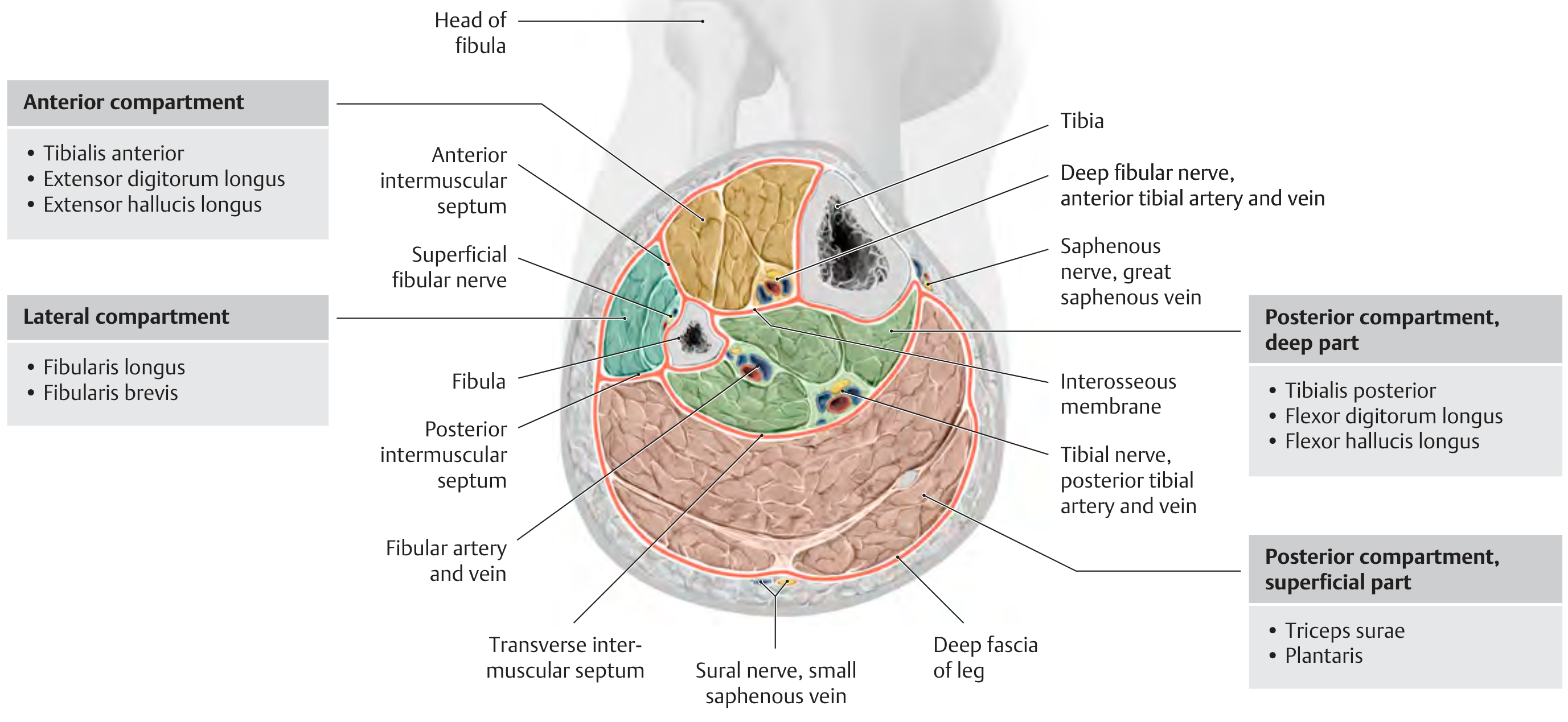
Source: THIEME Atlas of General Anatomy and Musculoskeletal System
The 4 Compartments
1. Anterior Compartment
| Muscles | Tibialis anterior, Extensor digitorum longus, Extensor hallucis longus, Peroneus (fibularis) tertius |
| Artery | Anterior tibial artery |
| Nerve | Deep fibular (peroneal) nerve |
| Function | Ankle/toe dorsiflexion |
| ACS sign | Numbness - 1st web space; weakness of ankle/toe extension |
2. Lateral Compartment
| Muscles | Fibularis (peroneus) longus, Fibularis brevis |
| Artery | No major named artery (supplied by perforators) |
| Nerve | Superficial fibular (peroneal) nerve |
| Function | Foot eversion |
| ACS sign | Pain on passive foot inversion; numbness - dorsum of foot; weakness of eversion |
3. Superficial Posterior Compartment
| Muscles | Gastrocnemius, Soleus, Plantaris (collectively = Triceps surae + Plantaris) |
| Nerve | Sural nerve |
| Function | Plantar flexion |
| ACS sign | Pain on passive ankle extension; numbness - dorsolateral foot; weakness of plantar flexion |
4. Deep Posterior Compartment
| Muscles | Tibialis posterior, Flexor digitorum longus, Flexor hallucis longus |
| Artery | Posterior tibial artery + fibular (peroneal) artery |
| Nerve | Tibial nerve |
| Function | Toe flexion, foot inversion, plantar flexion |
| ACS sign | Pain on passive toe extension/foot eversion; numbness - sole of foot; weakness of toe/ankle flexion |
Acute Compartment Syndrome - Clinical Summary
The two compartments most vulnerable to ACS are the anterior (anterior tibial artery + deep fibular nerve) and deep posterior (posterior tibial artery + tibial nerve). As the THIEME atlas notes, the interosseous membrane and intermuscular septa create poorly distensible spaces - even a small rise in pressure from edema or hematoma (e.g. after tibial fracture) can cause irreversible neuromuscular ischemia within hours. Emergency fasciotomy is the only definitive treatment.
"Tibialis anterior syndrome is characterized in its acute stage by severe pain and an inability to dorsiflex the toes due to the unopposed action of the plantar flexors - causing the toes to 'claw up.'" - THIEME Atlas of Anatomy
Cutaneous Nerve Distribution of the Foot
The nerve diagram below shows how compartment nerves distribute to the foot surface - useful for localizing which compartment is affected by sensory loss:
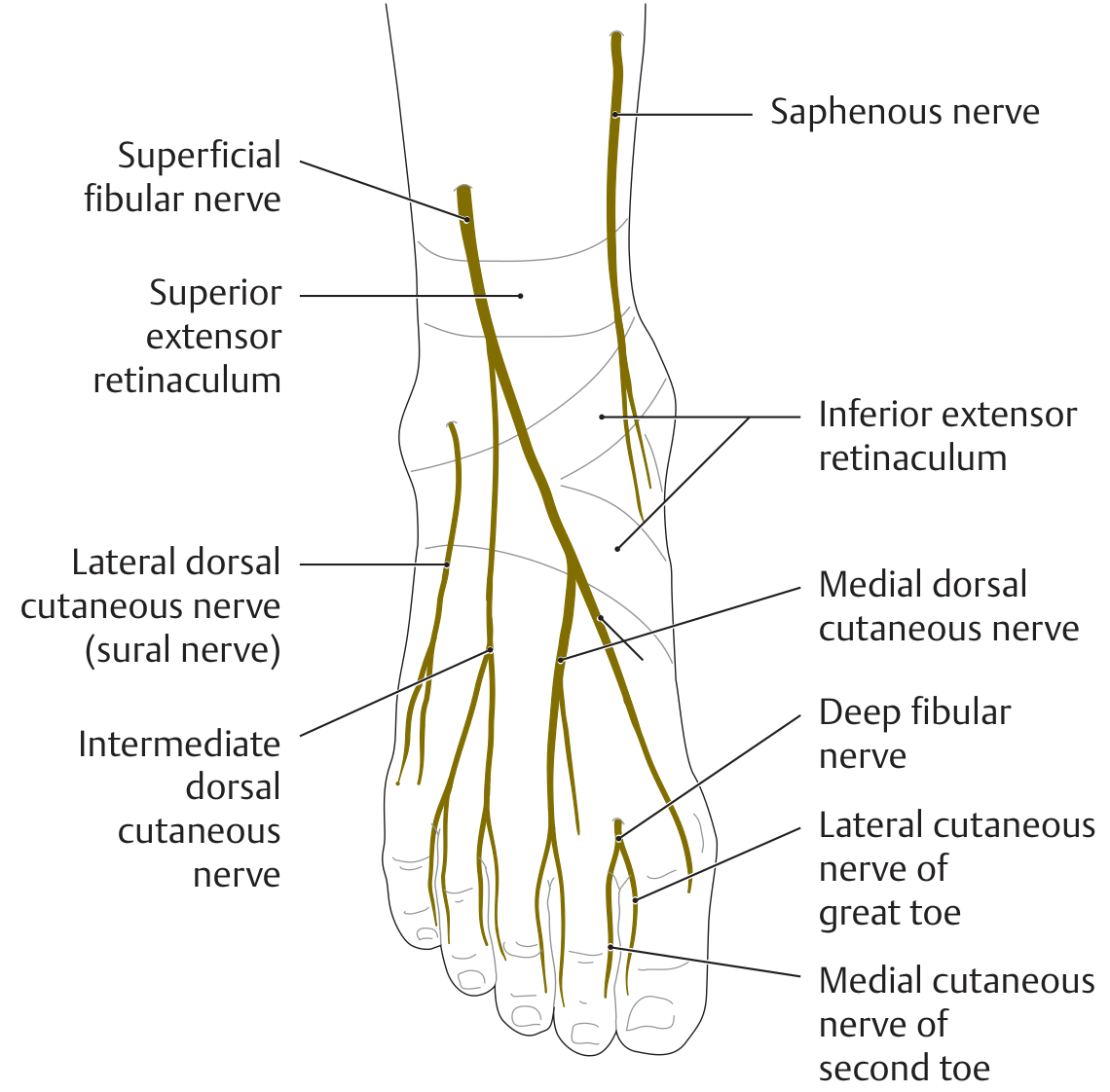
Source: THIEME Atlas of General Anatomy and Musculoskeletal System
| Nerve | Territory |
|---|---|
| Deep fibular nerve | 1st dorsal web space |
| Superficial fibular nerve | Dorsum of foot |
| Sural nerve | Lateral ankle and heel |
| Saphenous nerve | Medial ankle |
| Tibial nerve | Plantar aspect of foot |
Sources: THIEME Atlas of General Anatomy and Musculoskeletal System; Rockwood and Green's Fractures in Adults, 10th ed. (2025)
This is a shared conversation. Sign in to Orris to start your own chat.