
Stroke
Reading File
The topic "Stroke" is broad. Let me clarify before diving deep.
Asking for Preferences
What aspect of stroke are you interested in? · What is your purpose?
A comprehensive stroke overview for exam prep - let me pull from the medical library across all major areas in parallel.
Finding Sources
Finding Sources
Finding Sources
Good - I have the key sources. Let me now read the most comprehensive sections from Harrison's and Bradley's Neurology, plus the stroke management flowchart image.
Reading File
Reading File
Finding Sources
Good data coming in. Let me now read the critical sections on stroke types, clinical features, and acute management from Harrison's, plus the stroke management flowchart image.
Reading File
Reading File
Excellent. Let me now get the key sections on stroke types, etiology, TIA, and clinical features from Harrison's and read the intracerebral hemorrhage section.
Reading File
Reading File
Now let me read the core stroke overview section from the beginning of the block (definition, epidemiology, risk factors, TIA) and the thrombolysis/thrombectomy management section.
Reading File
Reading File
Let me now fetch the stroke management flowchart and read the acute treatment (thrombolysis/thrombectomy) section from Harrison's.
Loading Image
Finding Sources
I have the management flowchart. Now let me read the acute thrombolysis/thrombectomy section from Harrison's block 48 and the ICH/hemorrhagic section.
Reading File
Finding Sources
I now have very rich content from multiple authoritative textbooks. Let me also get the key thrombolysis/thrombectomy treatment details and TIA definition.
Finding Sources
Finding Sources
Finding Sources
Finding Sources
Reading File
Reading File
I now have all the information I need. Let me also quickly fetch the ischemic stroke pathophysiology diagram.
Loading Image
I now have comprehensive, authoritative content to compile a complete exam-ready stroke overview. Let me write it up.# Stroke - Comprehensive Exam Review
1. Definition & Epidemiology
A stroke (cerebrovascular accident, CVA) is a sudden-onset focal neurological deficit caused by interruption of blood supply to part of the brain, either from ischemia (85%) or hemorrhage (15%). It is the second leading cause of death and the leading cause of adult disability globally. "Time is brain" - approximately 1.9 million neurons are lost per minute during a large vessel occlusion.
A Transient Ischemic Attack (TIA) is a brief episode of focal neurological dysfunction with no infarction on imaging (tissue-based definition), with most episodes lasting <1 hour. It is a medical emergency - the short-term risk of completed stroke is high.
2. Classification
| Type | Subtype | Frequency |
|---|---|---|
| Ischemic | Thrombotic (large vessel, small vessel/lacunar) | ~85% |
| Ischemic | Embolic (cardioembolic, artery-to-artery) | included above |
| Hemorrhagic | Intracerebral hemorrhage (ICH) | ~10% |
| Hemorrhagic | Subarachnoid hemorrhage (SAH) | ~5% |
3. Pathophysiology of Ischemic Stroke
Three major mechanisms underlie ischemic stroke:
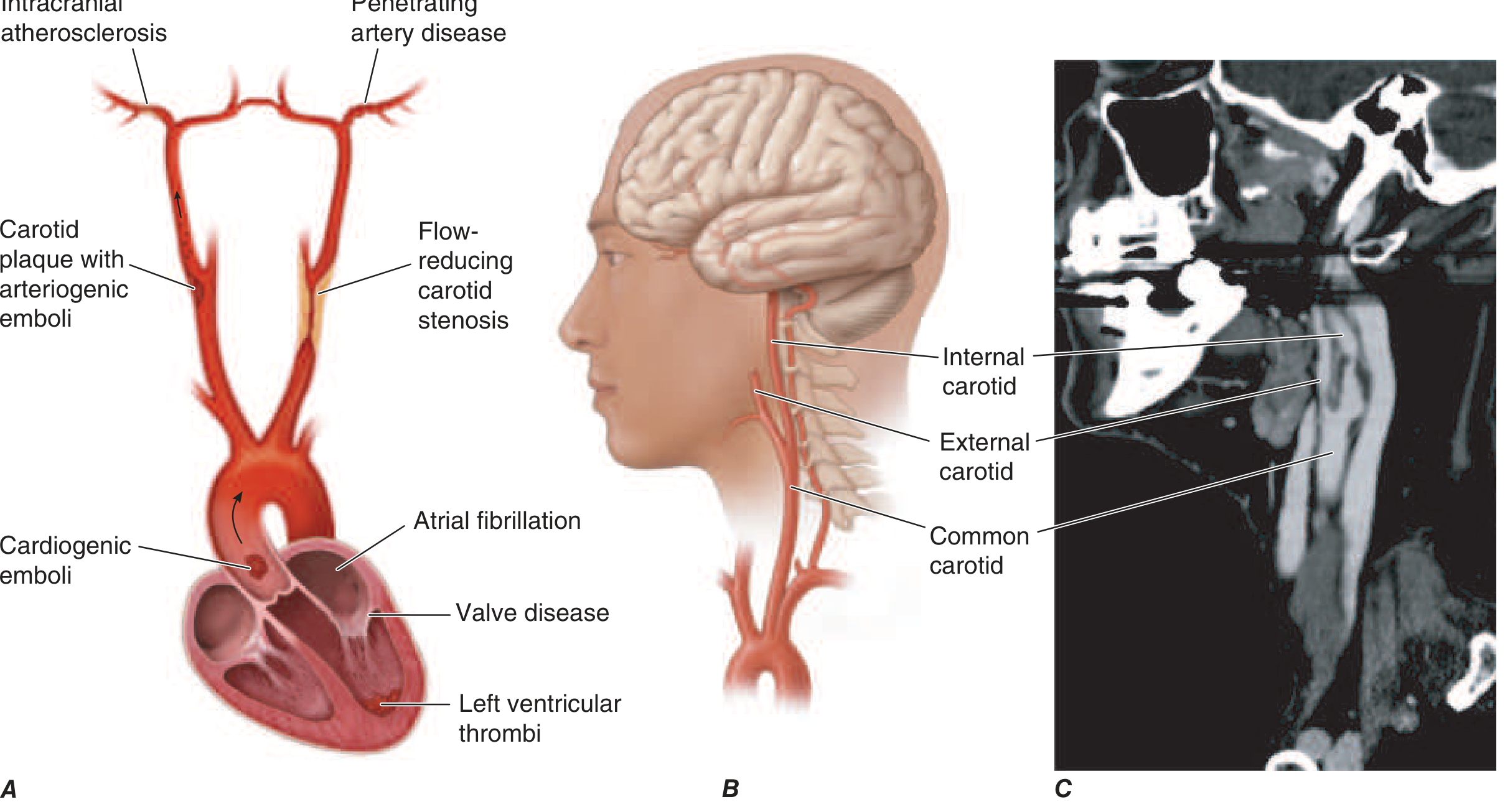
- Embolism - a thrombus from the heart (AF, mural thrombus, valve disease) or from an atherosclerotic plaque (carotid bifurcation, aortic arch) travels distally and occludes an intracranial vessel.
- In situ thrombosis - typically affects small penetrating arteries, producing lacunar infarcts in the basal ganglia, thalamus, internal capsule, and pons.
- Hypoperfusion - flow-limiting stenosis of a major extracranial vessel (e.g., internal carotid) produces "watershed" ischemia between arterial territories.
Collateral vessels play a key role: they determine the size of the infarct core and how much viable ischemic penumbra (salvageable tissue) exists around it. - Harrison's 22E, Fuster and Hurst's The Heart 15E
4. Etiology of Ischemic Stroke - High-Yield Table
| Common Causes | Uncommon Causes |
|---|---|
| Lacunar (small vessel) | Protein C/S deficiency |
| Large vessel atherothrombosis | Antiphospholipid syndrome |
| Atrial fibrillation (cardioembolic #1) | Factor V Leiden mutation |
| Mural thrombus (post-MI) | Sickle cell disease |
| Mitral stenosis / mechanical valve | Homocysteinemia |
| Carotid bifurcation atherosclerosis | CADASIL |
| Aortic dissection | Moyamoya disease |
| Paradoxical embolus (PFO) | Vasculitis (CNS) |
| Arterial dissection | Hypercoagulable states |
Source: Harrison's 22E Table 438-2
5. Risk Factors
Non-modifiable: Age, sex (M>F in middle age), race (African Americans have higher ICH rates), family history, prior TIA/stroke.
Modifiable: Hypertension (#1 modifiable), diabetes, hyperlipidemia, atrial fibrillation, smoking, obesity, physical inactivity, carotid stenosis, heart failure, illicit drugs (cocaine/amphetamines - both ischemic and hemorrhagic).
6. Clinical Features by Vessel Territory
Anterior Circulation (ICA/MCA/ACA)
MCA territory (most common):
- Contralateral face and arm > leg weakness and sensory loss (arm-face predominance)
- Dysphasia/aphasia (dominant hemisphere - usually left): Broca's = non-fluent; Wernicke's = fluent, incomprehensible
- Neglect, anosognosia, constructional apraxia (non-dominant hemisphere)
- Contralateral homonymous hemianopia
- Ipsilateral gaze deviation ("eyes look toward the lesion")
- Limb ataxia if large
ACA territory:
- Contralateral leg > arm weakness/sensory loss (leg predominance, reversed from MCA)
- Urinary incontinence
- Abulia, kinetic mutism (bilateral ACA occlusion)
- Contralateral grasp reflex
Internal carotid occlusion:
- Amaurosis fugax (monocular visual loss) - ischemia of ipsilateral retina via ophthalmic artery
- If circle of Willis is competent, may be asymptomatic
- Massive infarction (MCA + ACA territories) if circle of Willis is incompetent
Posterior Circulation (Vertebrobasilar)
Key rule: Crossed deficits (ipsilateral cranial nerve + contralateral limbs) = posterior circulation.
PICA occlusion - Lateral Medullary (Wallenberg) Syndrome:
- Ipsilateral: facial pain/temp loss, Horner's, dysarthria, dysphagia, ataxia
- Contralateral: limb/trunk pain/temp loss (spinothalamic)
- Preserved motor power (corticospinal tract spared)
- Vertigo, nausea, hiccups
Basilar artery occlusion:
- Bilateral pontine signs, "locked-in" syndrome
- High mortality if complete
Superior cerebellar artery: Ipsilateral cerebellar ataxia, contralateral pain/temperature loss
Posterior cerebral artery (PCA):
- Contralateral homonymous hemianopia with macular sparing
- Alexia without agraphia (dominant hemisphere)
- Thalamic sensory loss
7. Lacunar Syndromes (Small Vessel Disease)
Occur in deep structures from lipohyalinosis of small penetrating arteries (hypertension is the major cause):
| Syndrome | Location | Features |
|---|---|---|
| Pure motor hemiplegia | Posterior internal capsule or pons | Face+arm+leg weakness, no sensory loss, no cortical signs |
| Pure sensory stroke | Thalamus (VPL nucleus) | Contralateral numbness, no weakness |
| Sensorimotor stroke | Thalamo-capsular | Combined above |
| Ataxic hemiparesis | Pons or internal capsule | Ipsilateral arm ataxia + contralateral hemiparesis |
| Dysarthria-clumsy hand | Pons or genu of internal capsule | Slurred speech + hand clumsiness |
8. Transient Ischemic Attack (TIA)
Definition: Transient focal neurological deficit, no infarction on imaging; most last <1 h.
ABCD² Score (risk of stroke after TIA)
| Factor | Score |
|---|---|
| Age ≥60 yr | 1 |
| Blood pressure >140/90 mmHg | 1 |
| Clinical features - unilateral weakness | 2 |
| Clinical features - speech disturbance without weakness | 1 |
| Duration >60 min | 2 |
| Duration 10-59 min | 1 |
| Diabetes | 1 |
| Total: 0-7 |
- Score 0-3 = Low risk (1% stroke at 48h)
- Score 4-5 = Moderate risk (4.1% at 48h)
- Score ≥6 = High risk (8% at 48h; 22% at 3 months for score 7)
TIA patients should be treated with dual antiplatelet therapy (aspirin + clopidogrel) for 21 days, then aspirin alone. - Harrison's 22E, Rosen's Emergency Medicine
9. Diagnosis & Imaging
Noncontrast Head CT - First-line in Acute Stroke
- Primary role: Exclude hemorrhage before thrombolysis
- Findings in ischemic stroke: Hyperdense MCA sign (acute clot), loss of gray-white differentiation, subtle edema; frank hypodensity appears in 6-24h
- Findings in hemorrhagic stroke: Hyperdensity (bright white) of blood immediately
CT Angiography (CTA)
- Images entire arterial system from arch to vertex in one study
- Identifies large vessel occlusion (LVO) amenable to thrombectomy
CT Perfusion (CTP)
- Distinguishes infarct core (irreversibly damaged) from ischemic penumbra (salvageable)
- Critical for selecting thrombectomy candidates in the 6-24h window (DAWN/DEFUSE-3 trials)
MRI
- DWI (diffusion-weighted imaging): shows infarction within minutes of onset - most sensitive early
- FLAIR: shows infarction from days to years
- MRA: detects vessel stenosis/occlusion
- Superior to CT for posterior fossa infarcts (CT has poor sensitivity there)
Source: Harrison's 22E
10. Acute Management of Ischemic Stroke
Step-by-Step Pathway
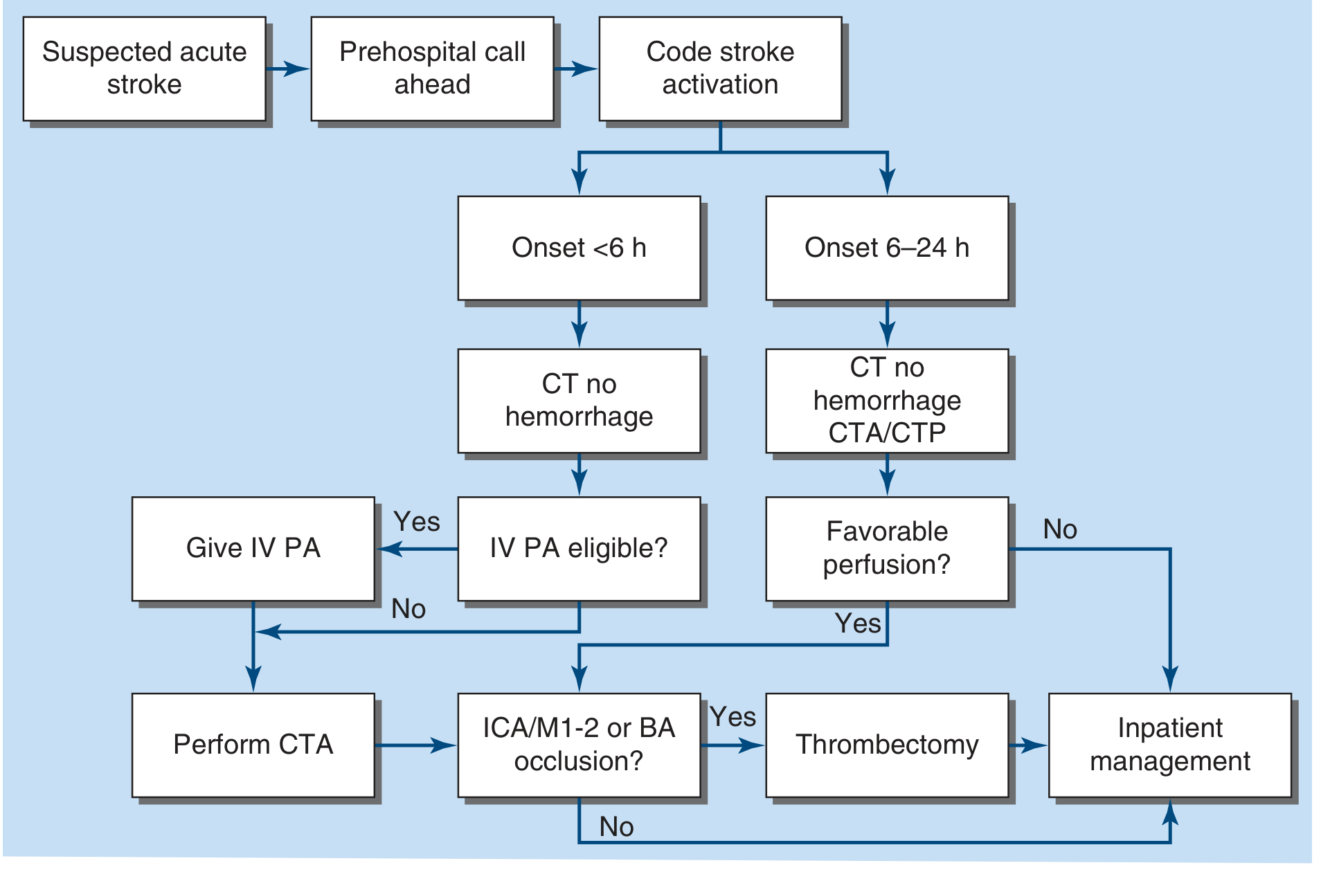
Harrison's 22E Figure 438-2
IV Thrombolysis (tPA / Alteplase)
- Dose: 0.9 mg/kg IV (max 90 mg); 10% as bolus, rest over 60 min
- Window: Within 4.5 hours of symptom onset (or last seen well time)
- Alternative agent: Tenecteplase (single IV bolus) is now preferred at many centers for its convenience
- Contraindications to IV tPA:
- Hemorrhage on CT
- Prior stroke or head trauma in last 3 months
- Major surgery within 14 days
- Active internal bleeding
- BP >185/110 (must treat first)
- Platelet count <100,000; INR >1.7
- Blood glucose <50 or >400 mg/dL
- Improving symptoms (TIA) - relative contraindication
Mechanical Thrombectomy (Endovascular)
- Indication: Large vessel occlusion (ICA terminus, M1/M2 MCA, basilar artery)
- Window: Within 24 hours of onset, if favorable perfusion imaging (selected by CTP/MR perfusion)
- Patients in the 0-6h window can get both IV tPA + thrombectomy ("bridging")
- Patients in 6-24h window: thrombectomy only (no IV tPA); selection based on perfusion mismatch (DAWN/DEFUSE-3 criteria)
- Source: Harrison's 22E, Bradley's Neurology in Clinical Practice
Blood Pressure Management in Acute Ischemic Stroke
- If giving IV tPA: Target BP <185/110 before and maintain <180/105 during infusion
- If NOT giving tPA: Permissive hypertension - treat only if BP >220/120 (collateral flow depends on elevated BP)
- Long-term: Target systolic <120 mmHg
Antiplatelet Therapy (Ischemic Stroke)
- Aspirin 325 mg should be started within 24-48h of stroke onset (not within 24h of tPA)
- Dual antiplatelet (aspirin + clopidogrel) for 21 days after minor stroke/high-risk TIA (POINT + CHANCE trials)
- Reduces early recurrence; major hemorrhage risk increased (0.2% → 0.9%)
Anticoagulation
- Atrial fibrillation: Start oral anticoagulation (apixaban preferred); timing depends on infarct size
- NOT recommended for routine atherothrombotic stroke (no benefit over aspirin; increased hemorrhage)
- May be used for dural sinus thrombosis, arterial dissection (limited evidence)
11. Hemorrhagic Stroke
Intracerebral Hemorrhage (ICH)
Causes: Hypertension (#1 - putamen, thalamus, pons, cerebellum); cerebral amyloid angiopathy (lobar, elderly); AVM; coagulopathy; illicit drugs (cocaine, amphetamines)
Classic presentation:
- Sudden onset of focal deficit + severe headache + elevated BP
- Progressive worsening over minutes to hours (unlike embolic = maximal at onset)
- Signs of raised ICP: vomiting, decreased consciousness, Cushing's response
Hypertensive ICH locations (in order of frequency):
- Putamen (55%)
- Thalamus (20%)
- Pons (10%)
- Cerebellum (10%)
- Lobar (5% - more often amyloid angiopathy)
ICH Score (prognosis): GCS score + ICH volume + intraventricular extension + infratentorial origin + age ≥80 years → predicts 30-day mortality.
Management of ICH:
- Reverse anticoagulation urgently (vitamin K + PCC for warfarin; idarucizumab for dabigatran; andexanet alfa for factor Xa inhibitors)
- Target BP ≤140/90 mmHg (INTERACT2, ATACH-II trials)
- No routine corticosteroids (do NOT use for cerebral edema in ICH)
- Surgical evacuation: cerebellum >3cm; superficial lobar; herniation
- ICP monitoring if GCS ≤8; osmotherapy (mannitol) for raised ICP
Subarachnoid Hemorrhage (SAH)
Cause: Ruptured berry aneurysm (#1); AVM; perimesencephalic nonaneurysmal SAH
Classic presentation:
- "Thunderclap headache" - worst headache of life, sudden onset
- Meningism (neck stiffness, photophobia)
- +/- third nerve palsy (posterior communicating artery aneurysm)
Diagnosis:
- CT head (sensitive 98% within 12h, decreases after)
- If CT negative + clinical suspicion: Lumbar puncture for xanthochromia (yellow CSF) - gold standard if CT negative
- CTA or DSA angiography to identify aneurysm
Complications:
- Rebleeding (highest risk first 24h; ~20% in 2 weeks)
- Vasospasm (days 3-14; treat with nimodipine, triple-H therapy historically)
- Hydrocephalus
- Hyponatremia (SIADH or cerebral salt wasting)
Treatment:
- Nimodipine 60 mg q4h for 21 days (reduces vasospasm and improves neurological outcome)
- Surgical clipping or endovascular coiling of aneurysm
12. Secondary Prevention
| Etiology | Treatment |
|---|---|
| Non-cardioembolic ischemic stroke | Antiplatelet: aspirin ± dipyridamole, OR clopidogrel |
| Atrial fibrillation | DOAC (apixaban preferred) or warfarin |
| Symptomatic carotid stenosis >70% | Carotid endarterectomy (same hospitalization); stenting alternative |
| Intracranial atherosclerosis >50% | Dual antiplatelet for ≥3 months |
| All ischemic stroke | High-intensity statin (atorvastatin 80 mg; LDL goal <70 mg/dL) |
| All stroke | BP control (SBP <120 mmHg long-term) |
13. Rehabilitation
- Stroke unit care reduces mortality and improves neurological outcomes significantly
- Early physical, occupational, and speech therapy
- Prevent complications: pneumonia (pneumatic compression/early mobilization), DVT (pneumatic compression stockings), pressure sores, contractures
- Constrained movement therapy (immobilizing the unaffected side) improves hemiparesis even years post-stroke - evidence for neural plasticity
- Selective serotonin reuptake inhibitors (SSRIs) may help post-stroke depression; motor benefit controversial
- Source: Harrison's 22E
14. High-Yield Exam Points
- FAST acronym: Face drooping, Arm weakness, Speech difficulty, Time to call emergency services
- "Time is brain": 1.9 million neurons lost per minute
- First investigation: Non-contrast CT head (to exclude hemorrhage)
- IV tPA window: 4.5 hours; dose 0.9 mg/kg
- Thrombectomy window: Up to 24 hours if perfusion imaging favourable
- Do not give tPA if: hemorrhage on CT, BP >185/110 (treat first), INR >1.7, or symptoms are improving
- Lacunar infarcts = small vessel; hypertension; pure motor/pure sensory most common
- Wallenberg = lateral medulla = PICA = crossed deficits, Horner's, dysphagia, no motor loss
- PCA = homonymous hemianopia with macular sparing
- ICH #1 cause = hypertension; #1 site = putamen
- SAH = worst headache of life; xanthochromia on LP; nimodipine prevents vasospasm
- TIA management = dual antiplatelet (aspirin + clopidogrel) × 21 days
- AF-related stroke = anticoagulation (DOAC first line)
- No heparin for routine atherothrombotic stroke
Sources: Harrison's Principles of Internal Medicine 22E (2025) | Bradley and Daroff's Neurology in Clinical Practice | Rosen's Emergency Medicine | Fuster and Hurst's The Heart 15E | Adams and Victor's Principles of Neurology 12E | Goldman-Cecil Medicine
This is a shared conversation. Sign in to Orris to start your own chat.