
Can u give me orientation of inner ear in skull image l
labeled diagram inner ear orientation temporal bone skull semicircular canals cochlea vestibule
"inner ear" AND anatomy
inner ear anatomy orientation temporal bone cochlea semicircular canals skull
https://www.ncbi.nlm.nih.gov/pmc/articles/PMC8335116/pdf/ajn…

How to orient it on a skull image
-
Find the external auditory canal
- The inner ear lies medial and slightly posterior to it.
- Move inward through the middle-ear region to reach the bony labyrinth.
-
Identify the cochlea
- It is the small snail-shaped structure.
- It lies anterior and inferior to the vestibule.
- Its base faces toward the internal auditory canal and its apex points anterolaterally.
-
Identify the vestibule
- This is the central chamber between cochlea and semicircular canals.
- It lies posterior to the cochlea and anterior to the semicircular canals.
-
Identify the semicircular canals
- They are posterior and superior to the vestibule.
- Superior (anterior) canal: vertical, arches upward.
- Posterior canal: vertical, more posterior.
- Lateral (horizontal) canal: roughly horizontal, but tilted about 30 degrees upward anteriorly.
-
Internal auditory canal
- Lies medial to the cochlea and vestibule.
- This is where CN VII and CN VIII enter the temporal bone.
Simple mental map
Lateral  ← external ear / middle ear

           Semicircular canals
                ↑ posterior-superior
                |
Cochlea  →  Vestibule  ← Internal auditory canal (medial)
anterior-inferior
Explain inner-ear orientation in coronal and sagittal views
coronal CT temporal bone inner ear cochlea vestibule semicircular canals labeled
coronal sagittal CT temporal bone inner ear cochlea vestibule semicircular canal orientation anatomy
"temporal bone" AND anatomy
- Cochlea = anterior and inferior
- Semicircular canals = posterior and superior
- Internal auditory canal (IAC) = medial
- Middle ear and mastoid = lateral
Coronal view
Anterior → posterior slice progression

Cochlea  →  Vestibule  →  Semicircular canals / mastoid
What you should see
- Cochlea: lower and more anterior. It appears as a compact spiral or “snail-like” bony structure.
- Vestibule: just posterior and superior to the cochlea, central in position.
- Superior semicircular canal: above the vestibule. It can appear as an arch or ring on more posterior coronal images.
- Lateral semicircular canal: posterior-lateral to the vestibule, near the epitympanum/aditus.
- Posterior semicircular canal: more posterior, often nearer the posterior temporal bone and mastoid.
Coronal shortcut
        Superior semicircular canal
                  ↑
            Vestibule
                  \
                   Cochlea
              anterior-inferior
Sagittal view
Anterior                                      Posterior

Cochlea  →  Vestibule  →  Semicircular canals → Mastoid
What you should see
- Cochlea: the most anterior inner-ear structure. It is also relatively inferior.
- Vestibule: immediately posterior to the cochlea, acting as the hub connecting cochlea and canals.
- Superior semicircular canal: rises above the vestibule, making a vertical loop.
- Posterior semicircular canal: lies posterior to the vestibule and is vertically oriented.
- Lateral semicircular canal: runs roughly horizontally, but its anterior end tilts upward, so it may not look perfectly horizontal on routine sagittal images.
- IAC: medial to the cochlea/vestibule. It carries CN VII and VIII toward the cerebellopontine-angle cistern.
Sagittal shortcut
             Superior canal
                 ∩
Cochlea → Vestibule → Posterior canal
  🐌         ○            ∩
 anterior                 posterior
Important imaging caveat
Show labeled coronal and sagittal inner-ear diagrams
labeled coronal temporal bone inner ear diagram cochlea vestibule semicircular canals
labeled sagittal temporal bone inner ear diagram cochlea vestibule semicircular canals
labeled coronal sagittal inner ear anatomy diagram cochlea vestibule semicircular canals
Labeled multiplanar inner-ear MRI
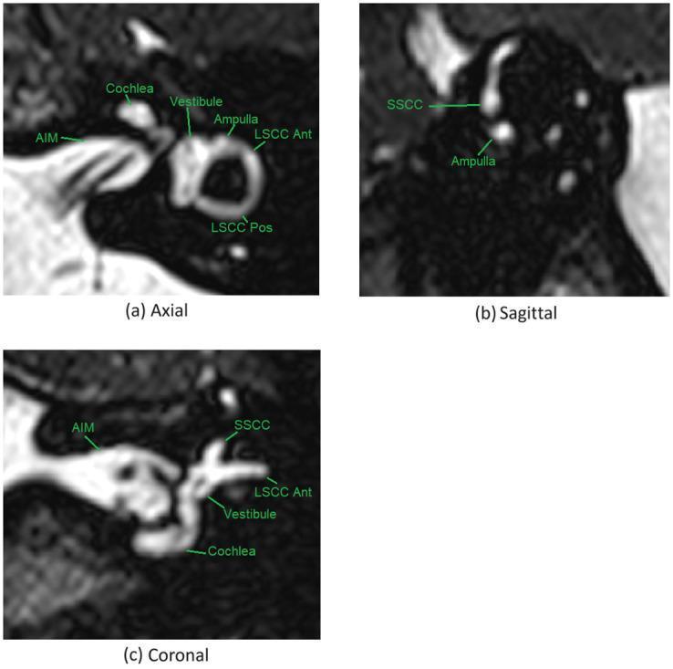
- Panel b, sagittal: cochlea is anterior, vestibule is central, and the superior semicircular canal arches above and posterior to the vestibule.
- Panel c, coronal: the superior semicircular canal is at the top, the vestibule is below it, and the cochlea is inferoanterior.
- IAM/IAC = internal auditory meatus/canal, medial route for the facial and vestibulocochlear nerves.
Labeled anatomic diagram
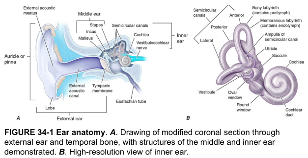
Cochlea anterior-inferior → vestibule central → semicircular canals posterior-superior.
Tnq
Posterior wall of middle ear
labeled posterior wall middle ear facial recess sinus tympani pyramidal eminence diagram
"middle ear" AND anatomy
posterior wall middle ear anatomy facial recess sinus tympani pyramidal eminence
Posterior wall of the middle ear
Main landmarks from superior to inferior
-
Aditus ad antrum
- A superior opening from the epitympanic recess into the mastoid antrum.
- This is why middle-ear infection can spread into mastoid air cells.
-
Fossa incudis
- A small depression in the upper posterior wall.
- Receives the short process of the incus.
-
Facial canal
- The descending or mastoid segment of the facial nerve lies in this region.
- It is a major surgical landmark.
-
Facial recess
- A recess lateral to the facial nerve, often used as a surgical route to the middle ear.
- Boundaries:
- Medial: facial nerve
- Lateral: chorda tympani
- Superior: fossa incudis / incus buttress
-
Pyramidal eminence
- A cone-shaped projection on the posterior wall.
- Contains the stapedius muscle.
- The stapedius tendon emerges from its apex to attach to the stapes neck.
-
Sinus tympani
- A deep recess medial to the pyramidal eminence.
- Lies between the posterior wall and medial wall and can hide cholesteatoma during surgery.
- Its superior boundary is the ponticulus and inferior boundary is the subiculum.
-
Posterior canaliculus for chorda tympani
- The chorda tympani enters the middle ear through the posterior wall, then runs forward between malleus and incus.
Easy diagram
              Superior
                 ↑
      Aditus ad antrum
           Fossa incudis
                 |
          Facial recess
     Chorda tympani | Facial nerve
                 |
       Pyramidal eminence
       (stapedius emerges)
                 |
          Sinus tympani
                 ↓
              Inferior
Show a labeled diagram of the posterior middle-ear wall
labeled diagram posterior wall tympanic cavity middle ear facial recess sinus tympani pyramidal eminence chorda tympani
labeled posterior wall middle ear diagram facial recess sinus tympani pyramidal eminence
"middle ear" AND anatomy
Posterior wall / retrotympanum of the middle ear

- Facial recess: lateral to the vertical (mastoid) segment of the facial nerve and medial to the chorda tympani. It is the route used for a posterior tympanotomy.
- Pyramidal eminence: bony projection containing stapedius muscle. The stapedius tendon emerges at its tip and attaches to the neck of stapes.
- Sinus tympani: deep recess medial to the pyramidal eminence, behind the oval-window region. It is a common hidden site for residual cholesteatoma.
- Ponticulus: ridge forming the superior border of the sinus tympani.
- Subiculum: ridge forming its inferior border.
- Facial nerve canal: runs in the posterior wall and is the key surgical landmark.
- Chorda tympani: crosses near the lateral aspect of the facial recess.

Lateral                          Medial
Chorda tympani → Facial recess → Facial nerve / pyramidal eminence → Sinus tympani