
Detail expalin of hiatus hernia.
Reading File
Now let me search the medical library for hiatus hernia content in parallel.
Finding Sources
Finding Sources
Searching PubMed
"hiatus hernia" OR "hiatal hernia"
Excellent! Now let me read the most comprehensive sources in parallel.
Reading File
Reading File
Reading File
Let me now read more sections for clinical features, diagnosis, complications, and treatment, plus fetch key images.
Reading File
Reading File
Reading File
Loading Image
Now let me fetch the remaining type illustrations and the antireflux barrier diagram, plus read diagnostic/treatment content.
Loading Image
Loading Image
Loading Image
Loading Image
Reading File
I now have rich, comprehensive content from multiple authoritative textbooks plus all four type illustrations. I have enough to write a thorough, well-illustrated answer.
Hiatus Hernia - A Detailed Explanation
Definition
A hiatus hernia (also called hiatal hernia) is a condition in which abdominal contents - most commonly the stomach - migrate upward through the esophageal hiatus of the diaphragm into the mediastinum (thoracic cavity). The esophageal hiatus is a normally tight, slit-like muscular opening in the right crus of the diaphragm through which the esophagus passes from the chest into the abdomen.
- Bailey and Love's Short Practice of Surgery, 28th Ed.
- Grainger & Allison's Diagnostic Radiology
Relevant Anatomy
The esophagogastric junction (EGJ) is normally anchored below the diaphragm by the phrenoesophageal ligament - a fibroelastic membrane that attaches the distal esophagus to the diaphragm at the hiatus. The EGJ's antireflux function depends on three components acting together:
- Lower esophageal sphincter (LES) - the intrinsic smooth muscle high-pressure zone (~2.0-2.5 cm long)
- Crural diaphragm (CD) - the external sphincter that augments LES pressure, especially during inspiration, coughing, and straining
- Phrenoesophageal ligament - keeps the LES in its intra-abdominal position (subject to positive abdominal pressure)
When a hiatus hernia develops, the crural diaphragm is separated from the LES, dismantling this composite antireflux barrier.
- Yamada's Textbook of Gastroenterology, 7th Ed.
- Sleisenger & Fordtran's GI and Liver Disease
Classification - Four Types
Type I: Sliding Hiatus Hernia (85-95% of all cases)
The most common type. The gastroesophageal junction (GEJ) and gastric cardia slide upward through a widened, weakened hiatus into the chest. The phrenoesophageal ligament becomes stretched and attenuated. There is no true hernia sac.
Key mechanism: The GEJ is displaced from its intra-abdominal position, resulting in:
- Loss of the intra-abdominal LES segment
- Decreased resting LES pressure
- Loss of the angle of His (esophageal flap valve)
- Separation of the LES from the crural diaphragm
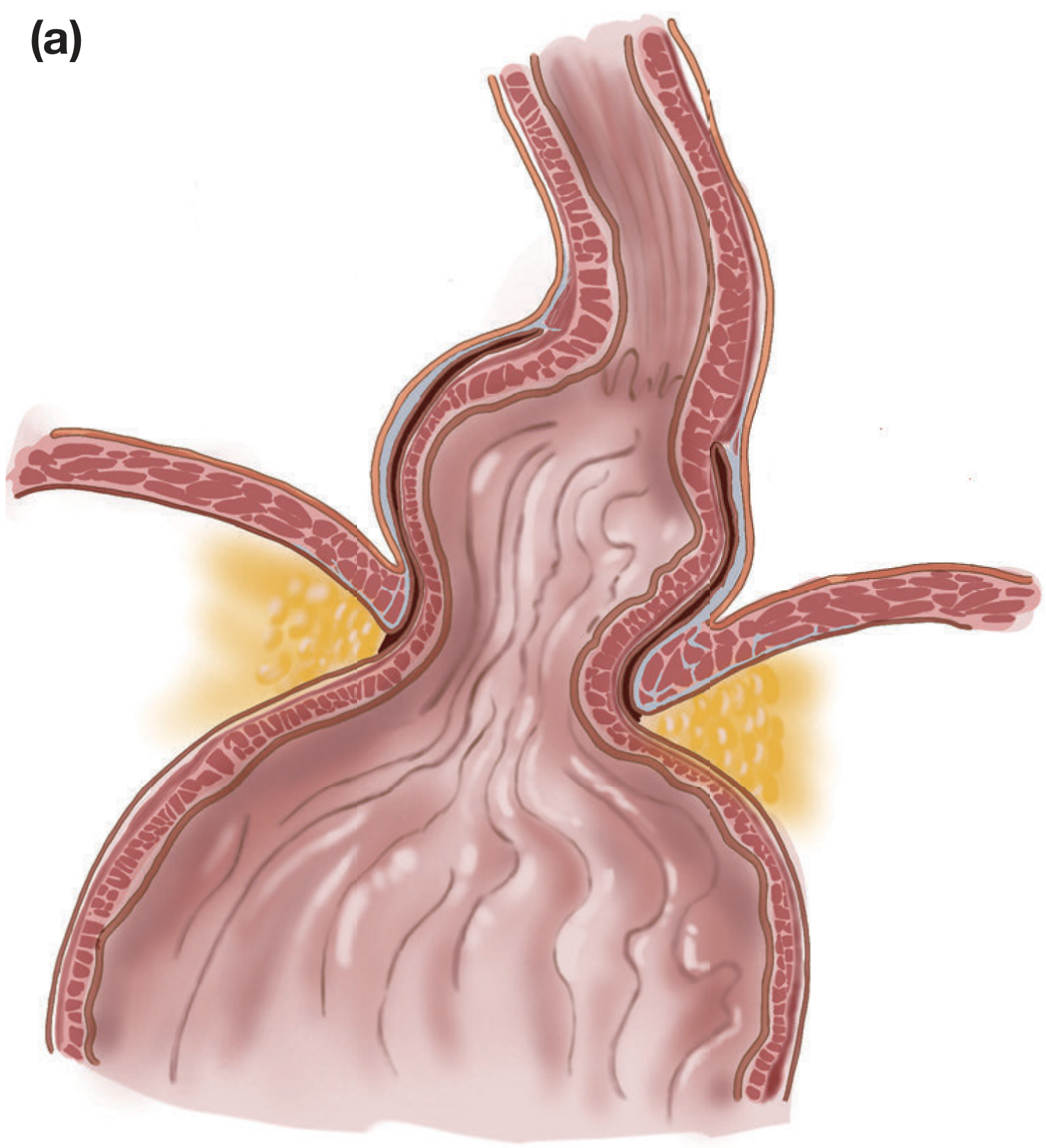
Sliding hernias are dynamic - they enlarge with increased intra-abdominal pressure, swallowing, and respiration. The hernia may "slide" back down at rest in early disease.
Type II: True Paraesophageal (Rolling) Hernia
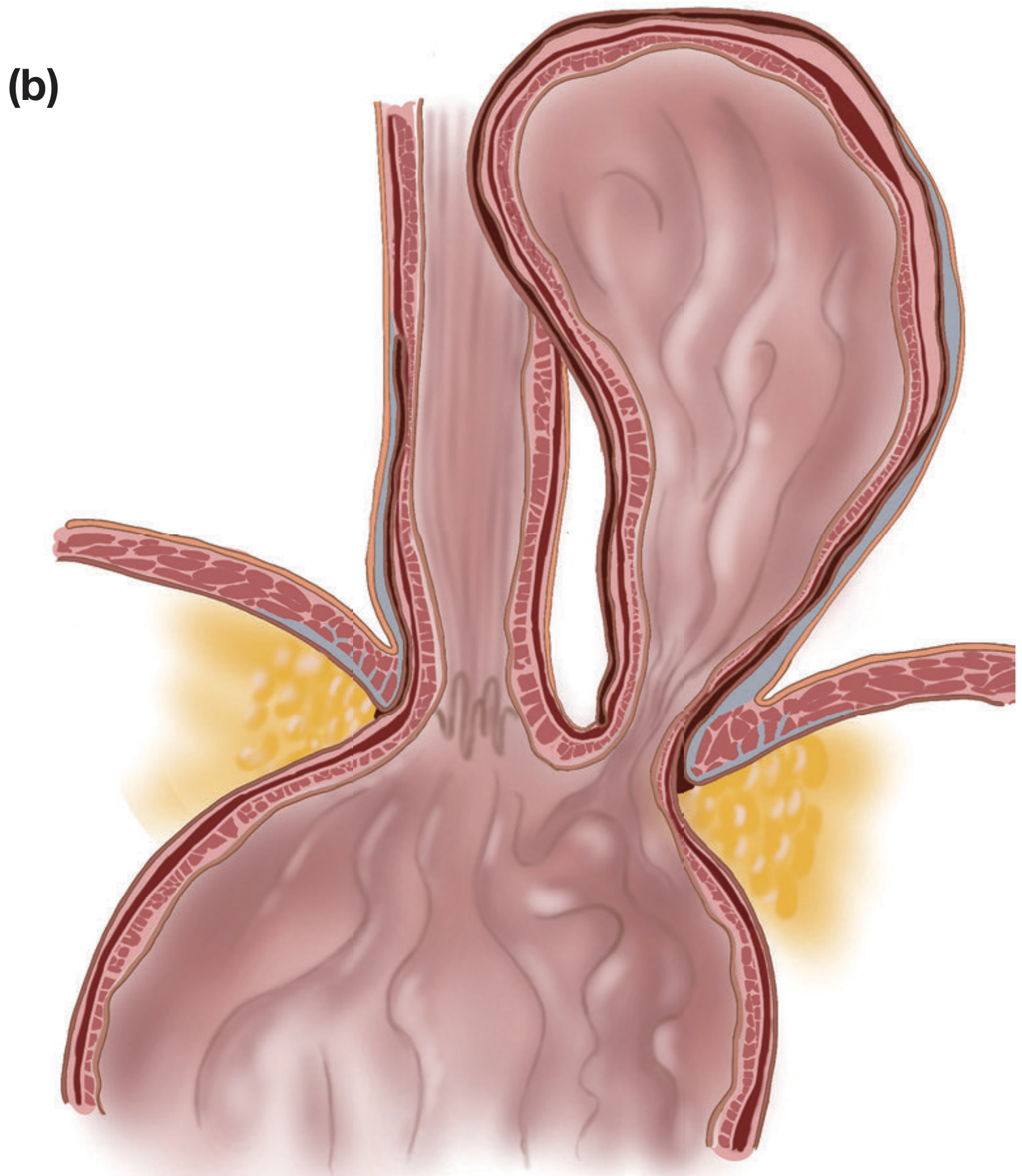
The least common paraesophageal type. The GEJ remains fixed at its normal anatomic position at the hiatus, but there is a focal defect in the phrenoesophageal membrane through which the gastric fundus herniates upward alongside the esophagus. All paraesophageal hernias (Types II-IV) have a true peritoneal hernia sac circumferentially covering the herniated contents.
Type III: Mixed (Combined) Hernia
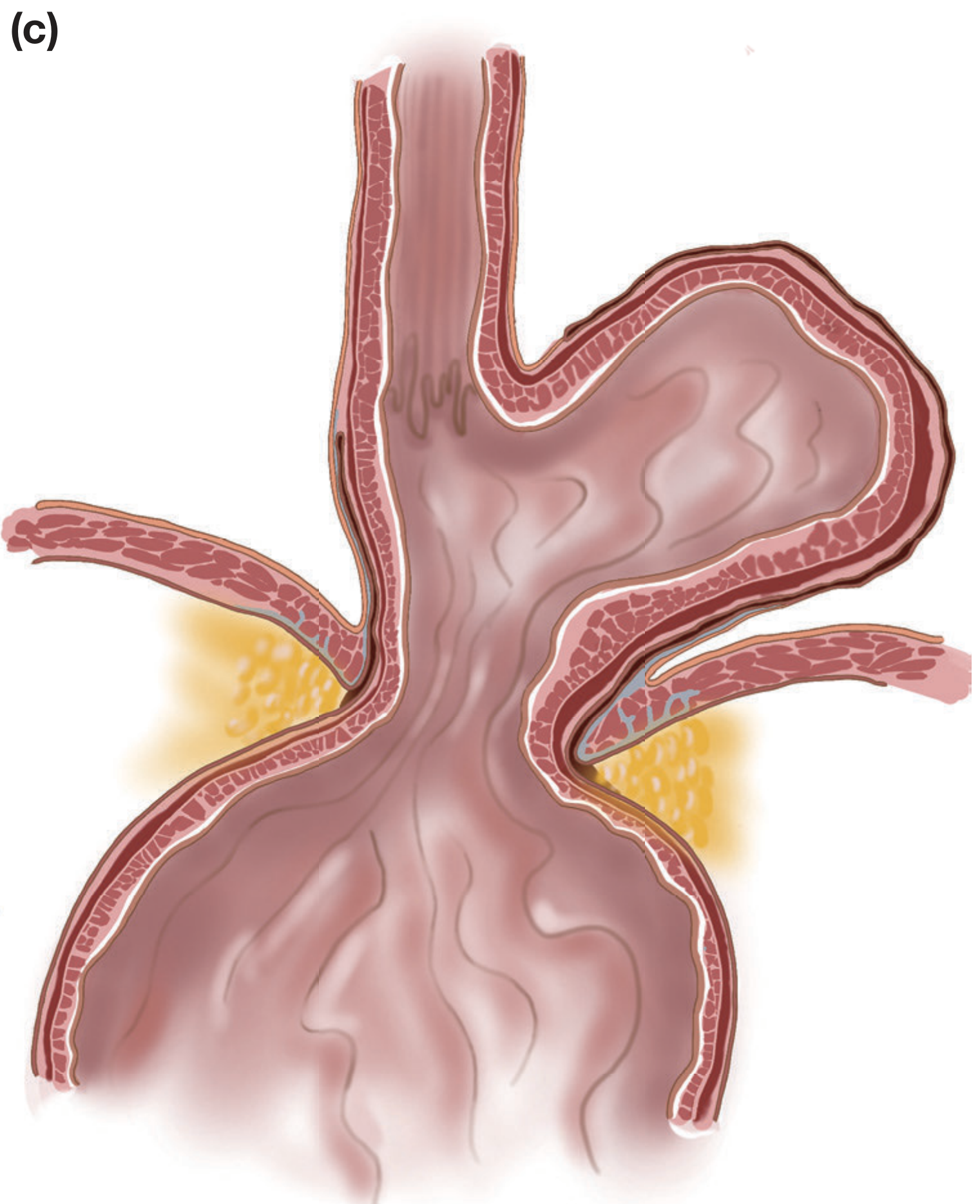
The most common paraesophageal type in clinical practice. Combines features of both Type I and Type II - both the GEJ and the gastric fundus are displaced above the diaphragm (at least 30% of the stomach above the diaphragm is the defining threshold). The stomach progressively inverts as it herniates, potentially creating an "upside-down stomach."
Type IV: Giant Hiatus Hernia
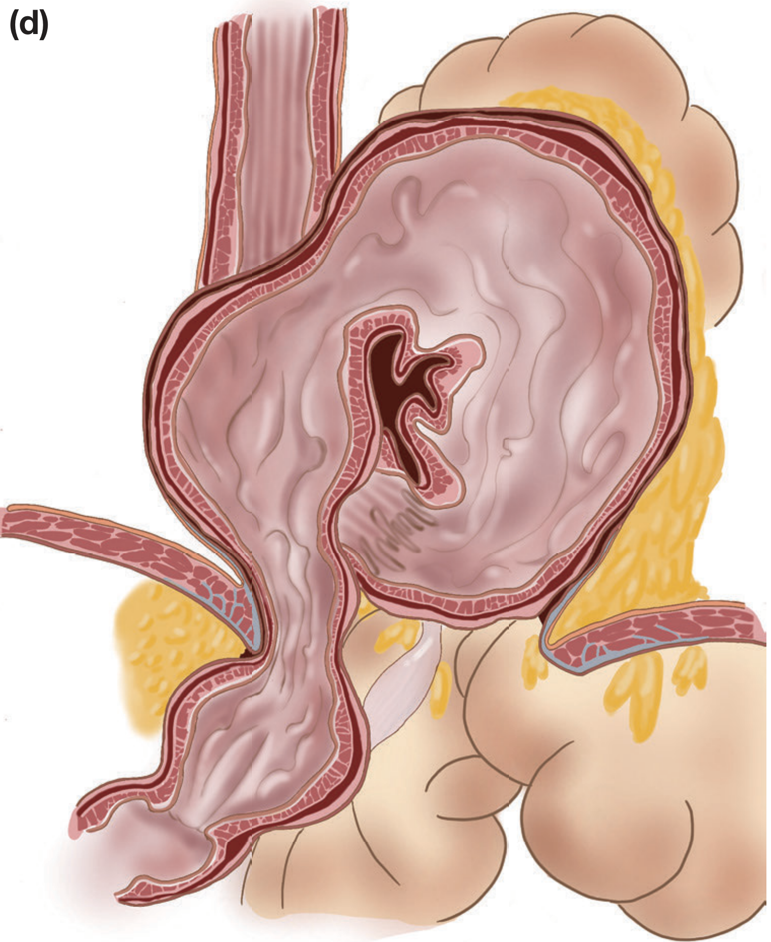
The most severe form, where other abdominal viscera migrate into the hernia sac in addition to the stomach. Organs involved can include: colon, small bowel, spleen, pancreas, liver, and omentum. The term "giant paraesophageal hernia" is used when more than half of the stomach has herniated into the thoracic cavity.
Pathophysiology - How Hiatus Hernia Causes Reflux
The diagram below shows the key mechanisms:
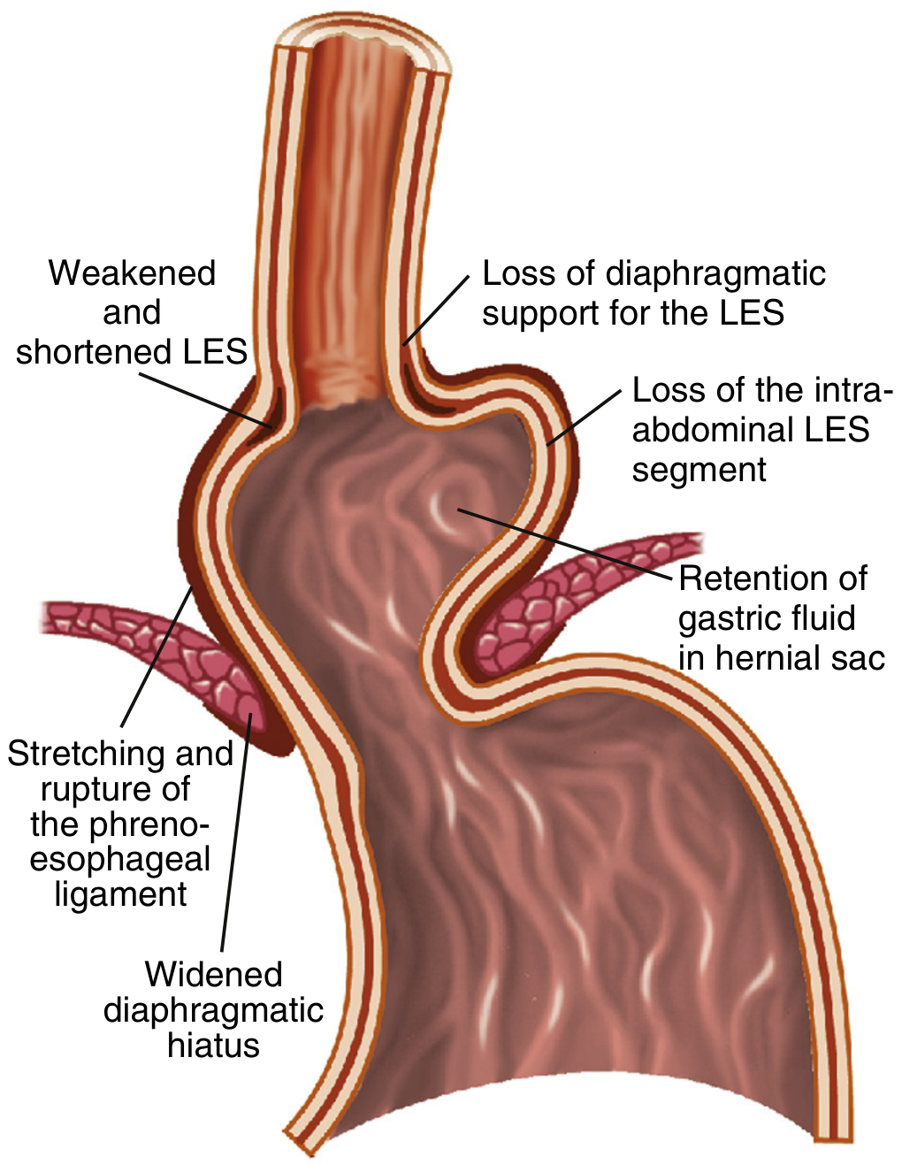
Five key mechanisms promote reflux in the presence of a hiatal hernia:
- Reduced basal LES pressure - loss of the intra-abdominal LES segment removes positive abdominal pressure contribution
- Loss of crural diaphragm augmentation - the CD can no longer add its pinchcock effect to the LES
- Acid pocket - the hernia sac acts as a reservoir of unbuffered gastric acid; during transient LES relaxations (tLESRs), this trapped acid refluxes back up
- Increased tLESR frequency - gastric distension with gas triggers more tLESRs in hernia patients
- Impaired esophageal clearance - retrograde flow from the hernia sac (re-reflux) during swallowing delays acid clearance
Large hernias (≥3 cm) and non-reducible hernias (gastric rugal folds remain above the diaphragm between swallows) carry the greatest reflux risk.
- Sleisenger & Fordtran's GI and Liver Disease
- Yamada's Textbook of Gastroenterology, 7th Ed.
Etiology and Risk Factors
The exact cause remains incompletely understood, but contributing factors include:
| Factor | Mechanism |
|---|---|
| Advanced age | Loss of elasticity and muscle tone around the hiatus; increased prevalence with each decade |
| Obesity | Raised intra-abdominal pressure weakens the hiatus over time |
| Pregnancy | Elevated intra-abdominal pressure |
| Chronic cough / heavy lifting | Repeated Valsalva-type maneuvers weaken the phrenoesophageal membrane |
| Hereditary factors | Familial clustering of GERD suggests possible smooth muscle disorder |
| GERD itself | Reflux may cause esophageal shortening (longitudinal muscle contraction), pulling the GEJ upward |
| Female sex | 75% of paraesophageal hernia patients are female |
- Maingot's Abdominal Operations
- Sleisenger & Fordtran's
Epidemiology
- Hiatus hernias are very common; prevalence rises sharply with age
- Type I accounts for ~90-95% of all cases
- Paraesophageal hernias occur most often in women; ~50% of PEH patients are over 70 years
- Hiatus hernia is found in 54-94% of patients with reflux esophagitis - far higher than in the healthy population
- About 40% of patients with paraesophageal hernia have chronic anemia (from mucosal venous engorgement or Cameron ulcers)
Clinical Features
Type I (Sliding) - Symptoms are primarily from GERD:
- Heartburn (pyrosis) - the cardinal symptom; worse after meals, lying down, bending
- Regurgitation of acid or food contents
- Dysphagia - usually mild, to solids; can be from associated peptic stricture or Schatzki ring
- Chest pain (may mimic cardiac pain)
- Chronic cough, hoarseness, laryngitis - from laryngopharyngeal reflux
- Often asymptomatic - discovered incidentally on CXR or CT as an intrathoracic gas bubble or fluid level
Type II/III/IV (Paraesophageal) - Symptoms are primarily mechanical/obstructive:
- Postprandial fullness, early satiety
- Dysphagia and chest pain - from gastric distension in the chest
- Breathlessness - compression of the lungs
- Chronic iron-deficiency anemia - from Cameron ulcers (linear erosions at the diaphragmatic level)
- Volvulus - if the stomach twists:
- Organoaxial volvulus - rotation around the longitudinal axis (most common)
- Mesentericoaxial volvulus - rotation around the transverse axis
- The classic Borchardt's triad of gastric volvulus: sudden epigastric pain, retching without vomiting, inability to pass a nasogastric tube
Complications
| Complication | Type Most At Risk | Features |
|---|---|---|
| GERD / Reflux esophagitis | Type I | Erosive esophagitis, peptic ulcer |
| Barrett's esophagus | Type I | Intestinal metaplasia; pre-malignant |
| Esophageal adenocarcinoma | Type I (long-standing GERD) | Risk from chronic Barrett's |
| Peptic stricture | Type I | Progressive dysphagia |
| Gastric volvulus | Types II/III/IV | Obstruction, strangulation |
| Strangulation / ischaemia | Types II/III/IV | Surgical emergency |
| Gastric perforation | Types II/III/IV | High mortality |
| Respiratory compromise | Types II/III/IV (large) | Lung compression, aspiration pneumonia |
| Cameron ulcers | Types III/IV | Iron-deficiency anemia |
- Bailey and Love's Short Practice of Surgery, 28th Ed.
- Maingot's Abdominal Operations
Diagnosis
1. Plain Chest X-ray (CXR)
- Incidental finding in asymptomatic patients
- Shows a retrocardiac gas bubble or air-fluid level behind the heart
- Indicates a large or giant hernia
2. Upper GI Barium Swallow (Fluoroscopy)
- Gold standard for demonstrating sliding hernias
- Diagnosis criteria: Gastric rugae traversing the diaphragm, or the esophageal B ring (Schatzki ring) - the squamocolumnar junction - seen above the diaphragm
- Small (<2 cm) sliding hernias are difficult to assess accurately; have historically led to overdiagnosis
- Rolling/combined-type hernias are well-demonstrated fluoroscopically and on CT
3. Upper Endoscopy (OGD)
- Best for identifying consequences of reflux (esophagitis, Barrett's, peptic stricture)
- Retroflexed view shows the stomach herniating through the hiatus
- Required before surgery to assess esophageal length, mucosal changes
4. High-Resolution Manometry (HRM)
- Current gold standard for diagnosing small sliding hernias
- Demonstrates a double hump EGJ pressure pattern - the LES pressure peak and the crural diaphragm pressure peak are separated by >1 cm
5. CT Scan
- Excellent for large/giant hernias and type IV hernias
- Defines hernia contents and detects complications (volvulus, ischaemia)
6. 24-hour pH Monitoring / Impedance-pH
- Gold standard for documenting GERD and correlating reflux with symptoms
- Mandatory before anti-reflux surgery
Treatment
Conservative / Medical Management (Type I, mild-moderate GERD)
Lifestyle modifications:
- Weight loss
- Head-of-bed elevation (30 degrees)
- Avoid large meals, late eating, lying down after meals
- Avoid triggers: caffeine, alcohol, fatty foods, citrus, chocolate, mint
Pharmacological:
- Proton pump inhibitors (PPIs) - first-line; omeprazole, lansoprazole, pantoprazole
- H2-receptor antagonists - as adjuncts
- Antacids / alginates - for symptomatic relief
- Prokinetics - metoclopramide or domperidone to improve gastric emptying
Note: Medical therapy controls symptoms of GERD but does not repair the structural defect of the hernia itself.
Surgical Management
Indications for Surgery:
- Symptomatic paraesophageal hernia (Types II/III/IV) - all should be repaired electively
- Large hernia with respiratory compromise
- Failed medical management of GERD from Type I hernia
- Complications: volvulus, obstruction, ischaemia, perforation (emergency surgery)
- Asymptomatic giant PEH can be considered for repair in fit patients (annual risk of acute complications ~<2%)
Emergency Surgery (Acute Presentation):
- Resuscitation first
- Nasogastric tube decompression
- Immediate surgery if ischaemia, perforation, or unresolved obstruction is suspected
Surgical Principles (Elective):
The modern laparoscopic (minimally invasive) approach is standard. Key steps:
- Reduction of herniated contents back into the abdomen
- Extensive mediastinal dissection to restore adequate intra-abdominal esophageal length (at least 2-3 cm below the hiatus)
- Excision of the hernia sac (prevents recurrence in PEH)
- Crural repair (hiatal closure) - the diaphragmatic crura are sutured together posterior to the esophagus, sometimes reinforced with a biosynthetic mesh for large defects
- Fundoplication - the gastric fundus is wrapped around the distal esophagus to recreate the anti-reflux valve:
- Nissen fundoplication (360° wrap) - most common, best for GERD
- Toupet fundoplication (270° posterior wrap) - preferred if esophageal dysmotility
- Dor fundoplication (180° anterior wrap)
- Gastropexy - fixation of the stomach in the abdomen to prevent re-herniation (especially for PEH without fundoplication)
Robotic-assisted surgery is increasingly used and a 2025 systematic review (PMID: 41107615) confirms comparable safety and efficacy to standard laparoscopy.
Summary Box
| Feature | Type I (Sliding) | Types II/III/IV (Paraesophageal) |
|---|---|---|
| Frequency | 85-95% | 5-15% |
| GEJ position | Above diaphragm | Normal (II) or above (III/IV) |
| Hernia sac | No | Yes (true peritoneal sac) |
| Main symptoms | GERD (heartburn, regurgitation) | Obstructive (dysphagia, chest pain, early satiety) |
| Main risk | Barrett's esophagus, adenocarcinoma | Volvulus, strangulation |
| Primary treatment | Medical (PPIs); surgery if failed | Surgery (all symptomatic cases) |
Sources:
- Bailey and Love's Short Practice of Surgery, 28th Ed., Ch. 66
- Maingot's Abdominal Operations, Ch. on Hiatal Hernia
- Sleisenger & Fordtran's Gastrointestinal and Liver Disease
- Yamada's Textbook of Gastroenterology, 7th Ed.
- Grainger & Allison's Diagnostic Radiology
- Harrison's Principles of Internal Medicine, 22nd Ed.
This is a shared conversation. Sign in to Orris to start your own chat.