
Cellities
cellulitis lower leg erythema swelling skin infection
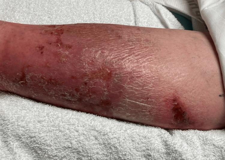
This clinical photograph displays the lower extremity of a patient, demonstrating classic physical examination findings of cellulitis. The skin exhibits diffuse erythema and swelling extending across the anterior and lateral aspects of the leg. The texture is notably compromised, showing signs of desquamation (peeling), xerosis (dryness), and lichenification, particularly in the mid-to-lower portion. Multiple discrete skin lesions are visible, including areas of darker, crusted scabbing and potential ulceration that may serve as a portal of entry for infection. Small petechiae or ecchymotic spots are scattered within the erythematous region. The presentation is highly characteristic of an acute bacterial skin and soft tissue infection (SSTI), emphasizing the visual transition from healthy proximal skin to distal inflamed, edematous, and scaling tissue. This image is an educational resource for identifying inflammatory dermatological conditions and assessing secondary skin changes associated with chronic or severe cellulitis.
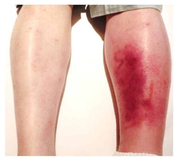
This clinical photograph shows a posterior view of a patient's lower legs, providing a side-by-side comparison between a healthy limb and a symptomatic one. The left lower leg (right side of the image) exhibits classic clinical signs of cellulitis, characterized by significant unilateral edema and diffuse erythema. The affected skin shows a spectrum of inflammatory discoloration, ranging from bright red to deep purplish hues, particularly over the calf region. The skin texture appears tense and glossy due to underlying swelling. A thin, vertical line of serous fluid exudate is visible on the posterior-lateral aspect of the left calf. In contrast, the right lower leg (left side of the image) appears normal with healthy skin tone and no evidence of swelling. This visual demonstrates the acute presentation of a soft tissue infection, useful for teaching the differential diagnosis of unilateral leg swelling and redness, which may include cellulitis, deep vein thrombosis (DVT), or localized vasculitis.
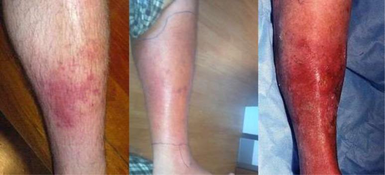
A sequence of three clinical photographs illustrating the progression of cellulitis on the lower leg, categorized by clinical severity. The 'Mild' stage (left) shows a localized, erythematous patch with poorly defined borders and scattered red papules. The 'Moderate' stage (center) demonstrates expanding confluent erythema and edema covering a larger surface area; notably, ink markings are visible on the skin to monitor the spread of infection. The 'Severe' stage (right) depicts intense, deep red to violaceous erythema involving the majority of the limb, accompanied by significant inflammatory swelling, a shiny skin texture suggestive of severe edema, and focal areas of potential skin breakdown or desquamation. This comparison highlights key diagnostic features of skin and soft tissue infections (SSTIs), including changes in color intensity, surface area involvement, and signs of tissue tension associated with worsening bacterial infection.
cellulitis skin infection treatment
Cellulitis
Definition
- Andrews' Diseases of the Skin, Ch. 14 - Rosen's Emergency Medicine, Ch. 126
Clinical Features
- Erythema - diffuse, rapidly spreading, typically with poorly defined and irregular borders
- Warmth, swelling (edema), and tenderness at the affected site
- Systemic features - fever, chills, malaise (not always present)
- Lymphangitis - linear streaks of inflammation extending proximally along vascular tracts, especially with Streptococcal or Pasteurella infections (e.g., animal bites)
- Vesicles or bullae - can form when localized edema becomes severe, causing epidermal layer separation
- Gangrene
- Metastatic abscesses
- Severe sepsis
- Necrotizing fasciitis (a distinct, life-threatening entity requiring urgent surgical debridement)

Causative Organisms
| Type | Common Pathogens |
|---|---|
| Nonpurulent cellulitis | Streptococci (~75%), especially Group A Strep (S. pyogenes) |
| Purulent cellulitis / abscess | Staphylococcus aureus, including CA-MRSA |
| Erysipelas | S. pyogenes |
| Diabetic foot | S. aureus, streptococci; chronic wounds - polymicrobial |
| Animal bites | Pasteurella multocida (cat/dog), polymicrobial (human) |
| Immersion injuries | Aeromonas, Vibrio species |
Risk Factors
- Disruption of the skin barrier (wounds, tinea pedis, insect bites, ulcers)
- Venous insufficiency / lymphedema / peripheral edema
- Obesity
- Diabetes mellitus
- Immunodeficiency / immunosuppression
- Prior history of cellulitis (recurrence is common)
- Saphenous phlebectomy, lymphadenectomy, or prior irradiation
Diagnosis
- Blood cultures: Not indicated in uncomplicated cases; reserved for immunocompromised patients or those with systemic sepsis
- Wound cultures: Only if an open wound is present; needle aspiration or biopsy are not recommended for typical nonpurulent cellulitis
- Imaging:
- Ultrasound: Useful to differentiate cellulitis from abscess; can also identify foreign bodies
- Plain X-ray / CT: Used when necrotizing infection is suspected (may reveal soft tissue gas along fascial planes) or to evaluate for osteomyelitis in diabetic foot
Differential Diagnosis ("Pseudocellulitis")
| Mimic | Distinguishing Features |
|---|---|
| Venous stasis dermatitis | Usually bilateral, above the ankle, no fever, does not worsen with limb elevation |
| Deep vein thrombosis (DVT) | Unilateral swelling, confirmed by Doppler ultrasound |
| Contact / allergic dermatitis | Itchy, not painful; follows exposure pattern |
| Erythema migrans (Lyme disease) | Targetoid, >5 cm, central clearing; in tick-endemic areas |
| Gout | Monoarticular, hyperuricemia, crystal deposits |
| Eosinophilic cellulitis (Wells syndrome) | Follows insect bite, eosinophilia (not neutrophilia), less pain |
| Fixed drug eruption | Linked to drug exposure, recurs at same site |
Management
Outpatient (mild, no systemic toxicity)
- Nonpurulent cellulitis: Dicloxacillin or cephalexin 500 mg every 6 hours for 5 days (covers streptococci and MSSA)
- If MRSA suspected (risk factors: prior MRSA infection, IV drug use, incarceration, competitive sports, recurrent furunculosis): trimethoprim-sulfamethoxazole (TMP-SMX) or doxycycline
Inpatient (systemic toxicity, immunocompromised, failure of oral therapy)
- IV penicillin or cephalosporins (e.g., cefazolin) for streptococcal infections
- IV vancomycin or linezolid if MRSA is a concern
Adjunctive measures
- Limb elevation
- Compression therapy for lymphedema (reduces recurrence)
- Marking the borders of erythema with a pen to monitor progression
Recurrent cellulitis
- Prophylactic penicillin (long-term low-dose) is used in patients with recurrent episodes due to lymphedema
- A 2026 systematic review (PMID 41275489) examines the evidence for secondary prevention strategies
- Treat any portal of entry (e.g., antifungal for tinea pedis)
Key Takeaways
- Cellulitis = diffuse, poorly-bordered skin infection; erysipelas = well-demarcated, raised borders
- Most cases are streptococcal (nonpurulent); purulent cellulitis suggests S. aureus / CA-MRSA
- The clinical mimicry rate is high (~30%) - always consider stasis dermatitis, DVT, and contact dermatitis
- Bilateral "cellulitis" is almost never cellulitis
- Necrotizing fasciitis is a surgical emergency; pain out of proportion to findings is a red flag
- A recent network meta-analysis (PMID 39240378) confirms the efficacy of first-line antibiotics (cephalosporins, penicillins) for cellulitis and erysipelas
Cellulitis with hand swelling in type 2 diabetes
hand cellulitis swelling diabetic patient erythema dorsum
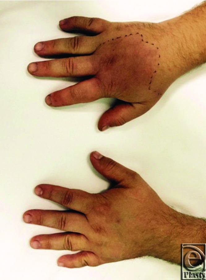
A clinical photograph showing a comparative dorsal view of two human hands. The top hand exhibits clinical signs of cellulitis or acute inflammation, characterized by diffuse swelling (edema) across the dorsum and extending into the digits, accompanied by confluent erythema. A black dotted ink line has been marked on the skin at the proximal aspect of the wrist to demarcate the advancing border of the erythema and monitor clinical progression. The digits on the affected hand appear significantly more bulbous compared to the bottom hand, which serves as a healthy control with normal anatomical contours and skin pigmentation. This image demonstrates the visual presentation of rapidly spreading soft tissue infection or a severe inflammatory response, highlighting the diagnostic importance of physical examination findings such as unilateral swelling, redness, and the use of clinical marking for progression tracking.
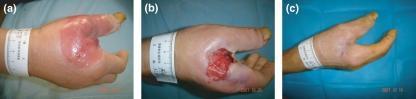
This sequence of three clinical photographs (a, b, and c) illustrates the progression and management of a hand abscess in a diabetic patient. Panel (a) shows the dorsum of the right hand at admission, characterized by significant soft tissue edema and a large, erythematous, fluctuant area consistent with a subcutaneous abscess. Panel (b) depicts the hand one week post-surgical debridement and drainage; it shows an open, granular ulcer bed with irregular borders and a reduction in surrounding cellulitis and swelling. Panel (c) shows the final outcome at six weeks following autologous platelet-rich gel therapy, demonstrating successful wound closure, epithelialization, and minimal residual scarring. The series highlights the clinical evolution from acute infection to surgical intervention and the subsequent role of regenerative therapies in healing complex diabetic hand ulcers.
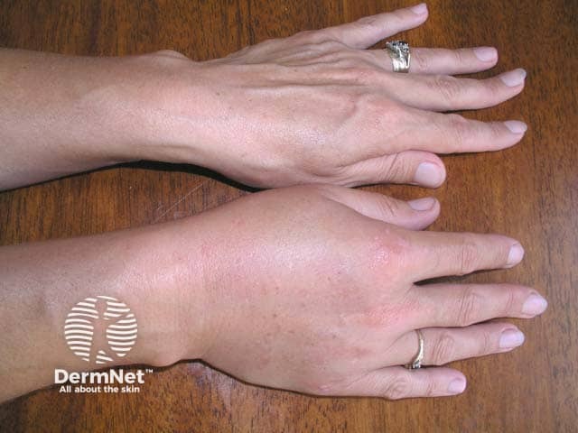
Clinical photography of the right hand illustrating acute soft-tissue edema consistent with angioedema. The dorsal and palmar aspects are swollen, with diffuse subcutaneous swelling around the digits and dorsum of the hand, mild to moderate erythema, and no discrete abscess, bullae, or skin necrosis. Visualized joints maintain normal alignment and there is no focal crepitus. The edema predominantly involves the palmar digits and dorsum, extending to the proximal interphalangeal joints while preserving skin folds and extensor surfaces. The appearance favors a vascular permeability–driven process rather than infectious cellulitis, though secondary inflammation cannot be excluded. In this context, clinical correlation with exposure history, recent medication use (notably ACE inhibitors or similar triggers), trauma, allergen exposure, or hereditary angioedema is essential. The image serves as a documentation tool for acute swelling, guiding differential diagnosis between angioedema, cellulitis, contact dermatitis, and other edematous conditions. It is useful for bedside assessment, patient education, and monitoring response to therapy (antihistamines, corticosteroids, epinephrine if indicated). This visual record supports triage decisions, risk stratification for airway compromise, and educational purposes for clinicians, residents, and students reviewing hand edema etiologies and radiology-lacking diagnostic clues in soft tissue swelling. The image thereby enhances recognition and comparative follow-up imaging decisions.
cellulitis hand infection diabetes mellitus
Cellulitis with Hand Swelling in Type 2 Diabetes
Why Diabetes Makes This Dangerous
- Immune dysfunction - Neutrophil and monocyte-macrophage function are directly impaired by hyperglycemia. Cellular immunity shows reduced proliferative response. These defects are worsened by poor glucose control and improve with tighter glycemic management.
- Vascular insufficiency - Microangiopathy and atherosclerosis reduce tissue perfusion, impairing antibiotic delivery and healing.
- Sensory neuropathy - Leads to wound neglect; patients may present late with advanced infection. They may have malaise without fever, or purulent discharge without pain - atypical presentations that delay diagnosis.
- Rosen's Emergency Medicine, Ch. 33 - diabetic immunocompromise
Clinical Presentation
- Diffuse erythema with poorly defined borders
- Dorsal hand swelling (key anatomical point below)
- Warmth and tenderness
- Possible lymphangitis - red streaks tracking proximally up the forearm
Why the Dorsum Swells
"The veins and lymphatics course through the softer tissues on the dorsum of the hand. Therefore, the dorsum of the hand often swells whenever there is an inflammatory or infectious process. For this reason, a deep space infection initially may be misdiagnosed as cellulitis over the dorsum of the hand."
- Tintinalli's Emergency Medicine, Ch. 283
Red Flag Features (Urgent / Admit)
| Feature | Concern |
|---|---|
| Pain with passive finger motion | Flexor tenosynovitis (surgical emergency) |
| Fluctuance on volar surface / web space | Deep space abscess |
| Crepitus or gas on X-ray | Necrotizing fasciitis / gas-forming organisms |
| Separation of adjacent digits | Collar-button abscess |
| Fever, rigors, hypotension, confusion | Systemic sepsis - IV antibiotics + ICU |
| Rapidly spreading erythema beyond wrist | Spreading infection up fascial planes |
| Pain out of proportion to findings | Necrotizing fasciitis |
"If deep structures of the hand are involved, emergently consult a hand specialist because treatment likely will involve inpatient care and operative drainage."
- Tintinalli's Emergency Medicine, Ch. 283
Microbiology in Diabetic Patients
| Setting | Likely Pathogens |
|---|---|
| Routine cellulitis (nonpurulent) | Group A Streptococcus, MSSA |
| Purulent / abscess | CA-MRSA (47-78% of ED hand infections), MSSA |
| Immunocompromised / AIDS-like | Mycobacterium spp., Candida albicans |
| Animal bite | Pasteurella multocida (rapidly spreading, suppurative cellulitis) |
| IV drug use | S. aureus + gram-negatives (polymicrobial) |
| Severe / chronic infection | Polymicrobial, gram-negatives, anaerobes, MDROs |
Severity Classification (IDSA / IWGDF)
| Clinical Findings | IDSA Severity | Action |
|---|---|---|
| No purulence or inflammation | Uninfected | No antibiotics |
| Erythema ≤2 cm, no systemic signs | Mild | Oral antibiotics, outpatient |
| Cellulitis >2 cm, lymphangitis, deep-tissue spread, or below-fascia involvement; systemically stable | Moderate | Parenteral antibiotics, likely admit |
| Systemic toxicity: fever, leukocytosis, tachycardia, hypotension, confusion, severe hyperglycemia, acidosis | Severe | IV antibiotics + surgical consult, ICU consideration |
- Fischer's Mastery of Surgery, Table 246.1 / Sabiston Textbook of Surgery, Table 35.5
Management
1. Glycemic Control
2. Antibiotic Therapy
- Nonpurulent: Cephalexin 500 mg every 6 hours OR TMP-SMX 160/800 mg twice daily
- If MRSA suspected: TMP-SMX 1-2 DS tabs q12h, doxycycline 100 mg q12h, or clindamycin 300-450 mg q8h
- Duration: 5-7 days (can extend if infection persists)
- Vancomycin IV (recommended given high CA-MRSA prevalence in most communities - up to 47-78% of hand infections)
- Add coverage for enteric gram-negatives (e.g., piperacillin-tazobactam) for immunocompromised patients or those with open trauma
- Broaden empirically for suspected polymicrobial infection in severe/chronic wounds
"Antibiotic therapy is needed for... comorbid diseases (diabetes mellitus); immunosuppression, location on face, hand, or genitalia." - Washington Manual of Medical Therapeutics
3. Immobilization and Elevation
- Apply a bulky hand dressing with splint in position of function: wrist 15-30° extension, MCP joints 50-90° flexion, IP joints 5-15° flexion
- Elevate above the level of the heart - on pillows or suspended in stockinette
- This reduces inflammation, prevents secondary injury, and limits fascial spread
4. Wound Care
- If purulent: incision and drainage (I&D) - superficial abscesses (≤5 cm) in ED; deep space infections require OR
- Avoid superficial wound swabs (unreliable, reflect colonization) - obtain deep tissue cultures at debridement
- Debride all necrotic tissue
5. Imaging
- Plain X-ray: first line - detects foreign bodies, bony involvement, soft tissue gas
- Ultrasound (point-of-care): differentiates cellulitis from abscess; assists foreign body localization
- CT: for suspected deep space infection, necrotizing fasciitis (tissue gas along fascial planes)
- MRI: most sensitive for osteomyelitis if bone involvement is suspected
6. Surgical Consultation
- Any deep space infection
- Flexor tenosynovitis
- Collar-button abscess
- Necrotizing fasciitis
- Failure to improve within 24-48 hours of parenteral antibiotics
Key Takeaways
- Dorsal swelling ≠ dorsal infection - the infection source is often volar; always examine the palmar surface carefully.
- CA-MRSA dominates hand infections (up to 78% in ED studies) - empiric vancomycin IV is appropriate for moderate-severe infection.
- Diabetics present atypically - absent fever or pain does not rule out serious infection; signs of hyperglycemia or metabolic instability indicate severity.
- Hand infections in diabetics need early hand surgery involvement - delays cost fingers.
- Tight glucose control is both treatment and prevention - hyperglycemia is the root cause of immune failure.
- Superficial wound swabs are unreliable - always culture deep tissue at debridement.
- The threshold for admission and parenteral antibiotics is lower in diabetic patients than in the general population.
Thyroid 50 mg tab
Propylthiouracil (PTU) 50 mg Tablet
Drug Overview
Mechanism of Action
- Inhibits thyroid peroxidase (TPO) - blocks oxidation and organification of iodide, preventing thyroid hormone (T3 and T4) synthesis. It also blocks coupling of iodotyrosines.
- Inhibits peripheral deiodination of T4 → T3 (type 1 deiodinase inhibition) - this is unique to PTU (methimazole does NOT have this effect), making PTU especially useful in thyroid storm where rapid T3 lowering is needed.
Note: PTU does not block iodide uptake into the gland.
- Katzung's Basic and Clinical Pharmacology, 16e - Goldman-Cecil Medicine
Pharmacokinetics
| Parameter | PTU | Methimazole (comparison) |
|---|---|---|
| Absorption | Rapid; peak at 1 hour; bioavailability 50-80% | Complete but variable rate |
| Half-life (plasma) | ~1.5 hours | ~6 hours |
| Protein binding | High (more protein-bound) | Lower protein binding |
| Duration of action | A single 100 mg dose inhibits organification by 60% for 7 hours | A single 30 mg dose works for >24 hours |
| Dosing frequency | Every 6-8 hours (divided doses required) | Once daily possible |
| Placental crossing | Less (due to higher protein binding) | More |
| Excretion | Renally, as inactive glucuronide within 24 hours | Renally, 65-70% in 48 hours |
- Katzung's Basic and Clinical Pharmacology, 16e
Indications (When PTU is Preferred Over Methimazole)
| Indication | Reason |
|---|---|
| First trimester of pregnancy | Less placental crossing (higher protein binding); methimazole is teratogenic (associated with congenital malformations - choanal atresia, aplasia cutis) |
| Thyroid storm | PTU's additional T4→T3 conversion block provides faster normalization of active T3 levels |
| Minor adverse reactions to methimazole | As an alternative when methimazole is not tolerated |
"Due to the hepatotoxicity of propylthiouracil, the FDA has limited indications for its use to the first trimester of pregnancy, the treatment of thyroid storm, and patients with minor adverse reactions to methimazole."
- Harrison's Principles of Internal Medicine, 22e
Dosing
Hyperthyroidism (Graves' disease) - Titration Regimen
| Phase | Dose |
|---|---|
| Initial (high) | 100-200 mg every 6-8 hours (total 300-600 mg/day) |
| Maintenance | 50-100 mg/day (in divided doses) once euthyroidism is restored |
| Duration | 12-18 months to achieve remission |
- Lower doses may suffice in areas of low dietary iodine intake.
- Thyroid function (free T4) is reviewed at 4-6 weeks; dose is titrated down as levels normalize.
- TSH remains suppressed for weeks after euthyroidism - do not use TSH alone to monitor early response.
Block-Replace Regimen (alternative)
- Full suppressive dose of PTU maintained + levothyroxine (LT4) supplementation added to avoid drug-induced hypothyroidism.
Thyroid Storm
- 500-1,000 mg loading dose, then 200-400 mg every 4-6 hours (much higher doses than routine hyperthyroidism)
Equivalence
- 400 mg PTU ≈ 30 mg methimazole (roughly 13:1 ratio)
Adverse Effects
Common (3-12% of patients)
- Nausea, GI distress - most common early effects
- Maculopapular pruritic rash (4-6%) - most common cutaneous effect; may respond to antihistamine or switching to methimazole
- Urticaria, fever, arthralgia (1-5%)
Serious / Rare
| Adverse Effect | Frequency | Details |
|---|---|---|
| Agranulocytosis | 0.1-0.5% | Granulocyte count <500/mm³; idiosyncratic, abrupt onset; usually within first 90 days; do NOT monitor CBC routinely - instead instruct patients to stop the drug immediately and seek urgent CBC if fever, sore throat, or mouth ulcers develop |
| Hepatotoxicity / Fulminant hepatic necrosis | Rare but serious | Black box warning for PTU; most common cause of drug-related liver failure from antithyroid drugs; avoid in children; monitor LFTs |
| Vasculitis / lupus-like reaction | Rare | |
| Hypoprothrombinemia | Rare | |
| Exfoliative dermatitis | Rare |
"An increased risk of severe hepatitis, sometimes fatal, is reported with propylthiouracil (black box warning), so it should be avoided in children and adults unless no other options are available."
- Katzung's Basic and Clinical Pharmacology, 16e
Special Populations
Pregnancy
- First trimester: PTU is preferred (methimazole carries teratogenic risk)
- Second/third trimester: Switch to methimazole (PTU hepatotoxicity risk outweighs teratogenic benefit at this stage)
- Both thionamides cross the placenta (FDA Pregnancy Category D); both can cause fetal hypothyroidism - use the lowest effective dose
- Both are considered safe during breastfeeding (low concentrations in breast milk)
Children
- PTU should be avoided due to high hepatotoxicity risk; methimazole is preferred
Elderly / Cardiac patients
- Lower threshold to use antithyroid drugs before radioiodine
- Adjunct propranolol (20-40 mg every 6 hours) or atenolol to control adrenergic symptoms (palpitations, tremor, heat intolerance) while waiting for PTU to take effect
Drug Interactions
- Warfarin: Thyrotoxicosis increases warfarin metabolism; as PTU corrects hyperthyroidism, warfarin dose may need reduction (increased bleeding risk)
- Digoxin: Higher doses needed in thyrotoxic state; reduce as euthyroidism is restored
- Radioiodine (¹³¹I): PTU has a prolonged radioprotective effect - must be stopped for a longer period before radioiodine than methimazole, or a larger radioiodine dose will be needed
Monitoring
| Parameter | Timing |
|---|---|
| Free T4 (unbound T4) | At 4-6 weeks after starting; primary monitoring marker |
| TSH | Not reliable early (remains suppressed for months); use once T4 normalizes |
| LFTs | Baseline and if symptoms of hepatitis develop |
| CBC | Only if fever, sore throat, or signs of infection appear - not routine |
| TRAb (TSH receptor antibodies) | At 12-18 months to predict remission likelihood in Graves' disease |
Remission and Long-Term Outcomes (Graves' Disease)
- Maximum remission rates: 30-60% after 12-18 months of treatment
- Higher remission rates when TRAb levels become undetectable
- Relapse is more likely in: younger patients, males, smokers, large goiters, severe hyperthyroidism at presentation, persistent TRAb
- If relapsed after antithyroid drugs: proceed to radioiodine (¹³¹I) or thyroidectomy
- Alternative: Continue low-dose antithyroid drug for up to 10 years
Key Summary
| Feature | PTU 50 mg |
|---|---|
| Drug class | Thionamide antithyroid |
| Primary use | Hyperthyroidism (Graves' disease, thyroid storm) |
| Available strength | 50 mg tablets |
| Usual maintenance dose | 50-100 mg/day in divided doses |
| Dosing frequency | Every 6-8 hours (cannot be given once daily) |
| Preferred over methimazole in | First trimester pregnancy, thyroid storm |
| Most dangerous adverse effect | Agranulocytosis + fulminant hepatic necrosis (black box) |
| Key patient instruction | Stop immediately if fever, sore throat, or mouth ulcers develop |
| Onset to euthyroidism | 4-8 weeks |
If potisum 5.2.2 means what we have to do
Potassium 5.2 mEq/L - What Does It Mean and What To Do?
Interpretation
| Level | Classification |
|---|---|
| 3.5 - 5.0 mEq/L | Normal |
| 5.1 - 5.5 mEq/L | Mild hyperkalemia ← your patient is here |
| 5.6 - 6.0 mEq/L | Moderate hyperkalemia |
| > 6.0 mEq/L | Severe hyperkalemia (emergency) |
First - Rule Out Pseudohyperkalemia
- Hemolysis in the blood tube (most common) - blood left too long, tight tourniquet, difficult venepuncture
- Thrombocytosis (platelet count >1,000,000)
- Leukocytosis (WBC >100,000)
- Prolonged fist clenching during blood draw
If the sample was haemolysed or there are no clinical symptoms, repeat the test with a fresh sample before taking any action.
What To Do at 5.2 mEq/L (Mild Hyperkalemia)
"Mild hyperkalemia (potassium level 5 to 5.5 mEq/L) generally requires no treatment other than education to avoid foods with large concentrations of potassium."
- Goldman-Cecil Medicine
Step 1 - Get an ECG
| ECG Finding | Significance |
|---|---|
| Tall, peaked T waves | Earliest sign (typically at K+ >5.5) |
| Prolonged PR interval | Moderate hyperkalemia |
| Wide QRS / sine wave | Severe - emergency |
| Ventricular fibrillation | Life-threatening |
Step 2 - Find and Treat the Cause
| Category | Common Causes |
|---|---|
| Reduced excretion (most common) | Chronic kidney disease (CKD), acute kidney injury, diabetic nephropathy |
| Drugs | ACE inhibitors, ARBs (e.g., ramipril, losartan), potassium-sparing diuretics (spironolactone, amiloride), NSAIDs, beta-blockers, TMP-SMX, tacrolimus, cyclosporine |
| Transcellular shift | Acidosis, insulin deficiency (uncontrolled diabetes), beta-blockade |
| Excess potassium intake | Potassium supplements, salt substitutes (KCl), potassium-rich IV fluids |
| Tissue breakdown | Rhabdomyolysis, chemotherapy, extensive trauma |
| Hormonal | Aldosterone deficiency (Addison's disease, hypoaldosteronism) |
- Tintinalli's Emergency Medicine, Table 17-17
Step 3 - Dietary Potassium Restriction
| Food Group | Examples |
|---|---|
| Fruits | Banana, oranges, avocado, dried fruits, kiwi |
| Vegetables | Potatoes (especially baked/fried), tomatoes, spinach, sweet potato |
| Legumes | Beans, lentils, dal |
| Dairy | Milk, yogurt (in large amounts) |
| Salt substitutes | Most contain potassium chloride - stop immediately |
| Juices | Orange juice, tomato juice, coconut water |
| Nuts | Almonds, cashews |
- Rice, white bread, pasta
- Apples, grapes, berries
- Cabbage, cauliflower, cucumber
- White chicken, egg white
Step 4 - Review and Adjust Medications
- Stop or reduce potassium supplements (including multivitamins with potassium)
- Stop salt substitutes (most are potassium chloride)
- If on an ACE inhibitor / ARB: discuss with the prescribing doctor - may need dose reduction or monitoring (do not stop independently without medical advice as these drugs protect the kidneys and heart)
- If on potassium-sparing diuretics (spironolactone, amiloride): review dose
- If on NSAIDs (ibuprofen, diclofenac): stop if possible (impair renal K+ excretion)
Step 5 - Repeat the Potassium Level
- Recheck potassium in 1-2 weeks after dietary changes and medication review
- Monitor more closely if there is CKD, diabetes, or ongoing use of ACE inhibitor/ARB
When to Escalate (Medical Emergency)
| Threshold | Action |
|---|---|
| K+ > 5.5 mEq/L | Pharmacological treatment needed (sodium bicarbonate, insulin-glucose, fludrocortisone) |
| K+ > 6.0 mEq/L | Urgent - IV calcium gluconate to protect the heart, insulin-dextrose, consider hospital admission |
| K+ > 6.5 mEq/L or ECG changes | Emergency - may need dialysis |
| Symptoms at any level: muscle weakness, paralysis, palpitations, chest pain | Go to hospital immediately |
Summary - What to Do at K+ 5.2 mEq/L
- Repeat the test to confirm (rule out haemolysis)
- Get an ECG - to confirm no cardiac changes
- Dietary education - avoid high-potassium foods
- Review medications - stop K+ supplements, salt substitutes, unnecessary NSAIDs
- Check for underlying cause - CKD? Diabetes? ACE inhibitor/ARB?
- Recheck potassium in 1-2 weeks
- No urgent drug treatment needed at this level unless ECG changes are present
Heavy infection and cellulitis case heavy antibiotics
Heavy Infection & Severe Cellulitis - Antibiotic Guide
Severity Classification - Choose Your Tier First
| Severity | Clinical Features | Setting |
|---|---|---|
| Mild | Localized, no systemic signs, hemodynamically stable | Outpatient, oral antibiotics |
| Moderate | ≥2 SIRS criteria OR failed outpatient oral antibiotics | Hospital admission, IV antibiotics |
| Severe | ≥2 SIRS + rapid progression, hypotension, end-organ damage, sepsis | ICU consideration, broad-spectrum IV |
| Necrotizing | Gas in tissue, crepitus, pain out of proportion, bullae, skin necrosis | Surgical emergency + IV antibiotics |
TIER 1 - MILD (Outpatient, Oral)
Nonpurulent Cellulitis (Strep-dominant)
| Drug | Dose | Duration |
|---|---|---|
| Cephalexin (1st choice) | 500 mg every 6 hours | 5-7 days |
| Dicloxacillin | 500 mg every 6 hours | 5-7 days |
| Penicillin V | 250-500 mg every 6 hours | 5-7 days |
| Clindamycin (if PCN allergy) | 300-450 mg every 8 hours | 5-7 days |
| Azithromycin / Erythromycin | Standard dosing | 5-7 days |
Purulent Cellulitis / Abscess (MRSA-dominant)
| Drug | Dose | Duration |
|---|---|---|
| TMP-SMX (1st choice for MRSA) | 1-2 DS tabs (160/800 mg) twice daily | 7-10 days |
| Doxycycline | 100 mg twice daily | 7-10 days |
| Clindamycin | 300-450 mg four times daily | 7-10 days |
Plus incision and drainage (I&D) for any abscess - this alone may be sufficient for small abscesses (<5 cm) without systemic signs.
TIER 2 - MODERATE (Hospital Admission, IV Antibiotics)
Nonpurulent Cellulitis (IV)
| Drug | Dose | Notes |
|---|---|---|
| Cefazolin (1st choice) | 1-2 g IV every 8 hours | Excellent for MSSA/Strep |
| Ceftriaxone | 1-2 g IV every 12-24 hours | Convenient once-daily dosing |
| Penicillin G | 1-2 million units IV every 6 hours | Pure streptococcal infections |
| Clindamycin | 600 mg IV every 8 hours | If penicillin allergy |
| Vancomycin | 15 mg/kg IV every 8-12 hours | Add if MRSA risk (IVDU, known MRSA) |
TIER 3 - SEVERE (ICU-Level, Broad-Spectrum IV)
Nonpurulent Severe Cellulitis / Sepsis from Cellulitis
| Drug | Dose | Coverage |
|---|---|---|
| Vancomycin | 15 mg/kg IV every 8-12 hours | MRSA, Strep, gram-positives |
| + Piperacillin-tazobactam | 4.5 g IV every 6 hours | Gram-negatives, Pseudomonas, anaerobes |
| Alternative | Dose |
|---|---|
| Vancomycin + Meropenem | Meropenem 500 mg-1 g IV every 8 hours |
| Vancomycin + Cefepime | Cefepime 1-2 g IV every 8 hours |
- Ciprofloxacin 400 mg IV every 12 hours + Metronidazole 500 mg IV every 8 hours (+ vancomycin for MRSA)
- Ciprofloxacin + Clindamycin (anaerobic coverage)
Purulent Severe Cellulitis / Sepsis
| Drug | Dose |
|---|---|
| Vancomycin | 15 mg/kg IV every 12 hours |
| Linezolid (alternative) | 600 mg IV every 12 hours |
| Daptomycin (alternative) | 4 mg/kg IV every 24 hours |
| Telavancin (alternative) | 10 mg/kg IV every 24 hours |
| + Piperacillin-tazobactam (if sepsis) | 4.5 g IV every 6 hours |
| + Meropenem (if sepsis, MDR risk) | 500 mg-1 g IV every 8 hours |
- Fitzpatrick's Dermatology, Table 151-5 - Tintinalli's Emergency Medicine, Table 192-4 - Goldman-Cecil Medicine
TIER 4 - NECROTIZING FASCIITIS (Surgical Emergency + IV)
| Drug | Dose | Coverage |
|---|---|---|
| Vancomycin | 15 mg/kg IV every 8-12 hours | MRSA + gram-positives |
| + Piperacillin-tazobactam | 3.375-4.5 g IV every 6 hours | Gram-negatives, anaerobes |
| + Clindamycin | 600 mg IV every 8 hours | Critical - blocks toxin production (anti-toxin effect for Strep/Staph) |
- Vancomycin + Ciprofloxacin 400 mg IV q12h + Clindamycin 600 mg IV q8h
- Vancomycin + Meropenem 1 g IV q8h + Clindamycin
Mandatory surgical debridement - all necrotic tissue must be excised. Antibiotics alone cannot treat necrotizing fasciitis.
- Goldman-Cecil Medicine, Table 94-2 - Rosen's Emergency Medicine, Table 127.3
Special Situations - Targeted Coverage
| Situation | Add / Modify |
|---|---|
| Diabetic patient | Broaden to pip-tazo; lower threshold for admission; cover gram-negatives |
| Immunocompromised / neutropenic | Antipseudomonal + aminoglycoside OR carbapenem; add vancomycin |
| IV drug user (IVDU) | Vancomycin + aminoglycoside (MRSA + gram-negative endocarditis cover) |
| Freshwater exposure (Aeromonas) | Add doxycycline 100 mg IV q12h + ceftriaxone 1 g IV q24h |
| Saltwater / seafood (Vibrio) | Doxycycline + ceftriaxone (same as above) |
| Animal bite | Amoxicillin-clavulanate OR pip-tazo (covers Pasteurella, Eikenella) |
| Human bite | Pip-tazo or ampicillin-sulbactam (polymicrobial oral flora) |
| Perineum (Fournier gangrene) | Pip-tazo/carbapenem + vancomycin + clindamycin (toxin blocker) |
Stepwise Management of Severe Cellulitis
1. ASSESS severity → SIRS criteria, ECG, blood cultures, CBC, CMP, CRP/ESR
2. RULE OUT necrotizing fasciitis → X-ray (gas?), CT, surgical review
3. START IV antibiotics IMMEDIATELY → Vancomycin + Pip-Tazo
4. MARK THE BORDERS → outline erythema with pen, photograph it
5. ELEVATE + IMMOBILIZE the limb
6. CULTURE → blood cultures x2, wound swab/deep tissue if purulent
7. DRAIN any abscess (I&D or surgical)
8. GLYCEMIC CONTROL → critical in diabetics
9. REASSESS at 24-48 hours → not improving? Broaden, check for abscess, rule out NF
10. NARROW antibiotics once culture sensitivities available
Duration of Antibiotic Therapy
| Scenario | Duration |
|---|---|
| Mild outpatient | 5-7 days |
| Moderate/severe IV (then step down oral) | 7-14 days total |
| Diabetic foot infection (mild-moderate) | 1-2 weeks |
| Necrotizing fasciitis (post-debridement) | Until no further debridement needed + clinically improving |
| Osteomyelitis complication | 4-6 weeks |
Quick Reference Summary
| Scenario | Go-To Antibiotic |
|---|---|
| Mild, nonpurulent | Cephalexin 500 mg PO q6h |
| Mild, purulent / MRSA | TMP-SMX DS PO BD |
| Moderate, IV | Cefazolin 1-2 g IV q8h |
| Moderate, MRSA risk | Vancomycin 15 mg/kg IV q12h |
| Severe / sepsis | Vancomycin + Pip-Tazo IV |
| Necrotizing fasciitis | Vancomycin + Pip-Tazo + Clindamycin + SURGERY |
Cellities leg scan
cellulitis leg ultrasound scan cobblestoning subcutaneous tissue
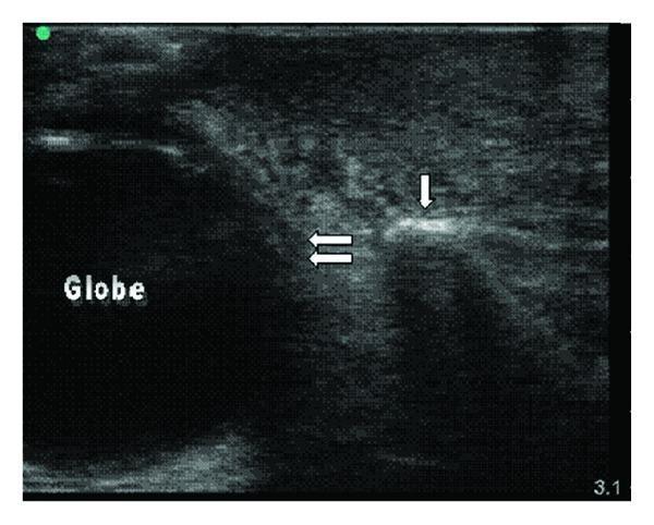
This diagnostic ultrasound image, captured in a transverse view using a high-frequency linear probe, demonstrates the bedside evaluation of periorbital and malar soft tissue. The left side of the image reveals the hypoechoic, circular structure of the ocular globe, with its posterior edge demarcated by double horizontal arrows. To the right, a bright, hyperechoic linear interface indicated by a single vertical arrow represents the cortical surface of the maxilla. The overlying subcutaneous soft tissue exhibits the classic sonographic appearance of cellulitis, characterized by 'cobblestoning.' This pattern is formed by hypoechoic streaks representing inflammatory fluid and edema that disrupt the normally organized, echogenic subcutaneous fat and connective tissue planes. The image serves as an educational example for differentiating bone, fluid-filled structures (globe), and inflammatory soft tissue changes (cellulitis) in the maxillofacial region, highlighting the utility of ultrasound in evaluating preseptal infections.
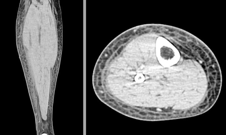
This diagnostic image displays a CT scan of the right lower extremity in two planes: a coronal view (left) and an axial cross-section (right). The primary pathology demonstrated is extensive subcutaneous edema, characterized by marked thickening and a diffuse reticular, honeycomb-like pattern within the subcutaneous fat layer. This 'cobblestoning' appearance is highly suggestive of an inflammatory or infectious process such as cellulitis. In both views, the deep muscle compartments appear enlarged and slightly hyperattenuating, likely due to reactive inflammation or lymphedema, though they remain well-demarcated from the subcutaneous layer by the deep fascia. The axial view confirms the circumferential nature of the soft tissue swelling. Key anatomical landmarks visible include the tibia and fibula, which show intact cortical bone without signs of osteomyelitis or periosteal reaction. No discrete fluid collections, abscesses, or pockets of subcutaneous gas are visualized, findings that are critical in differentiating simple cellulitis from necrotizing fasciitis or localized abscess formation in a clinical setting.
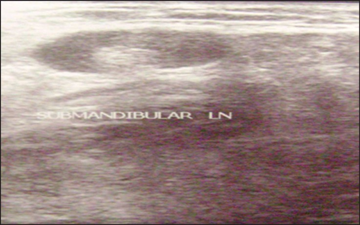
**Imaging Modality:** B-mode Ultrasound (Ultrasonography) **Anatomical Region:** Submandibular space of the neck. **Observed Pathology:** Cellulitis and reactive lymphadenopathy. **Visual Features:** - **Soft Tissue:** The image demonstrates increased echogenicity and thickening of the subcutaneous fat layers, characteristic of inflammatory "cobblestoning" or cellulitis. There is a loss of distinct fascial plane definition. - **Lymph Node:** A prominent, oval-shaped hypoechoic structure is labeled as a "SUBMANDIBULAR LN" (lymph node). It exhibits a well-defined border and a visible hyperechoic central fatty hilum, suggesting a reactive rather than malignant process. - **Echotexture:** The surrounding parenchyma shows a heterogenous, edematous appearance with diffuse low-level echoes, consistent with inflammatory fluid infiltration within the submandibular space. **Diagnostic Features:** The combination of diffuse soft tissue edema (cellulitis) and a reactive lymph node in the submandibular region is a key diagnostic indicator for localized orodontogenic or cervical infection. The absence of a clear, anechoic fluid collection suggests cellulitis rather than an organized abscess at this stage.
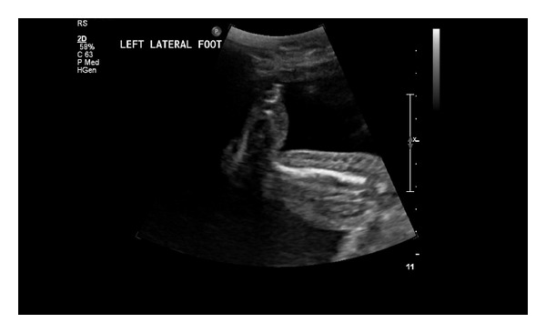
**Imaging Modality:** 2D Greyscale Ultrasound **Anatomical Region:** Left lateral foot and distal lower extremity. **Observed Pathology:** Significant subcutaneous soft tissue edema. The image demonstrates a prominent increase in the thickness of the subcutaneous layer along the dorsal and lateral aspects of the foot. **Characteristic Visual Features:** * **Cobblestoning Pattern:** There is a diffuse, reticulated hyperechoic appearance of the subcutaneous fat interspersed with anechoic/hypoechoic fluid channels, characteristic of soft tissue edema or cellulitis. * **Skin Thickening:** The overlying epidermal and dermal layers appear thickened. * **Fluid Accumulation:** Poorly defined pockets of anechoic fluid are visible within the interstitial spaces, consistent with localized swelling. * **Anatomical Landmarks:** Cortical surfaces of the underlying tarsal or metatarsal bones are visible as hyperechoic linear structures with posterior acoustic shadowing. **Key Diagnostic Features:** The presence of the "cobblestone" sign in the subcutaneous tissue is a primary sonographic indicator of inflammatory edema or lymphedema, correlating with the clinical presentation of distal extremity swelling. No discrete abscess or focal fluid collection is visualized in this specific plane.
cellulitis leg MRI CT lower extremity soft tissue infection findings
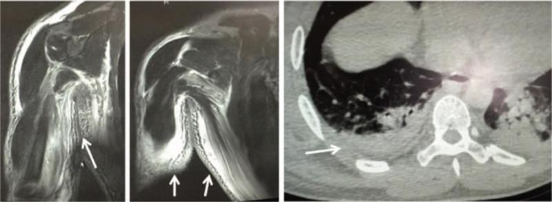
This composite diagnostic image includes magnetic resonance imaging (MRI) and computed tomography (CT) scans demonstrating extensive soft tissue pathology. The left and middle panels consist of MRI scans of the right upper extremity, showing significant soft tissue swelling and abnormal high-signal intensity (edema) within the subcutaneous and intermuscular fascial planes (white arrows). These findings are indicative of fluid accumulation and inflammatory changes consistent with necrotizing fasciitis or severe cellulitis. The right panel is an axial CT scan of the thoracic region, revealing substantial soft tissue thickening and edema along the right thoracic wall (white arrow). There is visible fluid collection in the muscle spaces and a notable loss of distinct fascial margins. The underlying lung parenchyma also shows some consolidative changes. The combined imaging highlights the rapid progression of soft tissue infection and inflammatory edema across multiple anatomical regions, including the forearm and trunk.
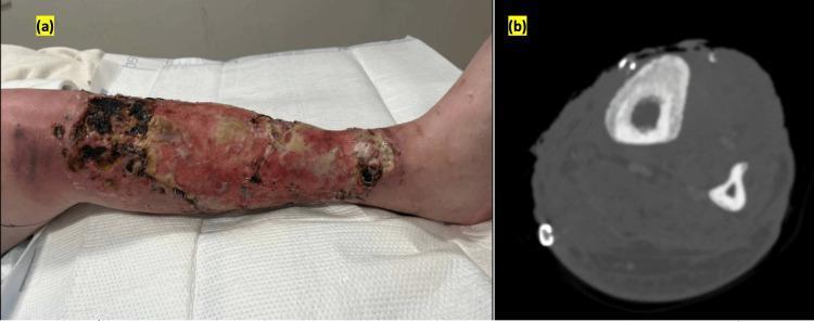
This composite educational figure illustrates the clinical and radiographic manifestations of chronic lower extremity infection. Panel (a) is a clinical photograph of a left lower leg demonstrating a extensive, circumferential ulcerated wound. The wound bed exhibits heterogenous tissue characteristics including dark black necrotic eschar, yellow-tan slough, and areas of reddish-pink granulation tissue with visible surgical staples from prior skin grafting. Significant circumferential erythema and swelling are present in the surrounding skin, consistent with cellulitis. Panel (b) is an axial contrast-enhanced CT scan of the same limb at the level of the mid-tibia and fibula. It reveals significant cortical thickening of the tibia, irregular bone erosions, and a periosteal reaction with new bone formation. Increased attenuation in the adjacent soft tissues suggests inflammation. Collectively, the images demonstrate a complex clinical scenario of chronic non-healing wounds complicated by secondary osteomyelitis, often seen in the context of intravenous drug use (IVDU) or severe skin and soft tissue infections (SSTIs).
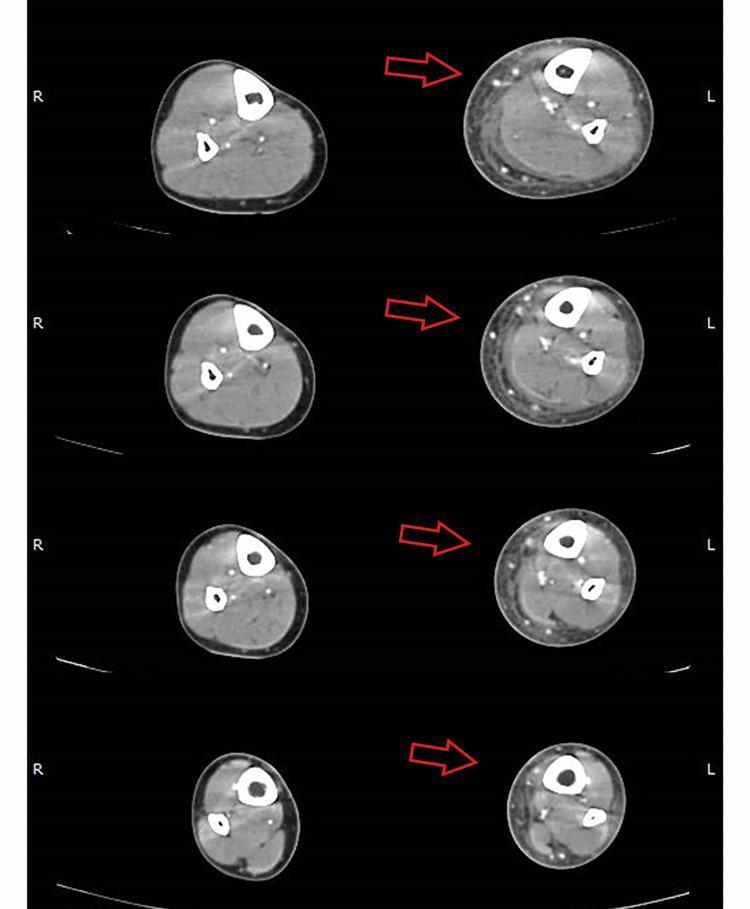
Arterial-phase axial computed tomography (CT) images of the bilateral lower legs demonstrate significant soft tissue pathology on the left side compared to the normal right side. The images show diffuse, circumferential edema of the subcutaneous tissue, characterized by increased thickness and decreased density. Key diagnostic features highlighted by red arrows include thickening of the crural fascia and a localized fluid flap (low-density collection) located on the medial aspect of the leg, adjacent to the gastrocnemius muscle belly. The transverse sections clearly delineate the tibia and fibula, allowing for comparison of the deep compartment and fascial planes. These radiological findings are highly characteristic of a severe necrotizing soft tissue infection (NSTI) or phlegmonous cellulitis, illustrating the progression of infection along fascial boundaries. This diagnostic image is intended for educational use in emergency medicine, radiology, and infectious disease specialties to aid in the identification of fascial plane involvement and subcutaneous fluid collections.
Cellulitis Leg - Imaging Guide
Key Principle
1. ULTRASOUND (First-Line Imaging)
Classic Ultrasound Finding: "Cobblestoning"
What Ultrasound Shows
| Finding | Significance |
|---|---|
| Cobblestoning (reticular fluid in subcut fat) | Cellulitis / inflammatory edema |
| Anechoic/hypoechoic fluid collection with posterior enhancement | Abscess - needs I&D |
| No discrete fluid collection | Cellulitis only - no drainage needed |
| Loss of fascial plane definition | Spreading infection / early NF |
| Reactive lymph node | Lymphadenopathy secondary to infection |
| Foreign body (hyperechoic structure with shadowing) | Retained foreign body as source |
Ultrasound Key Uses
-
Cellulitis vs. Abscess - the most common reason to scan; abscess appears as a walled-off fluid collection vs. the diffuse cobblestoning of cellulitis
-
Locating foreign bodies - more sensitive than plain X-ray for radiolucent objects
-
Guiding drainage - real-time abscess aspiration/I&D
-
Point-of-care (POCUS) - can be done immediately at the bedside in the ED
-
Rosen's Emergency Medicine, Ch. 126
2. CT SCAN (Computed Tomography)
CT Findings in Cellulitis
| CT Finding | Interpretation |
|---|---|
| Diffuse subcutaneous thickening with reticular/honeycomb pattern | Cellulitis - edema in subcutaneous fat |
| Well-demarcated fluid collection | Abscess |
| Gas/air in soft tissues | Necrotizing fasciitis / gas-gangrene - surgical emergency |
| Fluid/inflammation tracking along fascial planes | Necrotizing fasciitis (NF) |
| Intact fascial planes, no gas | Cellulitis, not NF |
| Cortical bone erosion / periosteal reaction | Osteomyelitis |
| Loss of fascial plane margins | Deep spreading infection |
When to Use CT
- Suspected necrotizing fasciitis (gas in tissue, crepitus, skin necrosis, severe systemic toxicity)
- Post-surgical wound infections (extension into deeper structures)
- Suspected deep abscess or undrained collection not seen on ultrasound
- Osteomyelitis (though MRI is more sensitive)
- Diabetic leg infections with concern for deep spread
"Plain radiographs or computed tomography (CT) may reveal soft tissue gas or inflammation along fascial planes, but cannot rule out necrotizing infection."
- Rosen's Emergency Medicine
3. CT WITH IV CONTRAST - Necrotizing Fasciitis vs. Cellulitis
CT Differentiation: Cellulitis vs. Necrotizing Fasciitis
| Feature | Cellulitis | Necrotizing Fasciitis |
|---|---|---|
| Subcutaneous edema | Yes | Yes (more extensive) |
| Fascial plane involvement | No (above fascia only) | Yes - fluid tracks along fascia |
| Gas in soft tissue | No | Yes (pathognomonic) |
| Muscle involvement | No | May involve muscle |
| Deep fluid collections | Absent | Along fascial layers |
| Bone involvement | No (unless osteomyelitis) | Possible |
4. MRI (Magnetic Resonance Imaging)
MRI Findings
| MRI Sequence | Cellulitis Finding |
|---|---|
| T2 / STIR | High signal (bright) in subcutaneous tissue - edema and inflammation |
| T1 | Low signal in affected subcutaneous fat (edema replacing fat) |
| T2 with fascial involvement | High signal tracking along deep fascia = NF |
| Post-gadolinium T1 | Enhancement of inflamed tissue; rim enhancement = abscess |
| T2 bone marrow signal | High signal = osteomyelitis (most sensitive test) |
When to Use MRI
- Suspected osteomyelitis (most sensitive modality - detects before plain films show changes)
- Deep infection extent assessment
- Uncertain diagnosis after ultrasound/CT
- Spinal / pelvic extension concern
5. PLAIN X-RAY
| Indication | What to Look For |
|---|---|
| Suspected foreign body (wood, metal) | Radiopaque foreign body visible |
| Chronic infection, diabetic foot | Bone erosion, periosteal reaction (osteomyelitis) |
| Gas-forming organisms | Subcutaneous gas = emergency |
| Trauma | Fracture, cortical breach |
Note: Plain X-ray cannot rule out NF or osteomyelitis (both may be normal in early stages).
Imaging Pathway Summary
CLINICAL CELLULITIS - leg swelling, redness, warmth
         |
         ├── Typical, no systemic signs, no fluctuance
         │         → NO imaging needed (clinical diagnosis)
         │         → Treat empirically
         │
         ├── Fluctuance / Possible abscess?
         │         → ULTRASOUND (bedside POCUS)
         │         → Cobblestoning = cellulitis
         │         → Fluid collection = abscess → I&D
         │
         ├── Crepitus / Gas / Rapidly spreading / Bullae / Sepsis?
         │         → URGENT CT with IV contrast
         │         → Look for gas, fascial tracking
         │         → If positive → EMERGENCY SURGERY
         │
         ├── Post-surgical / Deep extension suspected?
         │         → CT with IV contrast
         │
         ├── Diabetic / Chronic ulcer / Exposed bone?
         │         → Plain X-ray FIRST
         │         → MRI if osteomyelitis suspected
         │
         └── Diagnostic uncertainty / NF staging?
                   → MRI (most sensitive for extent + osteomyelitis)
Quick Scan Reference
| Modality | Best For | Key Finding in Cellulitis |
|---|---|---|
| Ultrasound | Abscess vs. cellulitis, foreign body | Cobblestoning in subcut fat |
| Plain X-ray | Foreign body, gas, bone erosion | Soft tissue gas (NF), radiopaque FB |
| CT | NF, deep spread, post-surgical | Gas along fascia, subcutaneous reticular edema |
| MRI | Osteomyelitis, extent of NF | T2 high signal in subcutaneous tissue and fascia |
Cellities ointment
Cellulitis - Ointments and Topical Treatments
Important Principle First
- Small superficial entry-point wounds (impetigo-like lesions, minor abrasions)
- Wound care after drainage/debridement
- Preventing portal-of-entry infections (e.g., tinea pedis, minor cuts)
- Decolonizing MRSA carriers to prevent recurrence
"Topical antibiotics are typically reserved for superficial cutaneous infections such as impetigo."
- Dermatology 5e (Dermatology 2-Volume Set)
Topical Antibiotic Ointments - Full Reference Table
| Drug | Brand Names | Formulation | Dose Frequency | Organisms Covered | Prescription? |
|---|---|---|---|---|---|
| Mupirocin | Bactroban, Bactoderm, Centany | 2% ointment or cream | 3x daily (TID) | S. aureus (incl. MRSA), Streptococcus | Rx only |
| Retapamulin | Altabax | 1% ointment | 2x daily (BID) | S. aureus (MSSA), Strep pyogenes | Rx only |
| Ozenoxacin | Xepi | 1% cream | 2x daily (BID) | S. aureus, Strep | Rx only |
| Fusidic acid | Fucidin | 2% cream/ointment | 3x daily (TID) | S. aureus (incl. MRSA), Strep | Rx (not USA; used in Europe, Canada, India) |
| Bacitracin | Baciguent | 400-500 units/g ointment | 3x daily (TID) | Gram-positive (Staph, Strep) | OTC |
| Polymyxin B + Bacitracin | Polysporin | Ointment | 1-3x daily | Gram-positive + gram-negative (NOT MRSA) | OTC |
| Polymyxin B + Bacitracin + Neomycin | Neosporin | Ointment | 1-3x daily | Broad gram-positive + gram-negative | OTC |
| Neomycin | Myciguent | 0.5% ointment/cream | 1-3x daily | Gram-positive | OTC |
| Gentamicin | Garamycin | 0.1% ointment/cream | 3-4x daily | Gram-negative, gram-positive | Rx |
| Silver sulfadiazine | Flamazine, SSD | 1% cream | 1-2x daily | Broad-spectrum (incl. Pseudomonas) | Rx |
- Dermatology 5e, Table 127.3
Drug-by-Drug Guide
1. Mupirocin (Bactroban) 2% - THE KEY TOPICAL
| Feature | Detail |
|---|---|
| Mechanism | Inhibits bacterial protein synthesis by reversibly binding isoleucyl-tRNA synthetase - unique mechanism, no cross-resistance with other antibiotics |
| Coverage | S. aureus (MSSA and MRSA), Streptococcus pyogenes |
| Primary use | Impetigo, minor infected wounds, nasal MRSA decolonization |
| Cellulitis role | Applied to the portal-of-entry wound (e.g., an ulcer or abraded area where bacteria entered) |
| Pregnancy | Safe - very little systemic absorption; topical antibiotic of choice in pregnancy |
| MRSA decolonization | Mupirocin nasal ointment + chlorhexidine body wash x 5 days - used before surgery or for recurrent MRSA infections |
| Resistance | High-level resistance can develop with prolonged use - do not use long-term |
2. Fusidic Acid (Fucidin) 2% - Common in Asia, Europe, India
| Feature | Detail |
|---|---|
| Mechanism | Inhibits bacterial protein synthesis by interfering with elongation factor G |
| Coverage | S. aureus (including MRSA), Strep - gram-positive focus |
| Formulations | 2% cream, ointment, or impregnated gauze |
| Dose | 3 times daily |
| Use in India/UK | Widely used for infected eczema, impetigo, minor skin infections |
| Combination | Fusidic acid + hydrocortisone (Fucidin H) - for infected eczema with inflammation |
| Availability | Not available in USA; used across Europe, Canada, India, Australia |
| Adverse effects | Occasional allergic contact dermatitis |
3. Retapamulin (Altabax) 1% Ointment
- Novel pleuromutilin antibiotic - inhibits 50S ribosomal subunit
- Active against MSSA and Strep pyogenes (not MRSA)
- Used for impetigo; limited use for mild infected wounds
- Apply BID for 5 days
4. Bacitracin Ointment - Over-the-Counter
- Inhibits gram-positive cell wall synthesis
- Useful for preventing infection in small wounds, cuts, abrasions
- Applied after minor procedures (e.g., biopsy, suturing) to keep wound moist and prevent superficial infection
- Not effective against gram-negatives or MRSA
- Allergic contact dermatitis is common with bacitracin - one of the most common contact allergens in North America
5. Neosporin / Polysporin - Over-the-Counter Combinations
- Polysporin = Bacitracin + Polymyxin B (gram-positive + gram-negative cover)
- Neosporin = Bacitracin + Polymyxin B + Neomycin (broadest OTC cover)
- Useful for minor wound care but neomycin is a major allergen - avoid in sensitive patients
- Not suitable for treating established cellulitis
6. Silver Sulfadiazine (SSD) 1% Cream
| Feature | Detail |
|---|---|
| Use | Burns, large exudating wounds, chronic leg ulcers |
| Coverage | Broad: bacteria (including Pseudomonas), fungi |
| Mechanism | Interacts with bacterial cell walls and membranes |
| Caution | Can form pseudo-eschar that may delay wound healing; avoid in sulfonamide allergy |
| G6PD deficiency | Use with caution (risk of hemolysis) |
Antimicrobial Wound Dressings (for Cellulitis + Open Wounds)
| Dressing Type | Examples | Coverage | Best For |
|---|---|---|---|
| Silver dressings | Mepilex Ag, Acticoat, Aquacel Ag | Broad-spectrum incl. MRSA, VRE | Infected chronic wounds, diabetic ulcers |
| Chlorhexidine / PHMB | Kerlix AMD, PuraPly AM | Broad-spectrum + antifungal | Painful wounds (less irritating) |
| Povidone-iodine | Inadine, Betadine gauze | Bacteria, fungi, viruses | Acute infected wounds |
| Honey (Manuka) | MEDIHONEY | Broad-spectrum; also autolytic debridement | Chronic wounds, sloughy tissue |
Silver dressings release antibacterial Ag+ ions for 3-7 days, require less frequent changes, and are active against MRSA and VRE with very low resistance risk.
- Dermatology 5e
When to Use Each - Practical Guide
| Situation | Recommended Topical |
|---|---|
| Minor cut/abrasion - prevention | Bacitracin or Polysporin ointment + dressing |
| Impetigo (superficial crusted lesion) | Mupirocin 2% TID x 5 days |
| Infected eczema / minor skin infection | Fusidic acid 2% TID (or fusidic acid + hydrocortisone) |
| Entry wound in established cellulitis | Mupirocin + systemic antibiotics |
| Open wound / ulcer with cellulitis | Silver dressing or PHMB dressing + systemic antibiotics |
| Burns with infection risk | Silver sulfadiazine 1% cream |
| MRSA carrier (recurrence prevention) | Mupirocin nasal ointment + chlorhexidine wash x 5 days |
| After I&D of abscess | Wound left open; dressing with povidone-iodine or silver |
| Diabetic foot ulcer with cellulitis | Systemic antibiotics primary; silver dressing for wound |
Key Summary
| Agent | Role in Cellulitis |
|---|---|
| Mupirocin 2% | Best topical for wound/portal of entry; MRSA decolonization |
| Fusidic acid 2% | Good alternative (especially in India/Europe); combined with steroids for infected eczema |
| Silver dressings | Antimicrobial wound dressings for open wounds alongside IV antibiotics |
| Bacitracin / Neosporin | Minor wound prevention only; NOT for established cellulitis |
| Silver sulfadiazine | Burns and large wounds |
| Systemic antibiotics | ALWAYS required for actual cellulitis - topicals are supportive only |