
Ankle joint anatomy
ankle joint anatomy bones ligaments diagram
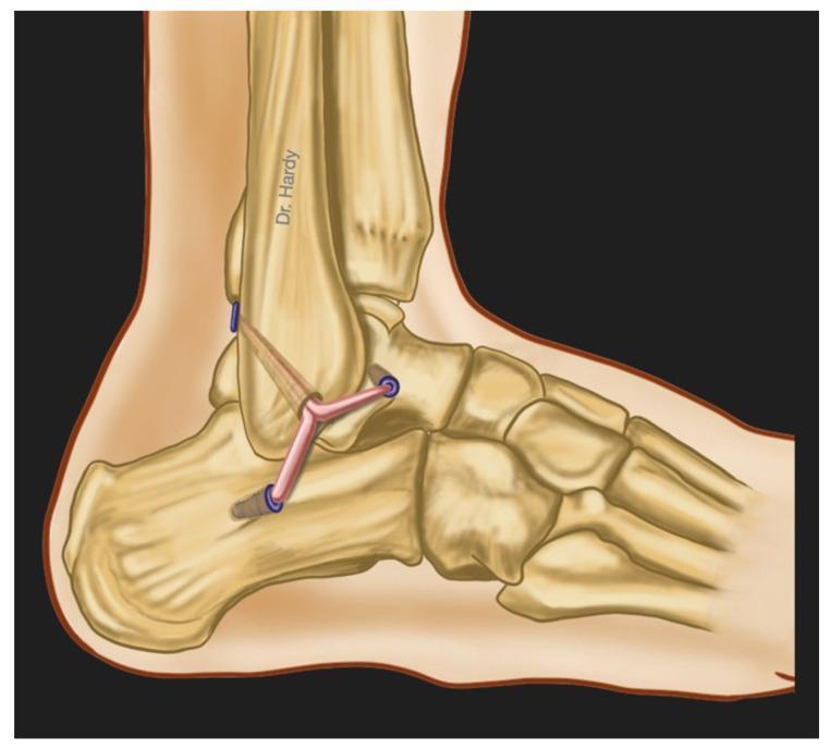
This medical anatomical diagram illustrates an anatomic surgical reconstruction of the lateral ankle ligament complex from a lateral view. The diagram depicts the skeletal structures of the distal fibula, talus, calcaneus, and tarsal bones. The primary focus is the surgical reconstruction of the three main lateral ligaments: the Anterior Talofibular Ligament (ATFL), the Calcaneofibular Ligament (CFL), and the Posterior Talofibular Ligament (PTFL). The reconstruction utilizes a split tendon autograft (such as a gracilis tendon) that is anchored to the distal fibula. The graft bifurcates and extends into three distinct limbs, which are secured into bone tunnels on the talus and calcaneus using blue-colored interference screws or anchors. This illustration demonstrates the surgical technique used to manage chronic lateral ankle instability by replicating the native anatomy of the lateral ligament complex to restore joint stability and biomechanical function.
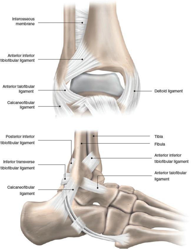
This medical illustration presents two views of the human ankle joint: an anterior-posterior (top) and a lateral (bottom) view, detailing the primary ligamentous structures and bony anatomy. Key anatomical structures include the distal tibia and fibula, talus, and calcaneus. The syndesmotic complex is represented by the interosseous membrane, anterior inferior tibiofibular ligament (AITFL), and posterior inferior tibiofibular ligament (PITFL). The lateral collateral ligament complex is clearly labeled, showcasing the orientation of the anterior talofibular ligament (ATFL) and the calcaneofibular ligament (CFL). The lateral view provides a detailed perspective on the diagonal course of the CFL from the fibular tip to the calcaneus, as well as the inferior transverse tibiofibular ligament. Medially, the deltoid ligament is visible in the anterior view. This educational diagram is designed for orthopedic and radiological study, illustrating the stabilization mechanisms of the ankle joint and the specific spatial relationships between ligaments and tarsal bones involved in inversion and eversion injuries.
ankle mortise tibiofibular joint talocrural
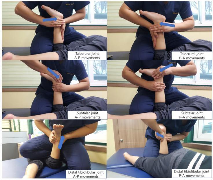
This clinical photographic series consists of six panels demonstrating manual joint mobilization techniques for the ankle complex, specifically targeting the talocrural, subtalar, and distal tibiofibular joints. The images depict a therapist performing both anterior-to-posterior (A-P) and posterior-to-anterior (P-A) glides on a patient in a supine position. The top row illustrates talocrural joint mobilization, focusing on the interface between the talus and the mortise to improve dorsiflexion and plantarflexion. The middle row displays subtalar joint mobilization, targeting the articulation between the talus and calcaneus. The bottom row demonstrates glides at the distal tibiofibular joint. Each panel features a blue directional arrow indicating the line of force applied by the therapist's hands, which are strategically placed to stabilize the proximal segment while mobilizing the distal bone. These techniques are characteristic of the Maitland manual therapy concept, frequently utilized in physical therapy to address joint hypomobility and improve range of motion in conditions like spastic diplegia or post-injury stiffness.
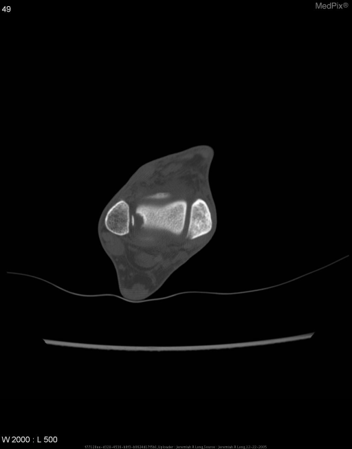
**Imaging Modality:** Computed Tomography (CT), bone window settings.
Ankle Joint Anatomy
Joint Type and Overview
- Dorsiflexion (~20°) - bringing the foot up
- Plantar flexion (~45°) - pointing the foot down
Bones and Articular Surfaces
Tibia
- The inferior articular surface articulates with the dorsal talus (talar trochlea)
- Concave in the sagittal plane, slightly convex in the coronal plane
- A smooth ridge divides it into a wider lateral and narrower medial segment
- The medial segment is continuous with the articular surface of the medial malleolus
- The medial malleolus has an anterior colliculus, posterior colliculus, and an intercollicular groove
- The posterior tibial border projects lower than the anterior border
Fibula
- The lateral malleolus extends further distally than the medial malleolus, reaching lower on the talus
- Its medial articular surface contacts the lateral facet of the talus (triangular shape)
- The malleolar fossa on the posterior fibula serves as the insertion point for the posterior talofibular ligament
- The fibula also forms part of the distal tibiofibular joint
Talus
- The talar trochlea (superior articular surface) is wider anteriorly - this is why the ankle is most stable in dorsiflexion (wider part locked in the mortise)
- The medial articular surface is comma-shaped; the lateral surface is triangular
- The malleoli of both tibia and fibula "envelop" the talus, providing medial and lateral stability - Firestein & Kelley's Textbook of Rheumatology
Ankle Ligament Anatomy
Medial (Deltoid) Ligament Complex
- Originates from the medial malleolus
- Resists eversion of the foot
- Is torn in eversion sprains
- Has both superficial and deep components attaching to the talus, navicular, and calcaneus
Lateral Ligament Complex
| Ligament | Origin | Insertion | Function |
|---|---|---|---|
| ATFL (Anterior Talofibular Ligament) | Anterior fibula (lateral malleolus) | Lateral talar neck | Resists internal rotation & inversion in plantar flexion - most commonly injured |
| CFL (Calcaneofibular Ligament) | Fibular tip | Lateral calcaneus (diagonal course) | Resists inversion; bridges both talocrural and subtalar joints |
| PTFL (Posterior Talofibular Ligament) | Malleolar fossa of fibula | Posterior talus (lateral tubercle) | Resists posterior displacement; strongest of the three |
Syndesmotic (Distal Tibiofibular) Ligament Complex
- Anterior inferior tibiofibular ligament (AITFL)
- Posterior inferior tibiofibular ligament (PITFL)
- Inferior transverse tibiofibular ligament (part of PITFL complex)
- Interosseous membrane (most proximal, continues the length of the leg)
Joint Capsule and Synovium
- The articular capsule is lax anteriorly and posteriorly, permitting dorsiflexion and plantar flexion
- It is tightly bound medially and laterally by the ligament complexes
- The synovial membrane lines the interior and does not communicate with adjacent joints, bursae, or tendon sheaths under normal conditions
- Synovial swelling tends to bulge anteriorly or anterolaterally (where the capsule is most lax) - Firestein & Kelley's Textbook of Rheumatology
Tendons Crossing the Ankle
Anterior Compartment
| Tendon | Action |
|---|---|
| Tibialis anterior | Dorsiflexion + inversion |
| Extensor hallucis longus | Extends great toe + dorsiflexes |
| Extensor digitorum longus | Extends toes + dorsiflexes |
| Peroneus tertius | Dorsiflexion + eversion |
Medial (Posterior to Medial Malleolus) - "Tom, Dick, And Nervous Harry"
| Tendon | Action |
|---|---|
| Tibialis posterior | Plantar flexion + inversion (supination) |
| Flexor Digitorum longus | Flexes toes + plantar flexes |
| (Posterior tibial Artery + Nerve) | Neurovascular |
| Flexor Hallucis longus | Flexes great toe (lies most posteriorly, under Achilles) |
Lateral (Posterior to Lateral Malleolus)
| Tendon | Action |
|---|---|
| Peroneus longus | Plantar flexion + eversion |
| Peroneus brevis | Eversion (inserts on 5th metatarsal base) |
Posterior
- Achilles tendon - the common tendon of gastrocnemius and soleus, inserting on the calcaneal tuberosity; the strongest tendon in the body, withstanding loads >12× body weight during running; its vascular watershed zone lies 2-6 cm above the calcaneal insertion
Neurovascular Supply
- Anterior tibial artery / dorsalis pedis supplies the anterior joint
- Posterior tibial artery passes behind the medial malleolus
- Peroneal artery contributes laterally
- Deep peroneal nerve (anterior) and tibial nerve (posterior medial malleolus) cross the joint
- Sural nerve runs laterally near the Achilles
Range of Motion Summary
| Motion | Primary Joint | Normal Range |
|---|---|---|
| Dorsiflexion | Talocrural | ~20° |
| Plantar flexion | Talocrural | ~45° |
| Inversion | Subtalar + intertarsal | ~30° |
| Eversion | Subtalar + intertarsal | ~20° |
Clinical Correlations
- Lateral ankle sprains (inversion injuries) - ATFL is torn first (most vulnerable), then CFL, then PTFL
- Deltoid ligament tear - eversion mechanism; often associated with fibular fractures
- High ankle sprain - syndesmotic injury (AITFL/PITFL); longer recovery than lateral sprains
- Maisonneuve fracture - fibular fracture at the proximal shaft from syndesmotic disruption
- Achilles tendon rupture - most common at the watershed zone 2-6 cm proximal to insertion
- Ankle stability is greatest in dorsiflexion (wider talus locked in mortise) and least in plantar flexion (narrow posterior talus in mortise) - this is why sprains commonly occur with a plantar-flexed foot
Erbs point
Erb's point brachial plexus C5 C6 upper trunk anatomy
Erbs point brachial plexus upper trunk C5 C6 nerve roots
brachial plexus anatomy diagram roots trunks divisions cords C5 C6 C7 C8 T1
Erb's Point
Erb's Point 1 - Cardiac Auscultation Point
-
Hearing aortic regurgitation (early diastolic murmur radiates here)
-
Detecting both aortic and pulmonary valve murmurs that may not be loudest at their "standard" areas
-
Complements the four classic auscultation areas (aortic, pulmonary, tricuspid, mitral/apex)
-
Color Atlas of Human Anatomy, Vol. 1 - Trunk
Erb's Point 2 - Brachial Plexus Point
-
The suprascapular nerve
-
The nerve to the subclavius
-
Color Atlas of Human Anatomy, Vol. 1; Campbell's Operative Orthopaedics 15th Ed
Why it matters clinically:
1. Erb-Duchenne Palsy (Erb's Palsy)
- Neonatal: excessive lateral traction on the infant's head during delivery (shoulder dystocia)
- Adults: forced shoulder depression with contralateral neck flexion (e.g., motorcycle accident, "stinger" injury in contact sports - compression between shoulder pad and superior medial scapula)
| Muscle Group | Muscle | Effect |
|---|---|---|
| Shoulder abductors | Deltoid, supraspinatus | Arm cannot be abducted |
| Lateral rotators | Infraspinatus, teres minor | Arm internally rotates |
| Elbow flexors | Biceps, brachialis, brachioradialis | Elbow cannot flex |
| Forearm supinators | Biceps, supinator | Forearm fully pronated |
| Wrist extensors | Extensor carpi radialis | Wrist drops |
- Arm hangs at the side
- Internally rotated at shoulder
- Elbow extended
- Forearm pronated
- Wrist flexed
- S Das Manual on Clinical Surgery; Harriet Lane Handbook; Bradley & Daroff's Neurology in Clinical Practice
Comparison with other brachial plexus injuries:
| Injury | Roots | Features | Prognosis |
|---|---|---|---|
| Erb-Duchenne | C5, C6 (±C4) | Waiter's tip; shoulder + elbow affected | Best |
| Total palsy | C5-T1 | Entire arm + hand; Horner if T1 | Worst |
| Klumpke | C8, T1 (±C7) | Claw hand; Horner syndrome (T1) | Poor |
- Miller's Review of Orthopaedics 9th Ed
2. Erb's Point as a Neurophysiology Electrode Site
- A delay between stimulus site and Erb's point = peripheral nerve disease
- A delay from Erb's point to C2 = nerve root or posterior column abnormality
- Preserved Erb's point potential but absent cortical potential = central (spinal cord/brainstem/cortex) lesion
Summary Table
| Erb's Point 1 | Erb's Point 2 | |
|---|---|---|
| Location | Left sternal border, 3rd ICS | 2-3 cm above clavicle, posterior to SCM |
| Domain | Cardiology (auscultation) | Neurology / Anatomy |
| Significance | Aortic regurgitation murmur | C5-C6 upper trunk of brachial plexus |
| Clinical relevance | Cardiac valve murmurs | Erb's palsy; SEP electrode site |