
Follow this
Reading File
Running Command
python3 -c " from docx import Document doc = Document('/tmp/workspace/attachments/ba19a3c6-a9a3-4a64-9bda-88354b0cd5d6/Universal_AI_Learning_Constitution.docx') for p in doc.paragraphs: style = p.style.name if p.style else 'None' if p.text.strip(): print(f'[{style}] {p.text}') "
Constitution loaded and adopted. Here is my acknowledgment:
Universal AI Learning Constitution v2.0 - Adopted
I have read and will follow this constitution for all teaching interactions going forward. Here is what it means in practice:
How I will now teach you:
- I will start by analyzing you - your prior knowledge, goal, depth needed, and time available. If I'm missing key information, I'll ask before diving in.
- I will map prerequisites before explaining any topic, and teach the missing ones first if needed.
- Every topic gets classified using PACER: Procedural, Analogical, Conceptual, Evidence, Reference - and taught in that priority order (concepts before facts, never rote memorization first).
- After every major concept, I will trigger active recall - asking you questions to test understanding, not just passive reading.
- I will adapt difficulty up or down based on your responses.
- Every substantial lesson ends with a summary + recall prompts.
- I will never fabricate facts and will flag uncertainty explicitly.
The workflow I'll follow every time:
Learner Analysis → Prerequisite Mapping → PACER Classification → Teach Concepts → Procedures → Evidence → Reference Facts → Summary → Recall Questions → Adapt
To get started: what do you want to learn?
Tell me the topic, and optionally:
- Your current level (beginner / some background / advanced)
- Your goal (understand it deeply / quick overview / prepare for an exam / apply it practically)
- How much time you have
Well i need to do triangle of auscultation which is for 5 Marks. Also make a exam ready answer in end
Searching sources...
Reading File
Finding Sources
Finding Sources
Reading File
Reading File
Reading File
Loading Image
Loading Image
Finding Sources
I now have all the information needed from authoritative textbooks. Let me teach this properly per the UALC constitution.
Triangle of Auscultation
Learner Analysis
This is an anatomy topic for an exam (5 marks), so you need: a clear definition, boundaries, floor, clinical significance, and the ability to write a structured answer. I'll teach concept first, then give you the exam-ready answer at the end.
Step 1 - What and Why (Conceptual)
The triangle of auscultation is a clinically important gap (window) in the musculature of the posterior thoracic wall where the muscles are thin or absent. Because fewer muscle layers overlie the chest wall here, breath sounds from the underlying lung are heard more clearly through a stethoscope - hence the name.
Think of it as a "thin spot" on the back through which you can listen to the lungs without the interference of thick overlapping muscles.
Step 2 - Boundaries (Reference - memorize these)
The triangle is formed by three borders:
| Border | Structure |
|---|---|
| Superomedial | Lower lateral border of Trapezius |
| Inferolateral | Upper border of Latissimus dorsi |
| Lateral | Medial (vertebral) border of the Scapula |
A simple way to remember:
"Trap - Lat - Scap" (Trapezius + Latissimus + Scapula border)
Step 3 - Floor (What lies underneath)
The floor of the triangle is formed by:
- The erector spinae muscles medially (deep back muscles)
- The 6th and 7th rib (or rhomboid minor depending on the source)
- Overlying these is the serratus posterior inferior muscle
The underlying lung (lower lobe) is what you are actually auscultating through this window.
Step 4 - Location and Position
- Located on the posterior thoracic wall
- The apex of the triangle points superolaterally toward the scapula
- It is best opened up (made larger) by asking the patient to:
- Fold their arms across the chest (crosses arms) AND
- Lean forward
This maneuver pulls the scapula laterally and anteriorly, widening the gap between all three borders and enlarging the triangle.
In surgical practice, dividing the lower trapezius and upper latissimus dorsi fibers increases the size of this triangle to improve access to posterior ribs.
Step 5 - Clinical Significance (Evidence)
- Auscultation: Best site on the back to hear lung breath sounds clearly, especially lower lobe breath sounds, because there is minimal muscle overlying the ribs here.
- Surgical access: Used as the approach for posterior rib fracture fixation - the erector spinae are reflected laterally through this interval to expose the fractured ribs.
- Posterolateral thoracotomy: Surgeons use this triangle as a landmark when planning posterior chest incisions.
- Pleural tapping / procedures: Awareness of this triangle helps in planning access to the posterior thorax.
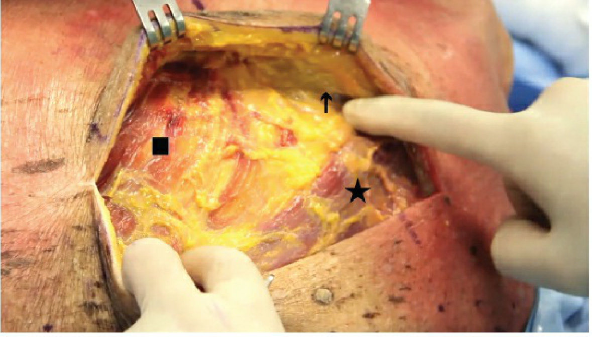
Step 6 - Diagram (from Rockwood & Green's Surgery textbook)
This is the surgical view of the triangle of auscultation opened up during a posterior approach - trapezius (star ★), latissimus dorsi (square ■), and the scapular border (arrow ↑):
Active Recall Check
Before you read the exam answer, try answering these:
- Name the three boundaries of the triangle of auscultation.
- What forms the floor?
- How do you enlarge the triangle clinically?
- Why is this triangle clinically important?
EXAM-READY ANSWER (5 Marks)
Triangle of Auscultation
Definition:
The triangle of auscultation is a triangular gap in the musculature of the posterior thoracic wall where the overlying muscles are absent or thin, allowing clearer auscultation of breath sounds from the underlying lung.
Boundaries:
| Border | Structure |
|---|---|
| Superomedial | Lower lateral border of Trapezius |
| Inferolateral | Upper border of Latissimus dorsi |
| Lateral | Medial (vertebral) border of the Scapula |
Floor:
Formed by the erector spinae muscles and the 6th and 7th ribs (with serratus posterior inferior beneath).
Enlargement:
The triangle is best opened by asking the patient to fold both arms across the chest and lean forward. This draws the scapula laterally, increasing the size of the gap.
Clinical Significance:
- It is the best site on the posterior chest wall to auscultate lower lobe breath sounds clearly, as fewer muscles overlay the ribs.
- Used as the surgical interval for the posterior approach to rib fracture fixation - the erector spinae are identified within this triangle and reflected laterally to expose the posterior ribs.
- Serves as a landmark for posterolateral thoracotomy incisions.
Sources: Rockwood and Green's Fractures in Adults, 10th ed., 2025; Fischer's Mastery of Surgery, 8th ed.
Summary in one line:
The triangle of auscultation is bounded by trapezius (superomedially), latissimus dorsi (inferolaterally), and the medial border of the scapula (laterally); its floor is the erector spinae / 6th-7th ribs; it is the best posterior auscultation site and is used surgically as the posterior rib approach.
Now lets do mammary gland/Breast which is for 10 marks
Searching sources...
Finding Sources
Finding Sources
Finding Sources
Reading File
Reading File
Reading File
Finding Sources
Reading File
Finding Sources
Finding Sources
Reading File
Reading File
Reading File
Reading File
Loading Image
Loading Image
Excellent - I have rich, comprehensive data from multiple authoritative textbooks. Now let me teach this properly following the UALC framework.
Mammary Gland / Breast - 10 Marks Teaching Session
Learner Analysis
10-mark question = you need: position, extent, structure, relations, blood supply, nerve supply, lymphatic drainage, and applied anatomy. I'll build from concept to detail, concept first, memorizable facts after.
PACER Classification
| Category | Content |
|---|---|
| C - Conceptual | What the breast IS and why it is structured that way |
| A - Analogical | Duct system = inverted tree; Cooper ligaments = tent poles |
| P - Procedural | How to systematically describe it in an exam |
| E - Evidence | Clinical consequences of the anatomy (peau d'orange, cancer spread) |
| R - Reference | Numbers, names, levels - memorize these last |
Step 1 - Concept: What is the Breast?
The breast is a modified sweat gland (apocrine type) that sits on the anterior chest wall. It is NOT a muscle or bone structure - it is a gland embedded in fat, held in shape by fibrous ligaments, and supplied by vessels from 3 directions.
Think of it like a dome-shaped cushion of fat + gland tissue, held up by tent-pole ligaments, with a tree of milk ducts draining to the nipple, all sitting on top of the pectoralis major muscle.
Step 2 - Position and Extent (Reference)
- Lies on the anterior thoracic wall between the skin and the superficial pectoral fascia overlying pectoralis major
- Vertically: 2nd to 6th rib
- Horizontally: Lateral border of sternum to mid-axillary line
- The axillary tail of Spence projects into the axilla through an opening in the deep fascia - this is the only part that extends beyond the anterior chest wall
- The nipple is at the level of the 4th intercostal space
Step 3 - Structure (Conceptual + Reference)
The breast has 3 principal tissue types:
- Glandular epithelium - the actual secretory tissue
- Fibrous stroma - connective tissue framework
- Adipose tissue - fat filling the spaces
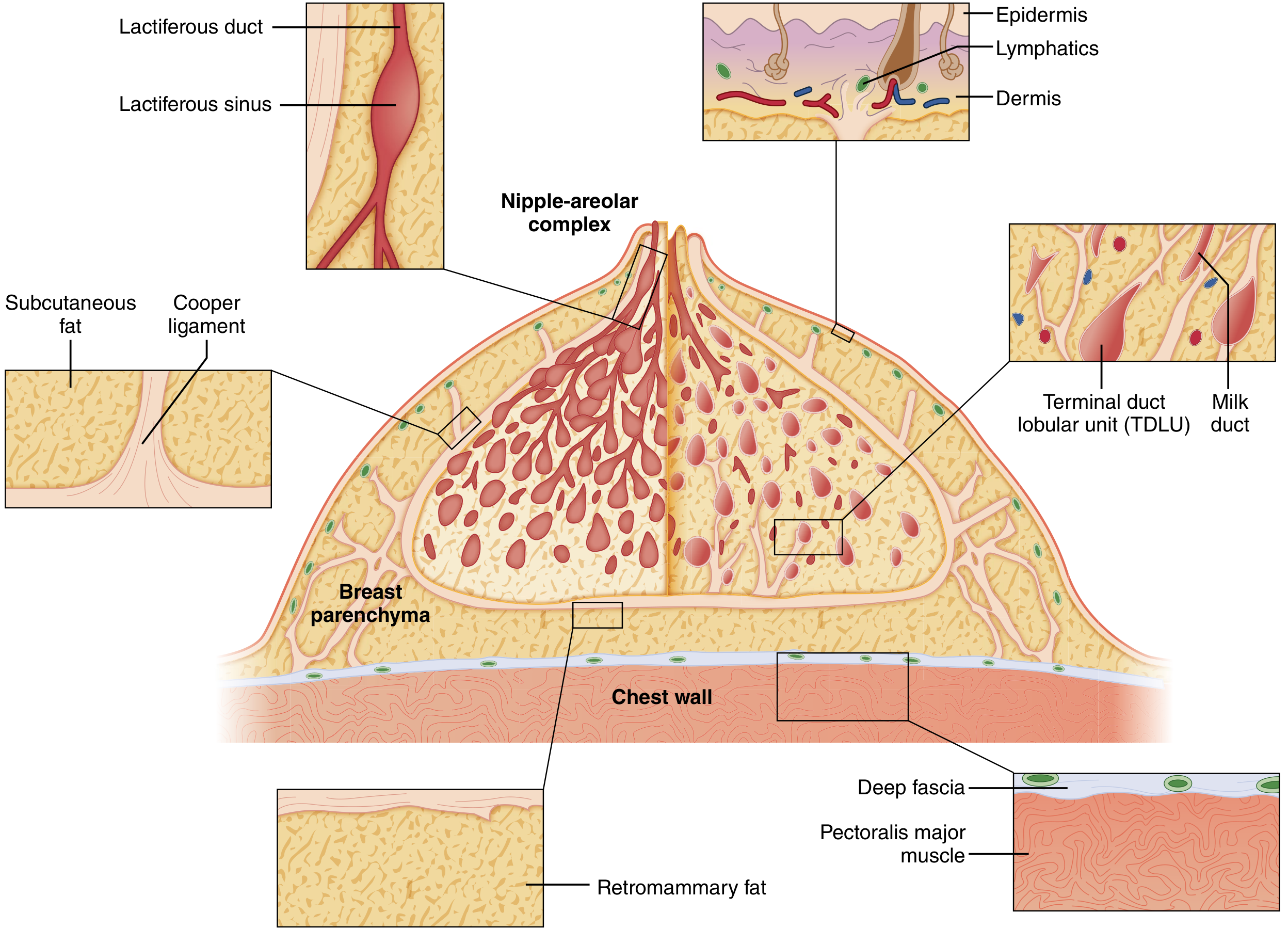
The Duct System (Inverted Tree analogy)
The glandular apparatus is arranged like an inverted tree, with the trunk (large ducts) at the nipple and branches spreading outward:
Nipple
└── 15-20 Lactiferous ducts (open at nipple apex)
└── Lactiferous sinus (dilated reservoir under nipple-areolar complex)
└── Segmental ducts
└── Terminal ductules / Acini (milk-forming units)
= Terminal Duct Lobular Units (TDLUs)
- Each breast has 15-20 lobes, each lobe being a separate compound tubuloacinar gland with its own lactiferous duct
- TDLUs are the milk-forming units and also the site of origin of most breast cancers
Cooper's Ligaments (Suspensory Ligaments)
- Fibrous bands running from the dermis of the skin down through the breast parenchyma to the deep fascia overlying pectoralis major
- They give the breast its shape and hold it up - think of them as tent poles
- More pronounced superiorly than inferiorly
- Clinical importance: When cancer infiltrates Cooper ligaments, they contract and pull the skin inward → producing skin dimpling (peau d'orange - "orange peel skin")
Here is the anatomy of the breast in cross-section:
Step 4 - Relations
| Surface | Relation |
|---|---|
| Anterior | Skin + subcutaneous fat |
| Posterior (base) | Pectoralis major (mainly), serratus anterior (laterally), external oblique (inferolaterally) |
| Deep to pec major | Pectoralis minor enclosed in clavipectoral fascia |
| Retromammary space | Loose areolar tissue between breast and pectoral fascia - allows the breast to move freely; rich with lymphatics |
Step 5 - Blood Supply (Reference)
Arterial supply from 3 sources:
| Source | Branches | Area Supplied |
|---|---|---|
| Internal thoracic (mammary) artery | 2nd, 3rd, 4th perforating branches (2nd is largest) | Anteromedial breast |
| Axillary artery | Thoracoacromial + Lateral thoracic arteries | Superolateral breast |
| Posterior intercostal arteries | 2nd, 3rd, 4th branches | Lateral breast |
Principal blood supply enters superolaterally (axillary) and superomedially (internal thoracic).
Venous drainage mirrors the arterial supply - to the axillary vein, internal thoracic vein, and intercostal veins.
Step 6 - Nerve Supply
- Lateral mammary branches: Lateral cutaneous branches of 4th-6th intercostal nerves
- Medial mammary branches: Anterior cutaneous branches of 2nd-6th intercostal nerves
- Upper breast: Supraclavicular nerves from the cervical plexus (C3, C4)
- The intercostobrachial nerve (lateral cutaneous branch of T2) runs in the axilla - cutting this during axillary surgery causes numbness of the medial upper arm
Step 7 - Lymphatic Drainage (HIGH YIELD for 10 marks)
Lymph first collects into the subareolar plexus of Sappey, then drains:
Main Pathway (75%): Axillary nodes
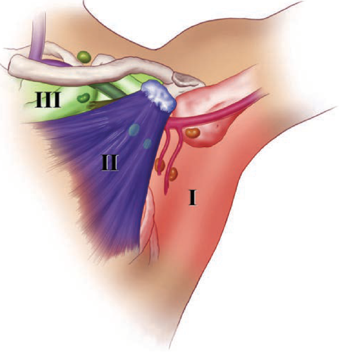
Axillary nodes are grouped into 3 levels based on their relation to pectoralis minor muscle:
| Level | Location | Node Groups |
|---|---|---|
| Level I | Lateral to pec minor | Anterior (pectoral/external mammary), Posterior (subscapular), Lateral (axillary vein) |
| Level II | Deep to pec minor | Central nodes + Rotter's (interpectoral) nodes |
| Level III | Medial to pec minor | Apical (subclavicular) nodes |
Cancer spreads Level I → II → III in sequence (sentinel node is usually Level I).
Rotter's nodes (interpectoral) = between pec major and pec minor - important in surgery.
Other Pathways (25%):
- Parasternal (internal mammary) nodes - medial breast lymphatics travel with perforating arteries
- Supraclavicular nodes - from upper breast
- Contralateral breast - small amount crosses midline
- Inferior phrenic / abdominal nodes - from lower breast
Here is the axillary lymph node levels diagram:
Step 8 - Applied Anatomy (Clinical significance - examiners love this)
| Anatomical Feature | Clinical Relevance |
|---|---|
| Cooper ligaments infiltrated by cancer | Skin dimpling / peau d'orange |
| Axillary tail of Spence | Breast lump can appear in the axilla |
| Axillary lymph node levels | Guides extent of axillary dissection in breast cancer |
| Intercostobrachial nerve in axilla | Preserved during sentinel node biopsy to avoid medial arm numbness |
| Retromammary space | Plane used in mastectomy |
| TDLUs | Site of origin of most carcinomas and fibroadenomas |
| Subareolar plexus of Sappey | Explains why all quadrant cancers drain to axilla |
| Nipple inversion | Caused by cancer pulling on the lactiferous ducts |
Active Recall Check
Try before reading the exam answer:
- What are the boundaries of the breast (extent on chest wall)?
- How many lobes does the breast have and what is a TDLU?
- What are Cooper's ligaments and what happens when cancer infiltrates them?
- Name the 3 arterial sources of the breast.
- What is the subareolar plexus of Sappey and where does most of its lymph drain?
- What are the 3 levels of axillary lymph nodes and their relation to pectoralis minor?
EXAM-READY ANSWER (10 Marks)
Mammary Gland (Breast)
1. Introduction
The mammary gland is a modified apocrine sweat gland located on the anterior thoracic wall, composed of glandular epithelium, fibrous stroma, and adipose tissue. It is the organ of milk production in females.
2. Position and Extent
- Located between the 2nd and 6th ribs vertically
- Horizontally: from the lateral border of sternum to the mid-axillary line
- The axillary tail of Spence projects superolaterally into the axilla through a gap in the deep fascia
- The nipple lies at the level of the 4th intercostal space
3. Structure
The breast contains 15-20 lobes of compound tubuloacinar type, each draining via its own lactiferous duct to the nipple. Each lactiferous duct has a dilated portion - the lactiferous sinus - just beneath the nipple-areolar complex (NAC). The ducts branch progressively, ending in terminal duct lobular units (TDLUs) which are the milk-secreting units and the most common site of origin of breast carcinoma.
Suspensory ligaments of Cooper are fibrous bands connecting the dermis to the deep pectoral fascia. They support breast shape. Their infiltration by malignancy produces skin dimpling (peau d'orange).
4. Relations
- Anteriorly: skin and subcutaneous fat
- Posteriorly: pectoralis major (main), serratus anterior (lateral), external oblique (inferolateral)
- A retromammary space of loose areolar tissue separates the breast from the pectoral fascia, allowing breast mobility and serving as the surgical plane in mastectomy
5. Blood Supply
Arterial:
- Perforating branches of the internal thoracic artery (2nd-4th spaces, 2nd is largest) - anteromedial supply
- Branches of axillary artery (thoracoacromial and lateral thoracic) - superolateral supply
- Posterior intercostal arteries (2nd-4th) - lateral supply
Venous: Drains to axillary, internal thoracic, and intercostal veins.
6. Nerve Supply
- Lateral mammary branches: Lateral cutaneous branches of 4th-6th intercostal nerves
- Medial mammary branches: Anterior cutaneous branches of 2nd-6th intercostal nerves
- Upper breast: Supraclavicular nerves (C3, C4) from cervical plexus
7. Lymphatic Drainage
Lymph first drains to the subareolar plexus of Sappey, then:
A. Axillary nodes (~75%) - three surgical levels:
- Level I (lateral to pec minor): pectoral (anterior), subscapular (posterior), lateral groups
- Level II (deep to pec minor): central nodes + Rotter's interpectoral nodes
- Level III (medial to pec minor): apical (subclavicular) nodes
B. Parasternal (internal mammary) nodes (~25%): drain the medial breast, traveling with perforating arteries
C. Other: supraclavicular nodes, contralateral breast, inferior phrenic nodes (lower breast)
8. Applied Anatomy
- Infiltration of Cooper ligaments by cancer → skin dimpling / peau d'orange
- Axillary tail → breast lump may present in the axilla
- Sentinel lymph node (first node receiving lymph) is typically in Level I axillary group
- The intercostobrachial nerve (T2) runs in the axilla and when divided causes numbness of the medial upper arm
- TDLUs are the site of origin of most carcinomas and fibroadenomas
Sources: Sabiston Textbook of Surgery, 21st ed.; Schwartz's Principles of Surgery, 11th ed.; Fischer's Mastery of Surgery, 8th ed.; Junqueira's Basic Histology, 17th ed.; Gray's Anatomy for Students.
https://youtu.be/RLJ8aUw468M
analyse_media
Searching the Web
youtube RLJ8aUw468M anatomy lecture topic
The video analysis came through - it's about the Brachial Plexus! The video covers the brachial plexus in detail. Here it is embedded for you:
The video is on the Brachial Plexus. Do you want me to teach this topic next using the UALC framework?
Here's what the video covers based on my analysis:
- Formation of the brachial plexus (roots, trunks, divisions, cords, branches)
- The "M" shape formed by musculocutaneous, median, and other nerves
- All 5 terminal branches and what they innervate
- Cord relationships and injuries
Just tell me:
- How many marks is this question for?
- Do you want a full teaching session + exam answer, or just the exam answer?
Next: Teach me Brachial Plexus - 10 marks
Searching sources...
Finding Sources
Finding Sources
Reading File
Finding Sources
Reading File
Reading File
Reading File
Loading Image
Reading File
Reading File
I have everything needed. Now let me teach this fully per the UALC constitution.
Brachial Plexus - 10 Marks Teaching Session
PACER Classification
| Category | Content |
|---|---|
| C - Conceptual | What is the brachial plexus and why it exists; how it is organized |
| A - Analogical | The "electrical junction box" analogy; the M-shape of terminal branches |
| P - Procedural | How to draw and describe it systematically |
| E - Evidence | Clinical injuries and their predicted deficits |
| R - Reference | Root values, trunk names, cord names, branch names - memorize last |
Step 1 - Concept First: What and Why?
The upper limb needs to receive signals from the brain (motor) and send signals back (sensory). These signals travel through spinal cord segments C5, C6, C7, C8, and T1. But rather than having 5 separate spinal nerves each going to different muscles, the body mixes and redistributes these fibers so that each peripheral nerve contains exactly the right combination of root fibers for its specific target.
Analogy: Think of the brachial plexus as an electrical junction box. Five cables (root wires: C5-T1) come in, get joined and redistributed inside the box (trunks → divisions → cords), and then 5 organized cables (terminal branches) come out to go to specific destinations in the upper limb.
Step 2 - Formation and Parts (The Road Map)
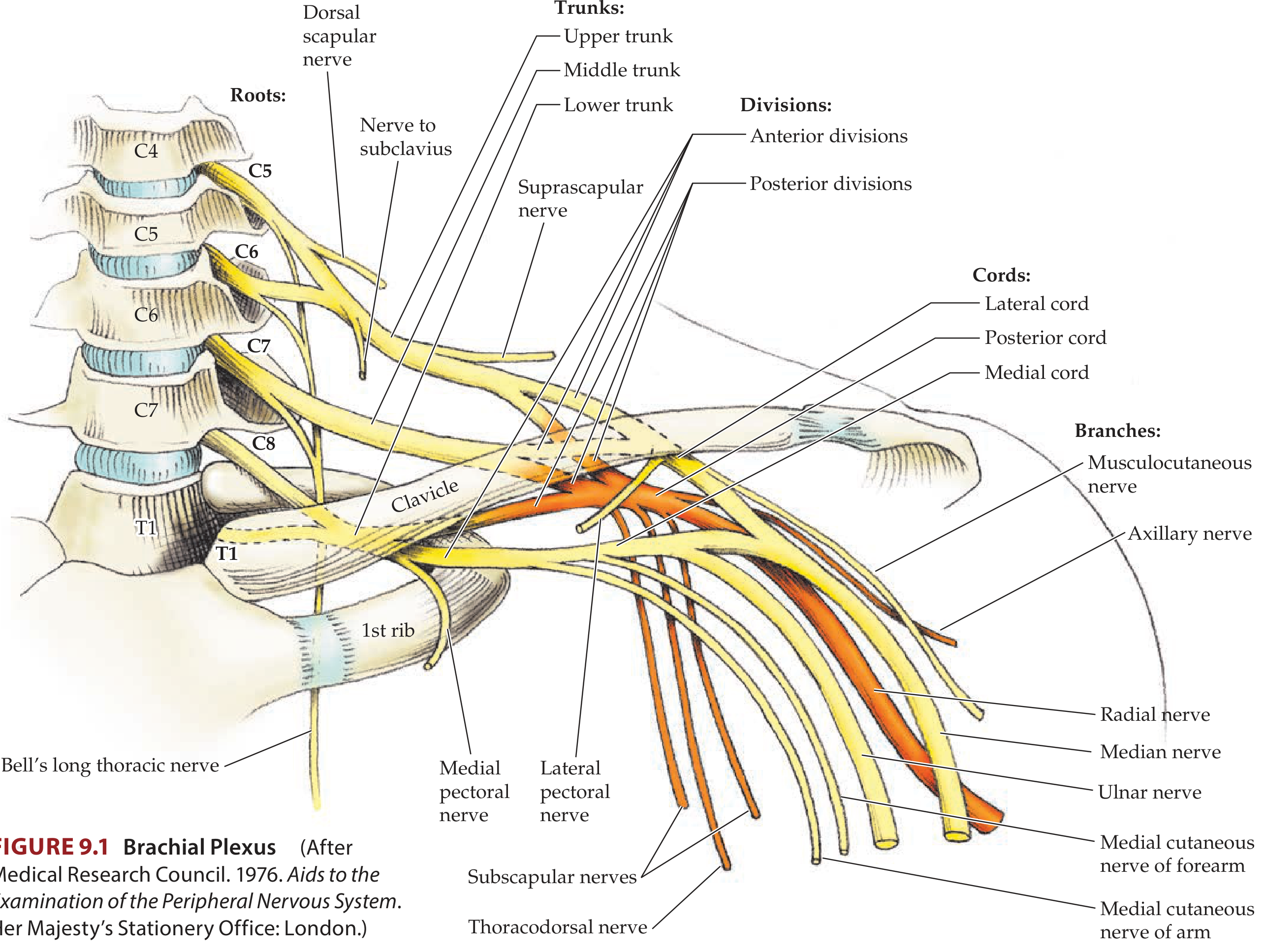
The brachial plexus is formed by the anterior rami (ventral rami) of spinal nerves C5, C6, C7, C8, and T1.
It has 5 parts - remember with the mnemonic:
"Robert Taylor Drinks Cold Beer" Roots → Trunks → Divisions → Cords → Branches
Here is the complete anatomy diagram from Neuroanatomy through Clinical Cases:
Step 3 - The 5 Parts in Detail
1. ROOTS (5 roots)
- Anterior rami of C5, C6, C7, C8, T1
- Pass between anterior scalene and middle scalene muscles
- Lie posterior to the subclavian artery in the neck
Branches from roots:
| Nerve | Origin | Supplies |
|---|---|---|
| Dorsal scapular nerve | C5 | Rhomboids major & minor |
| Long thoracic nerve (Bell's nerve) | C5, C6, C7 | Serratus anterior |
| Contribution to phrenic nerve | C5 | Diaphragm (partial) |
2. TRUNKS (3 trunks)
Roots join to form 3 trunks in the posterior triangle of the neck:
| Trunk | Roots | Position |
|---|---|---|
| Upper (Superior) trunk | C5 + C6 | Superior |
| Middle trunk | C7 alone | Middle |
| Lower (Inferior) trunk | C8 + T1 | Inferior - lies on rib I behind subclavian artery |
Branches from trunks (only from the UPPER trunk):
- Suprascapular nerve (C5, C6) - supplies supraspinatus + infraspinatus
- Nerve to subclavius (C5, C6) - supplies subclavius muscle
3. DIVISIONS (6 divisions)
- Each trunk splits into an anterior and a posterior division = 6 divisions total
- No peripheral nerves arise directly from divisions
- Anterior divisions → supply flexor (anterior) compartments
- Posterior divisions → supply extensor (posterior) compartments
4. CORDS (3 cords)
Divisions recombine to form 3 cords, named by their relationship to the 2nd part of the axillary artery:
| Cord | Formed by | Root values | Position |
|---|---|---|---|
| Lateral cord | Anterior divisions of upper + middle trunk | C5, C6, C7 | Lateral to axillary artery |
| Medial cord | Anterior division of lower trunk | C8, T1 | Medial to axillary artery |
| Posterior cord | All 3 posterior divisions | C5-T1 (all roots) | Posterior to axillary artery |
5. BRANCHES (Terminal + Collateral)
From the LATERAL CORD:
| Nerve | Supplies |
|---|---|
| Lateral pectoral nerve | Pectoralis major |
| Musculocutaneous nerve | Biceps brachii, Brachialis, Coracobrachialis (BBC mnemonic) + skin of lateral forearm |
| Lateral root of median nerve | (joins medial root to form median nerve) |
From the MEDIAL CORD:
| Nerve | Supplies |
|---|---|
| Medial pectoral nerve | Pectoralis major + minor |
| Medial cutaneous nerve of arm | Skin of medial arm |
| Medial cutaneous nerve of forearm | Skin of medial forearm |
| Ulnar nerve | Intrinsic hand muscles, flexor carpi ulnaris, medial FDP, skin of little + medial ring finger |
| Medial root of median nerve | (joins lateral root to form median nerve) |
From the POSTERIOR CORD: (mnemonic: STAR or ARTS)
| Nerve | Supplies |
|---|---|
| Subscapular nerves (upper + lower) | Subscapularis, teres major |
| Thoracodorsal nerve | Latissimus dorsi |
| Axillary nerve | Deltoid, teres minor + skin over deltoid |
| Radial nerve | All posterior compartment muscles of arm + forearm, skin of posterior arm/forearm/hand |
The "M" shaped median nerve:
The median nerve is the bridge between lateral and medial cords - the lateral root (C5-C7) + medial root (C8-T1) join in front of the axillary artery forming the letter "M" with the musculocutaneous and ulnar nerves on either side.
Step 4 - Where is the Plexus Located?
The brachial plexus runs a course in 3 regions:
- Neck (posterior triangle): Between anterior and middle scalene muscles - roots and trunks here
- Behind clavicle: Divisions cross the clavicle
- Axilla: Cords and terminal branches surround the axillary artery
Step 5 - Applied Anatomy / Injuries (HIGH YIELD)
Erb's Palsy (Upper trunk injury - C5, C6)
Cause: Forceful separation of head from shoulder (difficult childbirth, motorcycle accident - shoulder pushed down, head pulled up)
Muscles lost: Deltoid, supraspinatus, infraspinatus, biceps, brachioradialis
Classic posture - "Waiter's tip" / "Bellhop's tip" position:
- Arm: adducted + medially rotated (deltoid + lateral rotators lost)
- Elbow: extended + pronated (biceps lost)
- Wrist: flexed (brachioradialis lost)
- Hand is spared (C8, T1 intact)
Klumpke's Palsy (Lower trunk injury - C8, T1)
Cause: Forced abduction of arm (grabbing overhead to arrest a fall, birth injury with arm raised), Pancoast tumor of lung apex
Muscles lost: All intrinsic hand muscles (thenar, hypothenar, lumbricals, interossei), flexor carpi ulnaris
Classic posture - "Claw hand": Hyperextension at MCPs, flexion at IPs (lumbricals + interossei lost)
Horner syndrome may accompany Klumpke's (T1 carries sympathetic fibers - ptosis, miosis, anhidrosis on same side)
Other Important Injuries
| Nerve Injured | Cause | Deformity |
|---|---|---|
| Long thoracic (C5-C7) | Axillary surgery, stab wound | Winging of scapula (serratus anterior paralyzed) |
| Axillary nerve | Shoulder dislocation, surgical neck of humerus fracture | Loss of deltoid - flat shoulder; loss of sensation over "regimental badge area" |
| Radial nerve (posterior cord) | Fracture of shaft of humerus (spiral groove) | Wrist drop |
Active Recall - Test Yourself
- What is the mnemonic for the 5 parts of the brachial plexus?
- Which two roots form the UPPER trunk? Which is the middle trunk?
- Which cord gives rise to the RADIAL nerve?
- What forms the LATERAL cord? What are its branches?
- What is the posture in Erb's palsy and WHY?
- What is Klumpke's palsy and what causes the claw hand?
- Which nerve injury causes winging of scapula?
EXAM-READY ANSWER (10 Marks)
Brachial Plexus
1. Definition and Formation
The brachial plexus is a network of nerves formed by the anterior rami of C5, C6, C7, C8, and T1 spinal nerves. It provides the entire motor and sensory innervation of the upper limb (except the trapezius and the skin over the axilla).
2. Parts - "Robert Taylor Drinks Cold Beer"
Roots → Trunks → Divisions → Cords → Branches
3. Roots
- Anterior rami of C5-T1
- Pass between the anterior and middle scalene muscles
- Branches from roots:
- Dorsal scapular nerve (C5) - rhomboids
- Long thoracic nerve (C5, C6, C7) - serratus anterior
4. Trunks (in the posterior triangle of neck)
| Trunk | Roots | Branches |
|---|---|---|
| Upper | C5 + C6 | Suprascapular nerve; Nerve to subclavius |
| Middle | C7 | None |
| Lower | C8 + T1 | None |
5. Divisions
- Each trunk → anterior + posterior division (6 total)
- No peripheral nerves arise directly from divisions
- Anterior → flexor compartments; Posterior → extensor compartments
6. Cords (in the axilla, named relative to axillary artery)
| Cord | Formation | Root values |
|---|---|---|
| Lateral | Ant. div. of upper + middle trunk | C5-C7 |
| Medial | Ant. div. of lower trunk | C8-T1 |
| Posterior | All 3 posterior divisions | C5-T1 |
7. Terminal Branches
Lateral cord: Lateral pectoral nerve, Musculocutaneous nerve, Lateral root of median nerve
Medial cord: Medial pectoral nerve, Medial cutaneous nerve of arm + forearm, Ulnar nerve, Medial root of median nerve
Posterior cord (STAR): Subscapular nerves, Thoracodorsal nerve, Axillary nerve, Radial nerve
Median nerve = Lateral root (C5-C7) + Medial root (C8-T1) - formed in front of axillary artery
8. Applied Anatomy
Erb's Palsy (C5, C6 - upper trunk):
- Cause: Forceful head-shoulder separation (difficult delivery, motorcycle accident)
- Posture: "Waiter's tip" - arm adducted, medially rotated, elbow extended and pronated
- Hand movements spared
Klumpke's Palsy (C8, T1 - lower trunk):
- Cause: Forced arm abduction (grabbing a branch), Pancoast tumor
- Posture: Claw hand (intrinsic minus hand)
- Associated Horner's syndrome (ptosis, miosis, anhidrosis)
Long thoracic nerve injury (C5-C7):
- Cause: Axillary surgery, mastectomy, stab wound
- Result: Winging of scapula (serratus anterior paralysis)
Axillary nerve injury:
- Cause: Shoulder dislocation, fracture of surgical neck of humerus
- Result: Flat shoulder + loss of sensation over regimental badge area
Sources: Gray's Anatomy for Students; Neuroanatomy through Clinical Cases, 3rd ed.; Schwartz's Principles of Surgery, 11th ed.; Barash Clinical Anesthesia, 9th ed.
Now anatomical snuffbox - 5 marks
Searching sources...
Finding Sources
Finding Sources
Reading File
Finding Sources
Loading Image
Finding Sources
I have everything needed. Let me teach this efficiently - it's a 5-mark topic so tight and focused.
Anatomical Snuffbox - 5 Marks
PACER Classification
| Category | Content |
|---|---|
| C - Conceptual | What it is and why it exists |
| A - Analogical | A triangular pocket with a roof, walls, and floor - like a shallow valley |
| R - Reference | Boundaries, contents, floor - the memorizable facts |
| E - Evidence | Scaphoid fracture, radial pulse - clinical significance |
Step 1 - Concept: What is it?
The anatomical snuffbox is a naturally occurring triangular depression that appears on the posterolateral side of the wrist and base of the thumb when the thumb is fully extended and abducted. Historically, powdered tobacco (snuff) was placed in this depression before sniffing it - hence the name.
It is not just a surface landmark - it has a floor, walls (borders), roof, and important contents passing through it. This makes it examinable as a complete anatomical space.
Step 2 - Shape and Location
- Shape: Triangle
- Base: At the wrist (proximal end)
- Apex: Points toward the thumb (distal end)
- Location: Posterolateral aspect of the wrist
- Best seen when: Thumb is actively extended and abducted
Step 3 - Boundaries (the 3 walls)
This is the most important memorizable section:
| Border | Structures |
|---|---|
| Lateral (anterior) border | Tendons of Abductor Pollicis Longus (APL) + Extensor Pollicis Brevis (EPB) |
| Medial (posterior) border | Tendon of Extensor Pollicis Longus (EPL) |
| Proximal border (base/roof) | Styloid process of radius |
| Distal border (apex) | Base of 1st metacarpal |
Memory trick for borders:
Lateral = "AB" + "EB" (ABductor + Extensor Brevis) - these two come together Medial = "EL" (Extensor Longus) - alone on the other side
Step 4 - Floor
The floor is formed by two bones (proximal to distal):
- Scaphoid (proximal - most of the floor)
- Trapezium (distal)
Also on the floor are the distal ends of the tendons of extensor carpi radialis longus and extensor carpi radialis brevis, which pass deep to the other tendons here.
Step 5 - Contents (what passes through it)
| Structure | Layer | Clinical note |
|---|---|---|
| Radial artery | Deep - passes obliquely through the floor, deep to the extensor tendons | Pulse can be felt here |
| Superficial branch of radial nerve | Subcutaneous - passes over the roof | Sensory to dorsum of thumb |
| Cephalic vein | Subcutaneous - originates here from dorsal venous arch | Visible/palpable |
Here is the anatomy diagram:
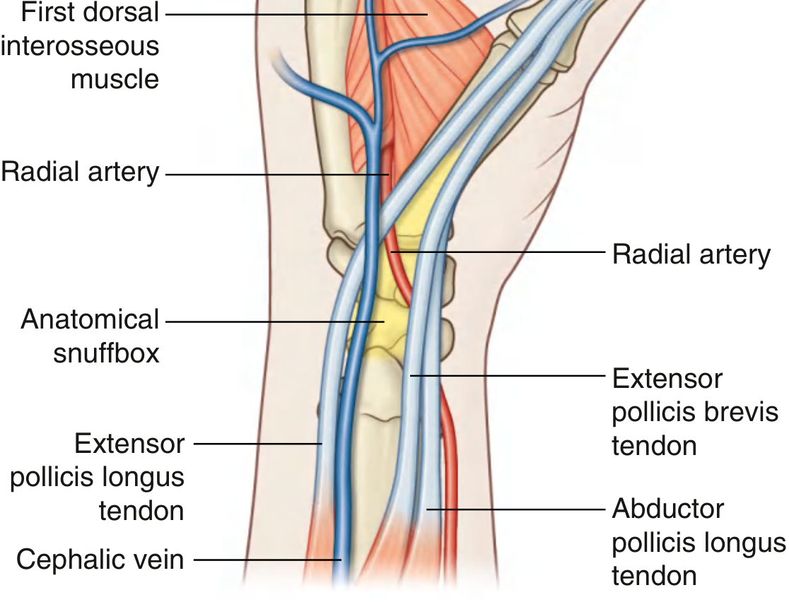
Step 6 - Clinical Significance (Applied - MUST know for exams)
1. Scaphoid Fracture (Most important)
- Mechanism: Fall on outstretched hand (FOOSH)
- Sign: Tenderness in the anatomical snuffbox (especially with the wrist in ulnar deviation, which brings the scaphoid into the snuffbox)
- Danger: Scaphoid has a poor blood supply (enters distally) - proximal fragment gets avascular necrosis if fracture is missed
- Exam trap: X-ray may be normal initially - if snuffbox tenderness is present, treat as scaphoid fracture and re-X-ray at 10-14 days
2. Radial Pulse
- The radial artery can be palpated in the snuffbox as an alternative site to the wrist (useful in surgeons during wrist operations, Allen's test)
3. Radial Nerve Injury
- The superficial branch of the radial nerve crosses subcutaneously over the snuffbox - vulnerable to injury by tight handcuffs or watch straps → painful paraesthesia over dorsum of thumb/first web space (Cheiralgia paraesthetica)
Active Recall - Try These
- Name the two tendons forming the lateral border of the snuffbox.
- What forms the medial border?
- Name the two bones forming the floor.
- Which artery passes through the snuffbox and at which depth?
- Why is snuffbox tenderness clinically important after a FOOSH injury?
EXAM-READY ANSWER (5 Marks)
Anatomical Snuffbox
Definition
The anatomical snuffbox is a triangular depression on the posterolateral aspect of the wrist, visible when the thumb is actively extended and abducted. Its base lies at the wrist and its apex points toward the thumb.
Boundaries
| Border | Structure |
|---|---|
| Lateral (anterior) | Tendons of Abductor Pollicis Longus + Extensor Pollicis Brevis |
| Medial (posterior) | Tendon of Extensor Pollicis Longus |
| Proximal | Radial styloid process |
| Distal | Base of 1st metacarpal |
Floor
Formed by the scaphoid (proximally) and trapezium (distally), along with the deep tendons of extensor carpi radialis longus and brevis.
Contents
- Radial artery - passes deep through the snuffbox (between floor and extensor tendons); radial pulse can be felt here
- Superficial branch of radial nerve - runs subcutaneously over the snuffbox; sensory to dorsum of thumb and first web space
- Cephalic vein - arises from the dorsal venous arch and passes subcutaneously over the snuffbox
Clinical Significance
-
Scaphoid fracture: Tenderness in the anatomical snuffbox (especially on ulnar deviation) after a fall on an outstretched hand strongly suggests scaphoid fracture. The initial X-ray may be normal; if tenderness persists, repeat at 10-14 days. Missed fracture risks avascular necrosis of the proximal fragment due to the distal-to-proximal blood supply of the scaphoid.
-
Radial pulse: The radial artery is palpable in the snuffbox - used as an alternative pulse site and for radial artery cannulation.
-
Superficial radial nerve injury: Tight straps or handcuffs can compress the superficial branch of the radial nerve as it crosses the snuffbox → painful numbness over the dorsum of the thumb (Cheiralgia paraesthetica).
Source: Gray's Anatomy for Students; Bailey and Love's Short Practice of Surgery, 28th ed.; Rockwood and Green's Fractures in Adults, 10th ed.
This is a shared conversation. Sign in to Orris to start your own chat.