
A 23-year-old man with a bleeding femoral fracture is hypotensive and receives a blood transfusion. Immediately after the transfusion starts, he develops fever, chills, severe itching, dyspnoea, stridor, neck swelling, SpO2 75% and bilateral polyphonic rhonchi. EXAM TASKS Identify and justify the event causing deterioration. List the immediate measures. Provide a treatment plan. Give me the best brief answer
Quick Answer:
This patient is having an acute transfusion reaction - specifically anaphylaxis triggered by the blood transfusion, superimposed on a trauma/haemorrhage context. The combination of fever, chills, urticaria (itching), stridor, neck swelling, bronchospasm (rhonchi), and severe hypoxia (SpO2 75%) immediately post-transfusion is classic anaphylaxis, not a simple febrile or haemolytic reaction.
Searching sources...
Reading File
Finding Sources
Finding Sources
Reading File
Finding Sources
Finding Sources
Reading File
Finding Sources
Reading File
Reading File
I now have enough high-quality textbook data to give a complete, authoritative answer.
Acute Anaphylactic Transfusion Reaction - Complete Management
1. IDENTIFICATION AND JUSTIFICATION
Diagnosis: Anaphylactic Transfusion Reaction (severe/grade IV)
This is NOT a simple febrile or haemolytic reaction. The clinical picture is anaphylaxis triggered by the blood transfusion, with features of angio-oedema + upper airway obstruction + lower airway bronchospasm + cardiovascular collapse + urticaria - all occurring within seconds to minutes of starting transfusion.
| Feature | This Patient | Significance |
|---|---|---|
| Fever + chills | Present | Immune activation |
| Severe itching | Present | IgE-mediated mast cell degranulation - urticaria |
| Stridor | Present | Laryngeal/supraglottic oedema - upper airway emergency |
| Neck swelling | Present | Angioedema |
| Dyspnoea + bilateral polyphonic rhonchi | Present | Bronchospasm - lower airway obstruction |
| SpO2 75% | Present | Critical hypoxaemia - life-threatening |
| Hypotension (pre-existing) | Present | Distributive + haemorrhagic shock |
| Onset: immediately after transfusion | Present | Classic anaphylaxis timing |
Pathophysiology: Anaphylaxis occurs most commonly in IgA-deficient patients who possess IgA-specific antibodies of the IgE class. Transfused IgA (present in donor plasma) cross-links these IgE antibodies on mast cells and basophils, triggering massive release of histamine, tryptase, and leukotrienes. This causes: (1) systemic vasodilation and capillary leak → hypotension; (2) laryngeal and supraglottic oedema → stridor and neck swelling; (3) bronchial smooth muscle contraction → wheezing and bronchospasm. Haptoglobin deficiency is an alternative mechanism (anti-haptoglobin IgG/IgE). - Roberts and Hedges' Clinical Procedures in Emergency Medicine, p. 607
Why not other reactions?
- Febrile non-haemolytic reaction: fever alone, NO stridor/bronchospasm/angioedema
- Haemolytic reaction: flank/back pain, haemoglobinuria, DIC - no urticaria or airway oedema
- TRALI: delayed pulmonary oedema, non-cardiogenic - no urticaria or stridor
2. IMMEDIATE MEASURES
Priority: STOP - CALL - AIRWAY - ADRENALINE
- STOP the transfusion immediately - first and non-negotiable step; do NOT resume
- Keep the IV line open with normal saline (new giving set)
- Call for help - activate resuscitation team / senior anaesthetist
- Airway assessment and positioning - sit the patient upright if tolerated; prepare for intubation
- High-flow oxygen - 15 L/min via non-rebreather mask targeting SpO2 >94%
- Epinephrine (Adrenaline) IM - first-line drug
- 1 mg/mL (1:1,000) solution, 0.5 mg IM into the anterolateral thigh
- Repeat every 5 minutes if no improvement
- If in shock with no response: IV epinephrine infusion 0.1-1 mcg/kg/min
- Aggressive IV fluid resuscitation - normal saline boluses (caution in context of ongoing haemorrhage)
- Monitor: continuous ECG, SpO2, BP, urine output
- Send blood sample back to blood bank + patient identification check
- Notify blood bank immediately - return unused blood product for investigation
3. TREATMENT PLAN
Phase 1: Immediate (0-5 minutes)
| Drug | Dose | Route | Rationale |
|---|---|---|---|
| Epinephrine | 0.5 mg (1:1,000) | IM (anterolateral thigh) | Reverses bronchospasm, oedema, hypotension - first-line |
| Oxygen | 15 L/min | Non-rebreather mask | Treat SpO2 75% |
| 0.9% NaCl | 500-1000 mL bolus | IV | Volume replacement for distributive shock |
Phase 2: Adjunct Pharmacotherapy (5-15 minutes)
| Drug | Dose | Route | Rationale |
|---|---|---|---|
| H1 antihistamine (chlorphenamine) | 10 mg | IV slow | Reduce urticaria, pruritus - adjunct only |
| H2 antihistamine (ranitidine) | 50 mg | IV | Complement H1 blockade |
| Corticosteroids (hydrocortisone) | 200 mg | IV | Prevent biphasic reaction, reduce oedema - delayed onset |
| Nebulised salbutamol | 2.5-5 mg | Inhaled | Epinephrine-resistant bronchospasm |
Critical note: Antihistamines and steroids do not act rapidly enough to treat acute anaphylaxis. Epinephrine is the only drug that works fast enough to reverse the life-threatening components. - Rosen's Emergency Medicine, p.3757
Phase 3: Airway Management
- If stridor persists or worsens despite epinephrine: early endotracheal intubation (do not delay - laryngeal oedema can progress to complete obstruction)
- Racemic epinephrine (nebulised) may help reduce supraglottic oedema as a bridge
- RSI with ketamine (maintains BP) + succinylcholine preferred in this haemodynamically unstable patient
- Have surgical airway (cricothyrotomy) kit at bedside
Phase 4: Haemorrhage Management (concurrent)
- This patient still has a bleeding femoral fracture - once airway and anaphylaxis are being controlled, orthopaedic/surgical team must address haemostasis
- Future blood transfusion (if still needed) must use washed red blood cells (plasma-depleted) or IgA-deficient blood products
- Pre-medicate with corticosteroids + antihistamines 30-60 minutes before any future transfusion
Phase 5: Investigation and Monitoring
- Serum tryptase: draw within 30 minutes - 6 hours of symptom onset (confirms anaphylaxis)
- Blood bank: ABO/Rh crossmatch verification, direct antiglobulin test
- Blood cultures (rule out septic transfusion reaction)
- FBC, U&E, LFTs, coagulation screen
- Admit to ICU/HDU for minimum 24 hours (risk of biphasic anaphylaxis)
- Allergy/immunology referral after discharge: IgA level, anti-IgA antibodies
Future Transfusion Safety
- Document reaction formally (haemovigilance reporting)
- Patient requires IgA-deficient blood products or washed cellular products for all future transfusions
- Provide medical alert bracelet
Summary mnemonic: SAEOC
- S - Stop transfusion
- A - Adrenaline (IM 0.5 mg, 1:1,000)
- E - Epinephrine infusion if shock persists + Ensure airway
- O - Oxygen + IV fluids
- C - Corticosteroids + antihistamines (adjuncts)
Sources: Roberts and Hedges' Clinical Procedures in Emergency Medicine, p. 607; Rosen's Emergency Medicine, p. 3755-3757; Goldman-Cecil Medicine, Transfusion Reactions chapter
Micro and macro vascular complication of DM2 in best shortest way..
Quick Answer: DM2 vascular complications split into microvascular (small vessel disease from chronic hyperglycaemia affecting retina, kidney, nerves) and macrovascular (accelerated atherosclerosis affecting coronary, cerebral, and peripheral arteries). Let me pull the best textbook content.
Searching sources...
Finding Sources
Reading File
Reading File
Finding Sources
Finding Sources
Reading File
Reading File
Loading Image
I have all the content needed. Here is the complete, concise answer.
Vascular Complications of Type 2 Diabetes Mellitus
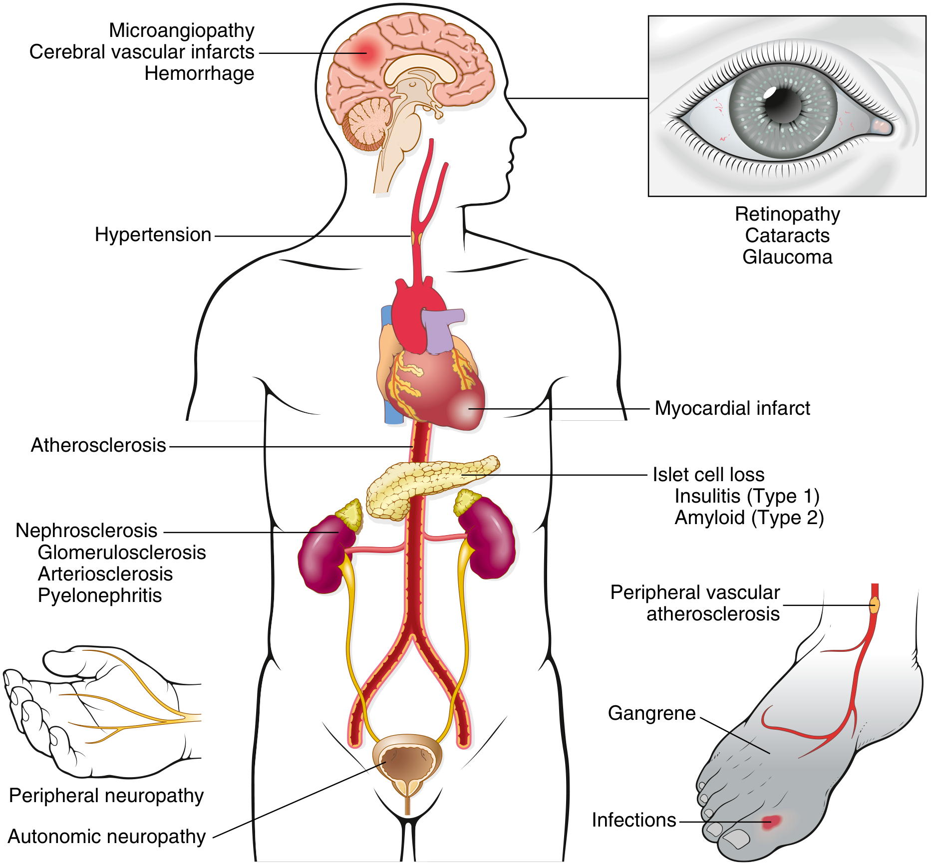
Pathogenesis (common root)
Chronic hyperglycaemia damages vessels through 3 main mechanisms:
| Mechanism | Effect |
|---|---|
| AGEs (Advanced Glycation End-products) | Cross-link basement membrane proteins; trap LDL in vessel walls; activate RAGE → cytokines, ROS, TGF-β, VEGF |
| PKC activation | Via DAG accumulation → VEGF (neovascularisation) + TGF-β (fibrosis/basement membrane thickening) |
| Polyol pathway (aldose reductase) | Sorbitol accumulation in nerves/lens/kidneys; NADPH depletion → oxidative stress |
MICROVASCULAR COMPLICATIONS
(Small vessel disease - diabetes specific)
1. Diabetic Retinopathy
- Most common cause of blindness in working-age adults
- Basement membrane thickening of retinal capillaries → pericyte loss → microaneurysms, haemorrhages, exudates
- Non-proliferative (NPDR): microaneurysms, dot/blot haemorrhages, cotton-wool spots, hard exudates
- Proliferative (PDR): VEGF-driven neovascularisation → vitreous haemorrhage, tractional retinal detachment
- Also: cataracts and glaucoma
2. Diabetic Nephropathy
- Leading cause of end-stage renal disease
- Glomerular hyperfiltration → basement membrane thickening → mesangial expansion → Kimmelstiel-Wilson nodular glomerulosclerosis (pathognomonic)
- Stages: microalbuminuria (30-300 mg/day) → macroalbuminuria → declining GFR → ESKD
- Also: nephrosclerosis (arteriolar hyalinosis), increased susceptibility to pyelonephritis
3. Diabetic Neuropathy
- Most common complication overall
- Distal symmetric polyneuropathy (most common): "glove and stocking" sensory loss, burning/tingling, loss of vibration and proprioception
- Autonomic neuropathy: gastroparesis, orthostatic hypotension, neurogenic bladder, erectile dysfunction, silent MI (no pain), sudomotor dysfunction
- Mononeuropathy: CN III palsy (pupil sparing), carpal tunnel syndrome
- Mechanism: sorbitol accumulation + oxidative stress + microvessel ischaemia of vasa nervorum
MACROVASCULAR COMPLICATIONS
(Large/medium vessel disease - accelerated atherosclerosis)
AGE-modified LDL trapped in vessel walls + endothelial dysfunction + dyslipidaemia + hypertension drive 2-4x faster atherosclerosis than the general population.
| Complication | Key Points |
|---|---|
| Coronary Artery Disease (CAD) | MI risk 2-4x higher; often silent (no pain due to autonomic neuropathy); leading cause of death in DM2 |
| Cerebrovascular Disease | Stroke risk 2-3x higher; both ischaemic stroke and lacunar infarcts |
| Peripheral Arterial Disease (PAD) | Prevalence 20-30% in DM2; lower limb ischaemia, claudication, critical ischaemia, gangrene |
| Heart Failure | Diabetic cardiomyopathy (independent of CAD/HTN); altered myocardial metabolism, fibrosis |
Quick Memory Summary
MICROVASCULAR → "3 N's" → retiNopathy, Nephropathy, Neuropathy
                            (Eyes, Kidneys, Nerves)

MACROVASCULAR → "3 C's" → Coronary, Cerebral, peripheral Circulation
                            (Heart, Brain, Legs)
Key distinction: Microvascular = hyperglycaemia alone is sufficient cause; Macrovascular = hyperglycaemia + dyslipidaemia + hypertension + insulin resistance all contribute. - Harrison's Principles of Internal Medicine 22E; Robbins Basic Pathology
This is a shared conversation. Sign in to Orris to start your own chat.