
Screenshot_20260519_083503.jpg
Please let me know diagnosis and changes seen in ECG
Reading File
Finding Sources
Finding Sources
Searching Images
right bundle branch block ECG changes
Reading File
Loading Image
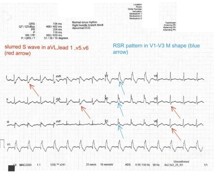
Based on the ECG image and the machine interpretation visible at the bottom of the tracing, here is a comprehensive analysis:
ECG Analysis
Patient Details (from the tracing)
- HR: 77 bpm
- P-R interval: 142 ms
- QRS: 141 ms (wide)
- QT/QTc: 420/477 ms (prolonged QTc)
- RV5/SV1: 0.13 / 0.28
- Date: 2025-04-30
Machine Interpretation (bottom of ECG)
The automated report clearly states:
- Sinus Rhythm
- Marked Right Axis Deviation
- Long QTc
- Right Bundle Branch Block (RBBB)
- ⚠️ "Report needs clinician review"
Diagnosis: Complete Right Bundle Branch Block (RBBB) with Marked Right Axis Deviation
ECG Changes Seen
1. Wide QRS (≥120 ms)
- QRS duration = 141 ms — confirms complete RBBB (threshold ≥120 ms)
2. RSR' (M-shaped) Pattern in V1–V3
- Classic "rabbit ears" or M-pattern in right precordial leads (V1, V2, V3)
- Represents delayed right ventricular depolarization after normal left ventricular activation
3. Broad, Slurred S Waves in Lateral Leads
- Wide terminal S waves in Lead I, aVL, V5, V6
- This is the hallmark mirror finding of RBBB in lateral leads
4. Secondary ST-T Wave Changes
- T-wave inversions in V1–V3 (discordant T waves) — these are expected secondary changes in RBBB, not necessarily ischemic
5. Marked Right Axis Deviation
- Axis deviation toward the right suggests right ventricular strain/hypertrophy or concurrent left posterior fascicular block (LPFB)
- RBBB + right axis deviation = Bifascicular Block pattern
6. Prolonged QTc (477 ms)
- QTc = 477 ms is prolonged (normal <440 ms in men, <460 ms in women)
- Increases risk of ventricular arrhythmias (Torsades de Pointes)
7. Sinus Rhythm
- Regular P waves preceding each QRS — normal sinus origin maintained
Summary Table
| Feature | Finding |
|---|---|
| Rhythm | Normal sinus rhythm |
| Rate | 77 bpm |
| QRS duration | 141 ms (widened) |
| Axis | Marked right axis deviation |
| V1–V3 | RSR' (M-shaped) pattern |
| Lateral leads I, V5, V6 | Broad slurred S waves |
| T waves | Inversions V1–V3 (secondary) |
| QTc | 477 ms (prolonged) |
| Overall diagnosis | Complete RBBB + Right Axis Deviation (Bifascicular block pattern) |
Reference ECG showing RBBB features:
Clinical Significance & Next Steps
- RBBB alone can be a normal variant, but RBBB + right axis deviation raises concern for:
- Pulmonary hypertension / chronic cor pulmonale
- Pulmonary embolism (acute)
- Right ventricular hypertrophy/strain
- Congenital heart disease (e.g., ASD)
- Ischemic heart disease (bifascicular block)
- Prolonged QTc warrants checking electrolytes (K⁺, Mg²⁺, Ca²⁺) and reviewing medications that prolong QT
- Clinician review is mandatory as flagged by the machine — correlation with clinical symptoms, echo, and troponin is recommended
⚕️ This analysis is for educational purposes. Clinical correlation by the treating physician is essential. — Goldman-Cecil Medicine, Table 42-3; Braunwald's Heart Disease
Do we need to give loding dose
Asking for Preferences
Reading File
Finding Sources
Finding Sources
Finding Sources
Finding Sources
Reading File
Reading File
Great question — given the ECG context (RBBB + prolonged QTc), here is a structured answer on loading doses relevant to this clinical scenario:
Loading Dose — Does This Patient Need One?
The answer depends on the drug in question and the clinical urgency. Here is what applies to this ECG:
⚠️ Key Concern First: Prolonged QTc (477 ms)
This ECG already has a prolonged QTc. Many antiarrhythmic drugs further prolong the QT interval and are relatively or absolutely contraindicated, including:
- Class Ia (quinidine, procainamide, disopyramide)
- Class III (sotalol, dofetilide, ibutilide)
These should be avoided or used with extreme caution in this patient.
Amiodarone — Most Relevant Drug Here
Amiodarone is the antiarrhythmic of choice in patients with structural heart disease, RBBB, and compromised LV function because it is the least proarrhythmic despite QT prolongation.
Does it need a loading dose? YES — always.
This is because amiodarone has an extremely long half-life (40–55 days) and takes 2–3 weeks to reach steady state. Without a loading dose, therapeutic effect would take ~265 days.
Loading Dose Regimens:
| Route | Regimen | When to Use |
|---|---|---|
| IV (emergency) | 150 mg over 10 min → 1 mg/min × 6 hrs → 0.5 mg/min × 18 hrs | Life-threatening arrhythmia (VT/VF) |
| IV (breakthrough) | Supplemental 150 mg bolus over 10 min | Breakthrough VT/VF during infusion |
| Oral (non-urgent) | 800–1200 mg/day × 1–3 weeks → 400–800 mg/day × 1–2 weeks → maintenance 200 mg/day | Stable arrhythmia (AF, SVT) |
| Oral (rapid) | 800–2000 mg/day × 5–7 days | Suppress ventricular arrhythmias urgently but non-emergently |
For This Patient Specifically (RBBB + Right Axis Deviation + QTc 477 ms)
| Clinical Situation | Decision |
|---|---|
| Asymptomatic RBBB with no active arrhythmia | ❌ No loading dose needed — RBBB alone requires no antiarrhythmic drug |
| AF with RBBB (rate control) | Beta-blocker or rate-limiting calcium channel blocker preferred; no loading dose unless converting rhythm |
| Sustained VT with RBBB pattern | ✅ IV Amiodarone loading required — 150 mg IV bolus → infusion |
| Symptomatic bifascicular block with syncope | Refer for permanent pacemaker — no drug loading indicated |
| QTc prolongation management | Avoid QT-prolonging drugs; correct electrolytes (K⁺, Mg²⁺) |
Bottom Line
- RBBB by itself does not require a loading dose of any drug
- If the patient has a symptomatic arrhythmia (VT, VF, AF), amiodarone is the drug of choice and always requires a loading dose (oral or IV depending on urgency)
- The prolonged QTc (477 ms) is a red flag — avoid Class I/III antiarrhythmics that further prolong QT
- If the bifascicular block (RBBB + right axis deviation) is causing syncope/hemodynamic compromise → cardiology referral for pacemaker rather than drug therapy
Please clarify the clinical presentation (symptomatic? active arrhythmia? syncope?) for a more specific recommendation.
— Braunwald's Heart Disease, 15th Ed; Goodman & Gilman's Pharmacological Basis of Therapeutics
This is a shared conversation. Sign in to Orris to start your own chat.