
Uterine Neoplasms
Reading File
Now let me search the medical library for uterine neoplasms comprehensively.
Finding Sources
Finding Sources
Finding Sources
Finding Sources
Finding Sources
Finding Sources
Now let me read the key sections in parallel - the main Robbins Pathologic Basis of Disease content on endometrial hyperplasia/carcinoma and the uterine tumors section from Berek & Novak's.
Reading File
Reading File
Reading File
Reading File
Reading File
Finding Sources
Reading File
Finding Sources
Now let me fetch the key images and also read the Robbins Pathologic Basis (larger Robbins) section on serous carcinoma and leiomyosarcoma.
Reading File
Loading Image
Loading Image
Loading Image
Searching PubMed
"endometrial cancer" AND "treatment"
I now have comprehensive information to write a thorough answer. Let me compile it all.
Uterine Neoplasms
Uterine neoplasms include a broad spectrum of tumors arising from the endometrium, myometrium, and stromal elements of the uterus. The most clinically important are endometrial carcinoma, endometrial hyperplasia (a precursor lesion), uterine leiomyoma, and leiomyosarcoma. All typically present with abnormal uterine bleeding as the earliest sign.
1. Endometrial Hyperplasia
Endometrial hyperplasia is defined as an abnormal proliferation of endometrial glands relative to stroma, resulting in an increased gland-to-stroma ratio compared with normal proliferative endometrium. It is an important precursor to the most common type of endometrial carcinoma.
Pathogenesis
The condition is driven by prolonged unopposed estrogenic stimulation, due to:
- Obesity (peripheral conversion of androgens to estrogens in adipose tissue)
- Anovulation / polycystic ovarian syndrome
- Menopause without progestin replacement
- Functioning granulosa cell tumors of the ovary
- Exogenous estrogen administration without progestin
Molecular basis: Inactivation of the PTEN tumor suppressor gene is the hallmark early event, found in >20% of hyperplasias (both with and without atypia) and 30-80% of endometrial carcinomas. PTEN normally inhibits the PI3K/AKT pathway; its loss leads to overactivation of this pathway and enhanced estrogen receptor-dependent gene expression. Patients with Cowden syndrome (germline PTEN mutations) have a high risk of endometrial carcinoma.
WHO Classification (2 categories)
| Category | Features | Risk of Progression to Carcinoma |
|---|---|---|
| Hyperplasia without atypia | Increased gland-to-stroma ratio, dilated glands, variable size/shape; no nuclear atypia | Low (1-3%) |
| Atypical hyperplasia / Endometrial Intraepithelial Neoplasia (EIN) | Complex crowded glands + nuclear atypia (rounded vesicular nuclei, prominent nucleoli); harbors clonal driver mutations | Significant (25-30%) |
Histology:
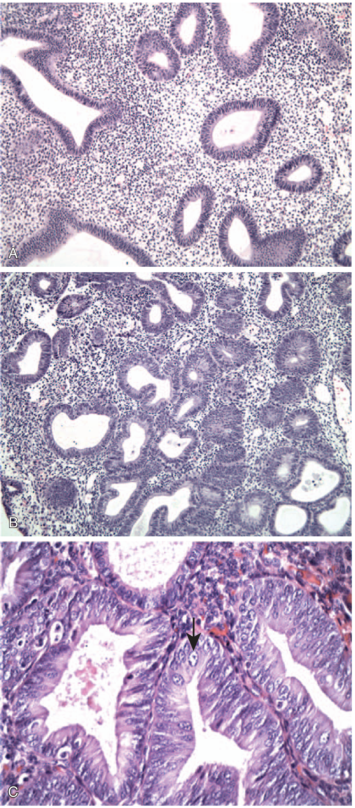
Endometrial hyperplasia. (A) Without atypia - architectural crowding. (B) With atypia - marked glandular crowding. (C) High power: vesicular nuclei with prominent nucleoli (arrow). - Robbins & Kumar Basic Pathology
Management
- Hyperplasia without atypia: Progestin therapy; low risk of progression
- Atypical hyperplasia (EIN): Hysterectomy (definitive); in younger patients desiring fertility, high-dose progestins may be attempted (must exclude co-existing invasion)
2. Endometrial Carcinoma
The most common cancer of the female genital tract in high-income countries. Peak incidence is in postmenopausal women aged 55-65 years, though the incidence in younger women is increasing.
Two Major Pathogenetic Types
| Feature | Type I: Endometrioid | Type II: Serous |
|---|---|---|
| Frequency | ~80-85% of cases | ~15% of cases |
| Setting | Estrogen excess, endometrial hyperplasia, perimenopausal | Endometrial atrophy, older postmenopausal women |
| Grade | Well to moderately differentiated | High-grade (always) |
| Key mutations | PTEN (30-80%), PIK3CA (~40%), KRAS (~25%), ARID1A (~33%), MMR defects (~20%) | TP53 (>90%), chromosomal instability |
| Molecular pathway | PI3K/AKT overactivation | p53 dysfunction + chromosomal instability |
| Precursor | Atypical hyperplasia (EIN) | Serous Endometrial Intraepithelial Carcinoma (SEIC) |
| Spread | Myometrial invasion → lymph nodes | Exfoliation via fallopian tubes → peritoneal implants |
| Prognosis | Good if early stage (5-yr survival ~90% for stage I) | Poor; often presents at high stage |
| Associated syndromes | Lynch syndrome (MMR defects); Cowden syndrome (PTEN) | - |
Molecular Subtypes (TCGA Classification)
- POLE ultramutated (<10%): mutations in proofreading domain of DNA polymerase ε → extremely high mutational burden → best prognosis despite high histologic grade
- MMR-deficient / MSI-high (~20%): mismatch repair defects (MLH1 promoter hypermethylation in sporadic; germline in Lynch syndrome)
- Copy-number low / no specific molecular profile (~40%): mostly endometrioid, intermediate prognosis
- Copy-number high / p53-mutant (~15%): mostly serous type; worst prognosis
Morphology
Endometrioid carcinoma: Closely resembles proliferative endometrium; may be exophytic or infiltrative. Graded 1-3 based on differentiation (grade 1 = mostly glandular; grade 3 = predominantly solid).
Serous carcinoma: Papillary growth pattern with marked cytologic atypia - high N:C ratio, atypical mitoses, hyperchromasia, prominent nucleoli. Diffuse, strong p53 immunostaining (due to mutant p53 accumulation).
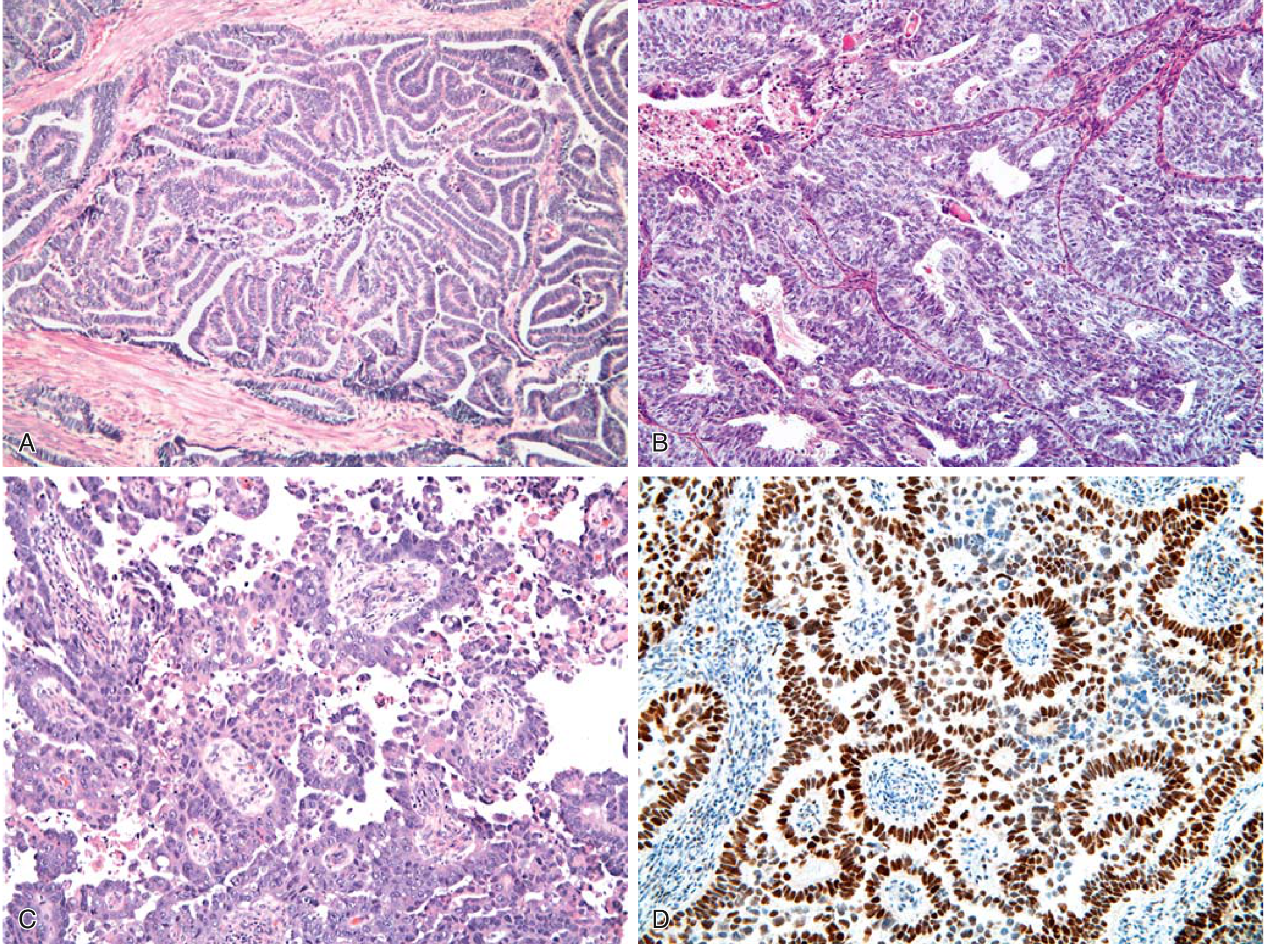
Endometrial carcinoma histology. (A-B) Endometrioid types, grades 1 and 3. (C-D) Serous carcinoma with papillae and strong p53 IHC staining. - Robbins & Kumar Basic Pathology
Clinical Features
- Presentation: Irregular or postmenopausal vaginal bleeding (usually leads to early diagnosis)
- Risk factors: Obesity, hypertension, type 2 diabetes, nulliparity, late menopause, estrogen-only HRT, tamoxifen use
- Diagnosis: Endometrial biopsy or curettage; all endometrioid carcinomas should be tested for MMR deficiency (3-5% have Lynch syndrome)
- Staging: FIGO surgical staging (hysterectomy + bilateral salpingo-oophorectomy + lymph node assessment)
- Treatment: Surgery ± radiation ± chemotherapy depending on stage and molecular subtype
Recent evidence update: A 2025 Cochrane review (PMID 40626388) and a 2025 network meta-analysis (PMID 40031426) confirm that immunotherapy combined with PARP inhibitors is emerging as a first-line strategy for advanced endometrial cancer, particularly in MMR-deficient/MSI-high tumors. A 2024 systematic review (PMID 39032722) supports fertility-sparing management for carefully selected stage IA cases.
3. Endometrial Polyps
- Sessile, 0.5-3 cm; project into the uterine cavity
- Composed of endometrium resembling the basalis with small muscular arteries and cystically dilated glands
- Neoplastic component is the stromal cell (clonal); the surface epithelium is reactive
- Incidence increases with age; may cause abnormal bleeding; malignant transformation is rare
4. Uterine Leiomyoma ("Fibroid")
The most common tumor in women overall. A benign smooth muscle neoplasm.
Key Features
- Epidemiology: Very common; more frequent in women of reproductive age; regress after menopause
- Hormonal dependence: Estrogen and progesterone stimulate growth
- Genetics: Chromosomal rearrangements involving chromosomes 6 and 12; MED12 gene mutations in up to 70% (MED12 regulates RNA polymerase II-mediated transcription)
Morphology
Sharply circumscribed, firm, gray-white masses with a characteristic whorled cut surface. May be:
- Intramural - within the myometrium
- Submucosal - beneath the endometrium (most likely to cause bleeding)
- Subserosal - beneath the serosa (may become pedunculated/"parasitic")
Histology: Bundles of bland smooth muscle cells resembling normal myometrium; may have foci of fibrosis, calcification, or degenerative softening.
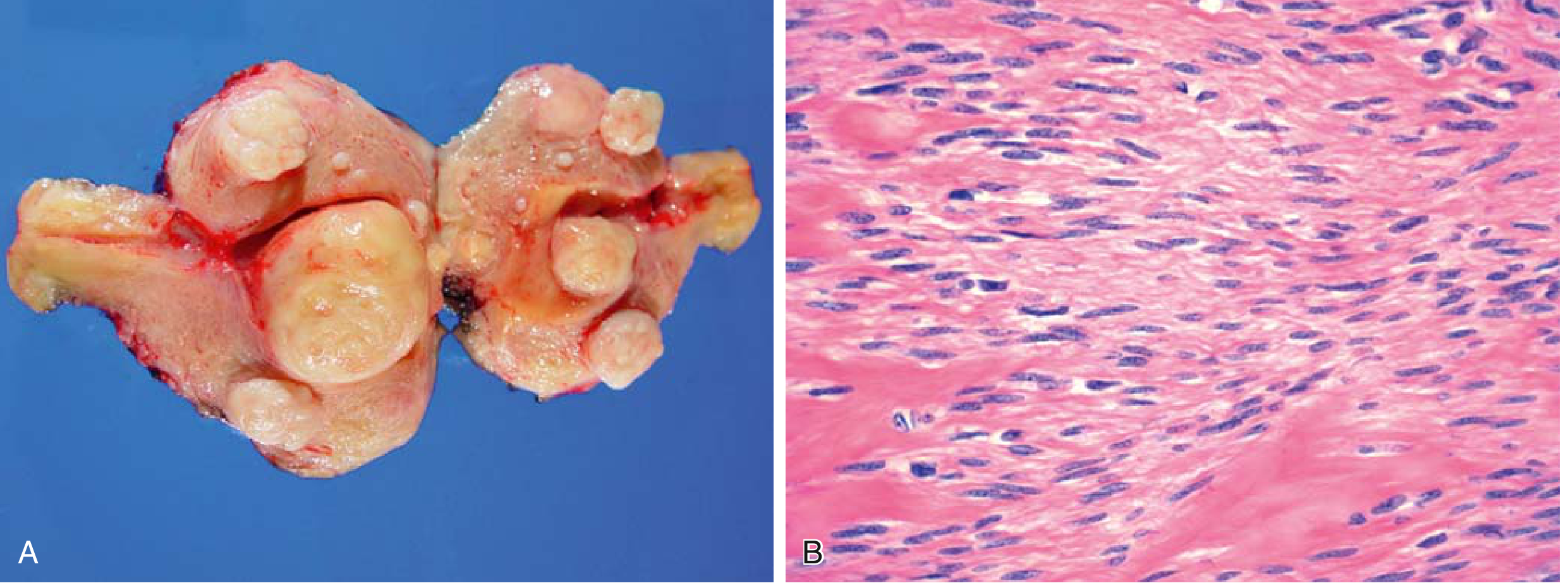
Uterine leiomyomas. (A) Multiple fibroids with characteristic whorled cut surface. (B) Bland smooth muscle bundles on histology. - Robbins & Kumar Basic Pathology
Clinical Features
- Often asymptomatic (found incidentally)
- Symptomatic cases: menorrhagia, pelvic pressure, urinary frequency, infertility (submucosal type)
- Treatment: Medical (GnRH agonists, progestins), interventional (uterine artery embolization, myomectomy), or hysterectomy
5. Leiomyosarcoma
An uncommon but aggressive malignant smooth muscle tumor.
| Feature | Leiomyoma (benign) | Leiomyosarcoma (malignant) |
|---|---|---|
| Occurrence | Multiple, common | Almost always solitary, uncommon |
| Age | Reproductive age (premenopausal) | Postmenopausal women |
| Karyotype | Simple, specific rearrangements | Complex, highly variable; chromosomal deletions |
| MED12 mutations | ~70% | ~30% |
| Cut surface | Firm, whorled, white | Soft, hemorrhagic, necrotic |
Diagnostic Criteria (all three must be assessed):
- Tumor necrosis (coagulative-type)
- Cytologic atypia
- Mitotic activity (elevated)
(Increased mitotic activity alone can be seen in benign smooth muscle tumors, especially in young women - all three features are needed to diagnose malignancy.)
Prognosis
Poor - these tumors often recur after surgery and >50% eventually metastasize hematogenously to lungs, bone, and brain. Abdominal/peritoneal dissemination also occurs.
Summary Table: Uterine Neoplasms
| Tumor | Origin | Benign/Malignant | Key Feature | Common Symptom |
|---|---|---|---|---|
| Endometrial hyperplasia | Endometrial glands | Premalignant | Increased gland:stroma ratio; PTEN mutation | Abnormal bleeding |
| Endometrioid carcinoma | Endometrial glands | Malignant | Estrogen-driven; PI3K/AKT mutations | Postmenopausal bleeding |
| Serous carcinoma | Endometrial surface epithelium | Malignant (high-grade) | TP53 mutations; peritoneal spread | Postmenopausal bleeding |
| Endometrial polyp | Endometrial stroma | Benign | Clonal stromal proliferation | Irregular bleeding |
| Leiomyoma | Smooth muscle (myometrium) | Benign | Whorled, MED12 mutations | Menorrhagia (submucosal) |
| Leiomyosarcoma | Smooth muscle | Malignant | Necrosis + atypia + mitoses | Mass, pain, bleeding |
Sources:
- Robbins & Kumar Basic Pathology, Chapter 17 (Female Genital System), pp. 690-693
- Robbins, Cotran & Kumar Pathologic Basis of Disease, Chapter 22, pp. 933-937
This is a shared conversation. Sign in to Orris to start your own chat.