
Laproscopy
Reading File
The query "Laparoscopy" is broad - let me clarify what you need before diving in.
Asking for Preferences
What aspect of laparoscopy are you interested in? · What is your context?
I'll provide a comprehensive overview of laparoscopy. Let me pull from the medical library.
Searching sources...
Finding Sources
Finding Sources
Good - I found several strong sources. Let me read the most comprehensive sections in parallel.
Reading File
Reading File
Reading File
Good content so far. Let me pull more detail on the technique, contraindications, and complications.
Reading File
Finding Sources
Excellent content retrieved. Let me fetch the diagram and also get specific contraindications/technique data.
Loading Image
Finding Sources
I have enough rich content to write a comprehensive answer. Let me now compile the full response.
Laparoscopy
Laparoscopy (also spelled laparoscopy) is a minimally invasive surgical and diagnostic technique that uses a rigid endoscope (laparoscope) inserted through small incisions in the abdominal wall to visualize and operate within the peritoneal cavity. It has largely replaced open surgery for a wide range of abdominal procedures.
1. Pneumoperitoneum - Creating the Working Space
The fundamental requirement of laparoscopy is lifting the abdominal wall away from the viscera to create a working space. Two approaches exist:
Carbon Dioxide (CO2) - Standard Choice
CO2 is the most widely used insufflation gas. It is rapidly absorbed across the peritoneal membrane, making it far safer than nitrogen in the event of vascular entry.
Nitrous Oxide (N2O)
N2O is physiologically inert, rapidly absorbed, and provides better analgesia when laparoscopy is done under local anesthesia. Controlled trials have confirmed its safety within the peritoneal cavity. However, its use in cancer surgery is cautioned due to unknown effects on port-site metastasis.
Inert Gases (Helium, Neon, Argon)
These cause no metabolic effects but are poorly soluble in blood, making gas embolism a serious risk if they gain venous access. They are rarely used clinically.
2. Physiologic Effects of CO2 Pneumoperitoneum
CO2 insufflation creates both local and systemic effects:
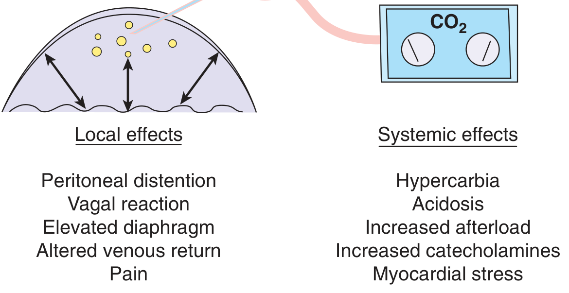
Respiratory Effects
- CO2 is absorbed across the peritoneum and generates carbonic acid, causing respiratory acidosis
- Body buffers (largely bone) absorb up to 120 L of CO2 before respiratory acidosis develops
- Once buffers are saturated, the anesthesiologist must increase ventilatory rate or tidal volume
- Severe respiratory acidosis can cause cardiac arrhythmias
- Hypercarbia causes tachycardia, increased systemic vascular resistance, elevated BP, and increased myocardial O2 demand
Cardiovascular Effects
- Increased intra-abdominal pressure compresses the inferior vena cava, reducing venous return from the lower extremities
- In reverse Trendelenburg position, venous stasis and DVT risk increase
- Increased intrathoracic pressure raises central venous pressure and cardiac filling pressures
- If intra-abdominal pressure is kept under 20 mmHg, cardiac output is usually well maintained
- DVT prophylaxis (sequential compression stockings, low molecular weight heparin) is recommended for longer procedures
Renal Effects
- Pneumoperitoneum decreases renal blood flow, GFR, and urine output
- This is partly from direct compression of kidneys and renal veins, and partly mediated by elevated renin and ADH
- Intraoperative oliguria is common and does not reflect intravascular volume status - IV fluids should not be driven by urine output alone
- Effects resolve within ~1 hour of desufflation
Gas Embolism
- Rare but life-threatening complication when gas accesses the venous system
- Suspect if hypotension develops during insufflation
- Diagnose with an esophageal stethoscope: characteristic "mill wheel" murmur
- Treatment: place patient in left lateral decubitus, head-down position to trap gas in the right ventricular apex, then aspirate via central venous catheter
3. Technique Overview
Access:
- A Veress needle is inserted through a small infraumbilical incision to insufflate CO2 and create the pneumoperitoneum
- Once adequate pneumoperitoneum is achieved, a primary trocar is introduced bluntly
- The laparoscope is passed through the trocar
- Additional working trocars are placed under direct vision for instruments
Alternative access:
- Direct trocar insertion (Hasson technique): trocar placed without prior pneumoperitoneum via an open mini-incision - reduces risk of Veress needle injury
Patient positioning:
- Trendelenburg (head-down): used for pelvic surgery (shifts bowel away from pelvis)
- Reverse Trendelenburg (head-up): used for upper abdominal surgery (shifts bowel caudally)
4. Types of Laparoscopy
| Type | Purpose |
|---|---|
| Screening | Excludes peritoneal penetration (e.g., in trauma) without full visceral evaluation |
| Diagnostic | Identifies pathology (e.g., staging cancer, evaluating abdominal pain, ectopic pregnancy) |
| Therapeutic | Treats the identified pathology (e.g., cholecystectomy, appendectomy, hernia repair) |
5. Indications
General surgery:
- Cholecystectomy (most common laparoscopic procedure)
- Appendectomy
- Hernia repair (inguinal, ventral)
- Anti-reflux surgery (Nissen fundoplication)
- Colectomy, splenectomy, adrenalectomy
- Bariatric surgery
Gynaecology:
- Sterilization
- Lysis of adhesions
- Endometriosis ablation (CO2 laser)
- Myomectomy, salpingectomy, oophorectomy
- Hysterectomy
- Evaluation of pelvic pain or infertility
Urology:
- Radical prostatectomy
- Nephrectomy
- Evaluation of nonpalpable testis (diagnostic)
Trauma:
- Screening/diagnostic in stable patients with penetrating abdominal trauma
- To detect diaphragmatic injury or peritoneal breach
- Not for hemodynamically unstable patients
6. Contraindications
Absolute:
- Hemodynamic instability (cannot tolerate pneumoperitoneum)
- Severe uncorrectable coagulopathy
- Inability to tolerate general anesthesia
Relative:
- Severe cardiac or respiratory disease (limited tolerance for CO2 physiologic effects)
- Prior multiple abdominal surgeries (extensive adhesions)
- Pregnancy (risk of uterine injury, decreased uterine blood flow, fetal acidosis from CO2, preterm labor risk)
- Increased intracranial pressure (pneumoperitoneum raises ICP - absolute contraindication in TBI)
- Morbid obesity (technical difficulty, increased physiologic demands)
- Large abdominal masses (>5 cm)
7. Complications
Related to Pneumoperitoneum
- Cardiac arrhythmias
- Subcutaneous emphysema
- Pneumothorax / Pneumomediastinum
- CO2 gas embolism
- DVT and pulmonary embolism
Related to Needle/Trocar Insertion
- Vascular injury (major vessels, trocar site bleeding)
- Bowel injury (small bowel, colon)
- Urinary tract injury (bladder, ureter)
- Incisional hernia (especially at 10-12 mm port sites)
Related to Surgical Technique
- Thermal/electrosurgical injury
- Bile duct injury (in cholecystectomy)
- Wound dehiscence
- Port-site metastasis (in oncologic cases)
8. Advantages Over Open Surgery
| Feature | Laparoscopy | Open |
|---|---|---|
| Incision size | 5-12 mm ports | Large laparotomy |
| Postop pain | Significantly less | More |
| Recovery time | Days-weeks | Weeks-months |
| Blood loss | Less | More |
| Wound infections | Less | More |
| Hospital stay | Shorter | Longer |
| Cosmesis | Excellent | Visible scar |
| Insensible fluid loss | Minimal | Substantial |
Disadvantages
- Steep learning curve
- Loss of tactile (haptic) feedback
- 2D visualization (standard laparoscopy; addressed by robotic 3D systems)
- Instrument degrees of freedom are limited
- Cannot perform in unstable patients or with extensive adhesions
- Equipment cost
9. Special Considerations
Gasless Laparoscopy
In patients who cannot tolerate CO2 (severe cardiopulmonary disease), an abdominal wall lift device can be placed through a 10-12 mm trocar to mechanically elevate the abdominal wall without pneumoperitoneum. This eliminates all gas-related physiologic effects.
Robotic-Assisted Laparoscopy
Adds 3D high-definition visualization and wristed instruments with multiple degrees of freedom, improving ergonomics and facilitating complex dissection and suturing. However, it adds cost and eliminates haptic feedback entirely.
Pregnancy
Laparoscopy during pregnancy carries additional risks: uterine injury during trocar placement, decreased uterine blood flow, preterm labor risk from increased intra-abdominal pressure, and fetal acidosis from CO2 absorption. It can be performed in pregnancy when necessary (e.g., acute appendicitis, ovarian torsion) with appropriate precautions.
Sources: Schwartz's Principles of Surgery 11e, p.483; Tintinalli's Emergency Medicine; Bailey & Love's Short Practice of Surgery 28e; Sabiston Textbook of Surgery; Barash Clinical Anesthesia 9e
This is a shared conversation. Sign in to Orris to start your own chat.