
Costochondritis
costochondritis diagnosis treatment
costochondritis chest wall anatomy costal cartilage diagram
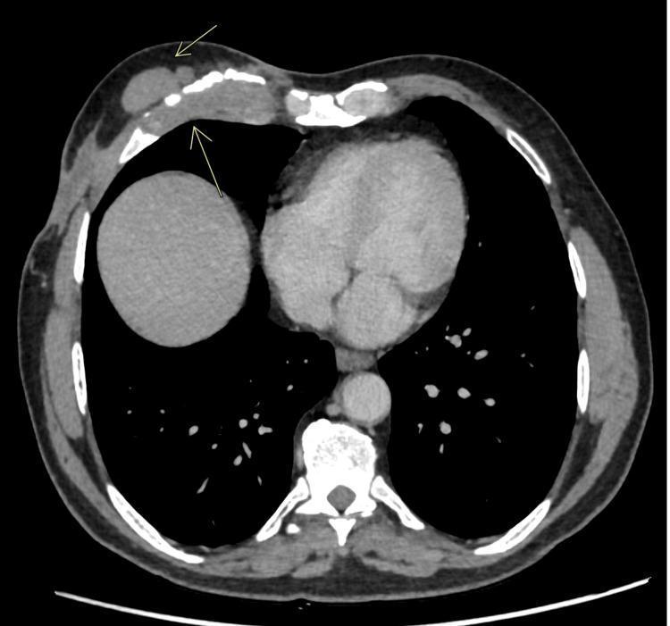
This diagnostic image is an axial contrast-enhanced computed tomography (CECT) scan of the thorax at the level of the lower chest and upper abdomen. The image demonstrates significant pathology of the anterior chest wall involving the right fifth costal cartilage. Key findings include a focal soft tissue mass and associated thickening at the costochondral junction. Evidence of bony erosion and fragmentation of the costal cartilage is visible, suggesting an aggressive inflammatory or infectious process, such as tubercular costochondritis. Additionally, multiple well-defined, ovoid soft tissue density nodules (indicated by arrows) are present within the subcutaneous tissues of the anterior abdominal and chest wall, which can be seen in conditions like neurofibromatosis or disseminated granulomatous disease. The internal anatomy shows the heart, descending aorta, and the superior aspect of the liver. This scan illustrates the radiological presentation of extrapulmonary tuberculosis manifesting as chest wall involvement with secondary subcutaneous nodules.
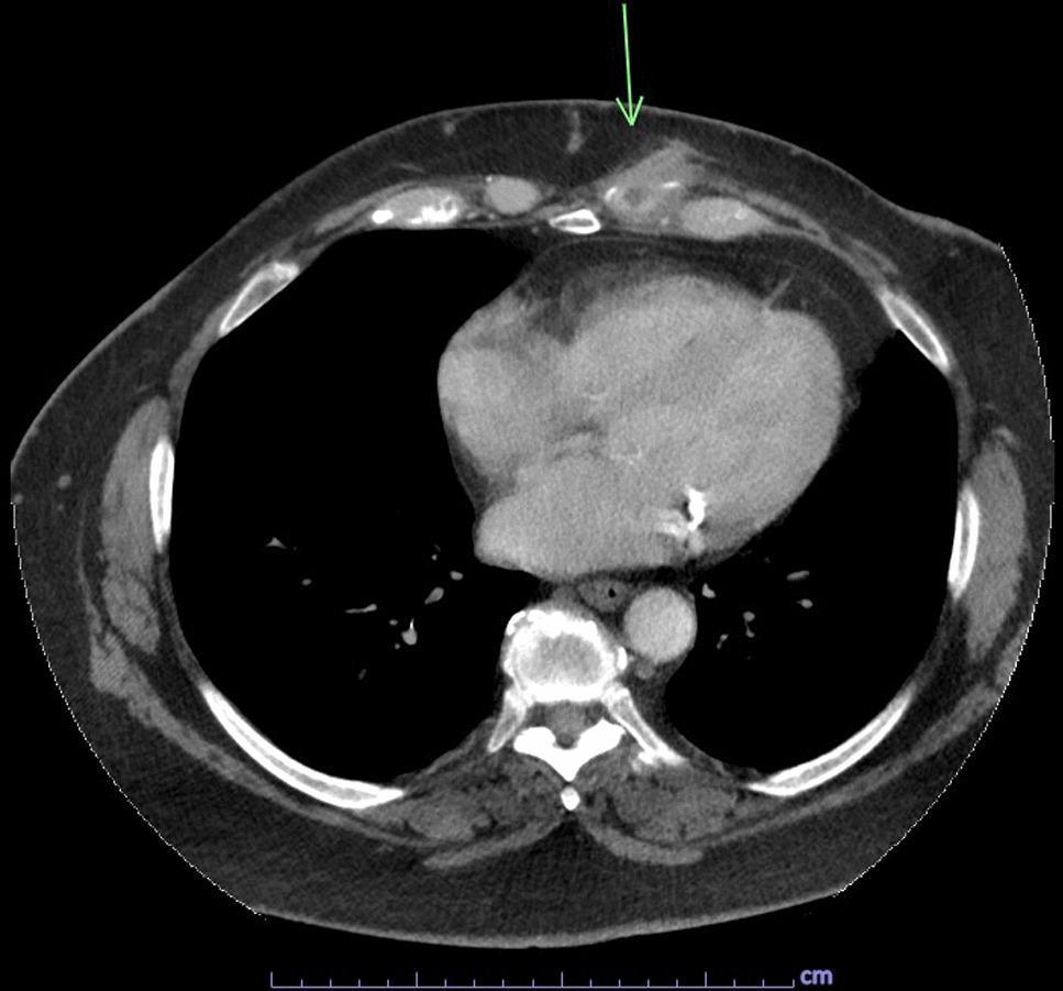
This diagnostic image is an axial chest computed tomography (CT) scan with intravenous contrast. The focus is on the anterior left chest wall at the level of the lower mediastinum. An arrow indicates a focal area of pathology involving the left seventh costal cartilage. Key findings include localized soft tissue thickening and a hypoattenuating fluid collection approximately 0.9 x 0.6 cm in size, characteristic of an abscess or persistent fluid collection associated with costochondritis. There is evidence of a sinus tract extending from this deep collection toward the skin surface. Centrally, the mediastinum contains the heart and great vessels, with visible calcifications within the coronary arteries. The lungs are clear without significant consolidation or pleural effusion. The vertebral bodies and posterior ribs appear intact. This scan is educationally significant for demonstrating the radiological appearance of infectious costochondritis and associated chest wall abscess formation, which in this clinical context was confirmed to be caused by Salmonella enterica serotype Choleraesuis.
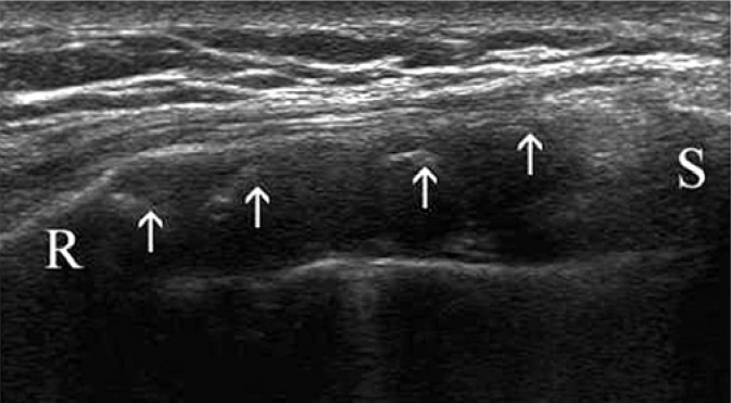
**Imaging Modality:** Greyscale Ultrasound (high-frequency linear transducer). **Anatomical Region:** Anterior chest wall at the costochondral junction. Key landmarks include the osseous rib (labeled 'R'), the sternum (labeled 'S'), and the intervening costal cartilage. **Observed Pathology:** Costal cartilage calcification. **Characteristic Visual Features:** The image displays a longitudinal view of the costal cartilage, which appears as a relatively hypoechoic, elongated structure. Within the cartilaginous matrix, multiple discrete, hyperechoic (bright) foci are visible (indicated by white arrows). These hyperechoic clusters represent areas of mineralization or calcification. The underlying cortical surfaces of the rib and sternum exhibit characteristic hyperechoic lines with distal acoustic shadowing. **Diagnostic Features:** The presence of scattered internal echogenicities within the normally hypoechoic costal cartilage is diagnostic of cartilaginous calcification. This finding is often associated with physiological aging or specific inflammatory conditions of the chest wall, such as costochondritis. The distribution is localized between the bony landmarks of the thoracic cage.
costochondritis chest pain musculoskeletal
Costochondritis
Definition and Overview
| Feature | Costochondritis | Tietze Syndrome |
|---|---|---|
| Age | Usually >40 years | Usually <40 years |
| Sex | More common in women | Equal sex distribution |
| Joints affected | 3rd, 4th, 5th costochondral joints | Typically 2nd or 3rd joint (one joint) |
| Swelling | Absent | Present (visible/palpable swelling) |
- Harrison's Principles of Internal Medicine, 22E (2025)
Pathophysiology
Clinical Features
- Anterior chest pain - can be sharp, aching, or pressure-like
- Pain radiates to the arms or shoulders in some patients
- Pain is aggravated by sneezing, coughing, deep inspiration, and twisting motions of the chest
- Onset can be sudden or gradual
- Tenderness on palpation of the affected costochondral joints - this is the hallmark finding
- Reproducibility of pain with palpation is key for diagnosis
- In Tietze syndrome specifically: visible or palpable swelling at the joint
Differential Diagnosis
- Cardiac: Acute coronary syndrome, pericarditis, aortic dissection
- Pulmonary-vascular: Pulmonary embolism, pneumothorax, pleuritis
- Inflammatory arthritis: Rheumatoid arthritis, psoriatic arthritis, ankylosing spondylitis, reactive arthritis (these involve costochondral joints but have other distinguishing features)
- Malignancy: Rib or chest wall invasion by tumor can mimic Tietze syndrome
- Infectious: Thoracic herpes zoster (look for dermatomal distribution and eventual rash)
- Metabolic: Osteomalacia with rib pain (check vitamin D levels; nuclear bone imaging may show microfractures)
- Other chest wall syndromes:
- Xiphoidalgia - pain at the xiphoid process
- Slipping rib syndrome - typically involves the 10th or lower ribs; pain below the rib cage, reproduced by hooking fingers around the rib and applying gentle downward traction
Important caution: Avoid overdiagnosing costochondritis in patients with acute chest pain. Many patients are tender to vigorous palpation of costochondral joints even without true costochondritis. Always rule out serious causes first.
Diagnosis
- Characteristic history of anterior chest pain with the features described above
- Reproduction of pain with direct palpation of the affected costochondral joint(s)
- No alternative serious cause identified
- Chest X-ray to exclude cardiopulmonary causes
- CT or MRI if malignancy, infection, or abscess is suspected
- Ultrasound can demonstrate costal cartilage changes (calcification, thickening)
Treatment
- Analgesics - paracetamol (acetaminophen) for mild pain
- NSAIDs - anti-inflammatory drugs (e.g., ibuprofen, naproxen) are first-line for pain and inflammation
- Local glucocorticoid injections - for refractory or severe cases, injection into the tender joint is effective
- Physical therapy - stretching, postural correction, and avoiding provocative activities
- Reassurance - important, as patients are often worried about cardiac causes
Key Clinical Pearls
- Costochondritis is one of the most common musculoskeletal causes of chest pain, accounting for up to ~30% of chest pain presentations in some series
- Always consider and exclude ACS, PE, and other life-threatening causes before settling on this diagnosis
- Tietze syndrome involves swelling; isolated costochondritis does not
- Inflammatory arthritides (RA, ankylosing spondylitis, psoriatic arthritis) can produce costochondral inflammation but will have other systemic or articular features
- Herpes zoster can produce severe anterior chest wall pain before the rash appears - keep this in mind in older or immunocompromised patients
- Vitamin D deficiency/osteomalacia should be excluded when rib pain is diffuse