
Spindle cell lesion
spindle cell lesion histology microscopy
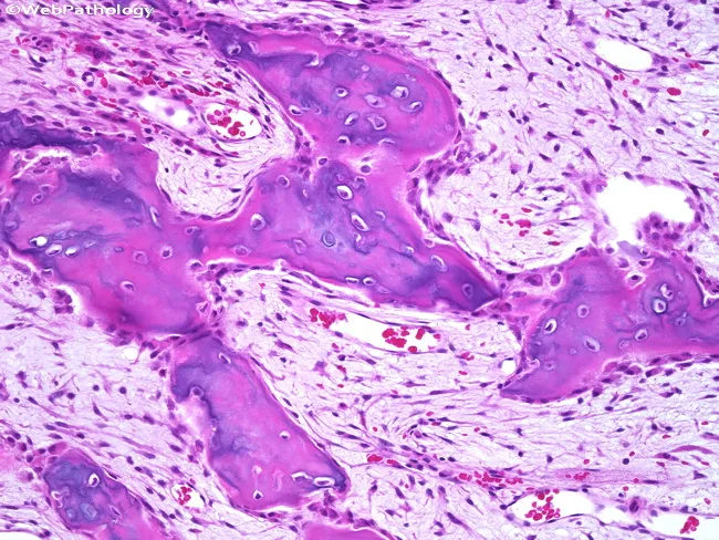
This is a histopathology image obtained from a periosteal bone lesion, analyzed by light microscopy after Hematoxylin and Eosin (H&E) staining. The specimen demonstrates a densely cellular spindle cell proliferation embedded in a fibrous stroma that interposes between irregular bone trabeculae at the periosteal surface. The bony trabeculae are variably mineralized, with woven and trabecular components intermixed with the soft tissue spindle cells, creating a biphasic pattern characteristic of BPOP (Bizarre Parosteal Osteochondromatous Proliferation), also known as Nora lesion. The spindle cells are uniform, spindle‑shaped mesenchymal cells with elongated nuclei and scant cytoplasm; mitotic activity is minimal or absent, and cytologic atypia is not evident. Surrounding cartilaginous nodules or a cartilaginous cap may be seen in some instances; there is no infiltration of adjacent marrow or invasion beyond the periosteal surface. The lesion remains largely confined to the surface with fibrous tissue separating osseous trabeculae. Diagnostic significance is that this histology supports a benign proliferative osseous lesion with a known tendency to recur after incomplete excision, but malignant transformation has not been reported. Clinically, recognition of BPOP guides surgical management toward complete excision with clear margins to reduce recurrence risk; differential considerations include osteochondroma and periosteal chondrosarcoma. This image is valuable for educational pathology discussions.
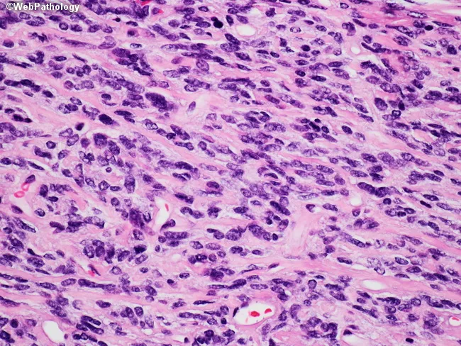
This image is a hematoxylin and eosin stained histology slide viewed under bright-field microscopy at high power, showing a densely cellular soft-tissue lesion comprised of interlacing fascicles of spindle-shaped cells with elongated, hyperchromatic nuclei and scant cytoplasm. The stroma appears collagen-rich and pink, with irregular, elongated mitotic figures scattered throughout; occasional inflammatory cells are present. There is mild-to-moderate cellular pleomorphism and a few multinucleated cells. The overall pattern is inconspicuous for glandular differentiation or epithelial components. No necrosis is conspicuous at this low-magnification region, though focal areas of increased mitotic activity suggest a neoplastic process rather than reactive scar. The cellular arrangement and stromal density resemble fibrosarcoma or other spindle cell sarcomas; however, without immunohistochemical studies, specific lineage cannot be confirmed. Differential diagnoses include malignant peripheral nerve sheath tumor, leiomyosarcoma, and synovial sarcoma, among others, particularly in deep soft tissues. This slide is typically obtained from formalin-fixed, paraffin-embedded tissue and prepared as a routine H&E section for initial histopathologic assessment. Clinically, this morphology may present as a painless soft-tissue mass with infiltrative growth. Diagnostic significance lies in identifying spindle cell neoplasia, guiding immunostaining panels, and correlating with radiologic features to determine extent, margins, and staging. This image is valuable for education, differential diagnosis practice, and pathology training.
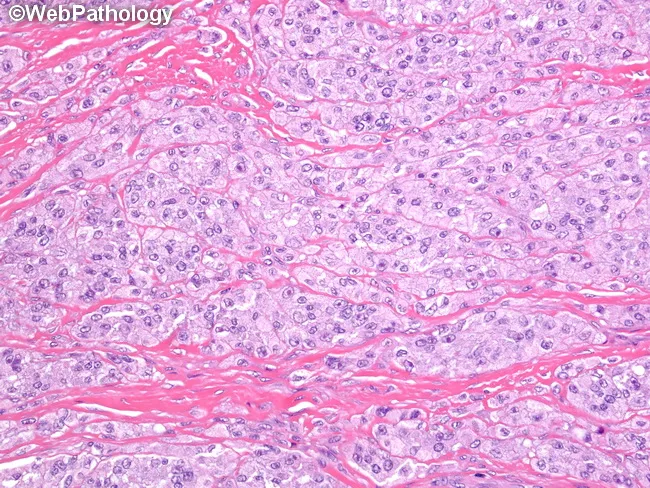
Histology: Light microscopy with Hematoxylin and Eosin stained section of soft tissue demonstrating a malignant spindle cell neoplasm. Tumor cells are fusiform to polygonal, organized in dense solid nests and broad fascicles separated by a conspicuous fibrous stroma. The fibrous septa extend between tumor nests and frequently appear to merge with adjacent tendons and aponeuroses, reflecting the tumor's intimate relationship with peri-tendinous structures. Cells show eosinophilic cytoplasm and elongated, ovoid nuclei with evenly distributed chromatin; occasional nuclear pleomorphism and mitotic activity may be present. Matrix is scant, with interstitial fibrous tissue creating a ribbon-like network that contributes to the characteristic appearance. In some fields, cells may contain clear cytoplasm due to glycogen-rich content, a feature that contributes to the name 'clear cell.' The overall pattern is consistent with a malignant spindle cell sarcoma with melanocytic differentiation. Immunophenotype (not shown here) often demonstrates S-100 positivity and sometimes HMB-45, helping distinguish from other soft tissue sarcomas; molecular testing typically reveals EWSR1-ATF1 fusion arising from t(12;22). Clinically, this lesion arises in extremities and may mimic melanoma, or other sarcomas. Definitive classification requires integrated histology, immunohistochemistry, and molecular genetics for definitive diagnosis and optimal management. Correlation with clinical presentation enhances diagnostic accuracy.
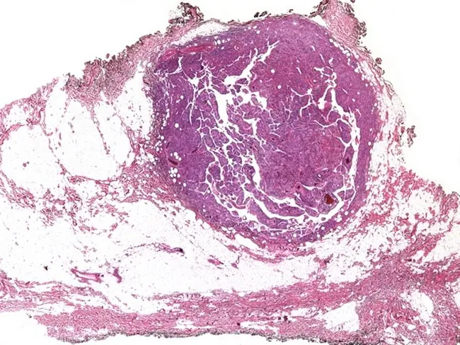
This histopathology image depicts a soft tissue lipomatous lesion illustrating a spindle cell/pleomorphic lipoma with a distinctive pseudoangiomatous pattern. The specimen is a subcutaneous adipose tissue fragment examined under bright-field light microscopy after Hematoxylin and Eosin staining. The architecture shows adipocytic components intermingled with alternating fibrous septa and spindle cells. Unusually, there are irregular, branching, slit-like spaces that mimic vascular channels; the spaces are lined by endothelial-like cells. In this case, subsequent immunohistochemistry demonstrates positivity of the lining cells for endothelial markers (e.g., CD31, CD34, ERG), consistent with a true angiomatous pattern, though in classic pseudoangiomatous lipoma these spaces reflect myofibroblastic or adipocytic stromal components rather than true neoplastic vessels. The adipocytes are mature, with minimal pleomorphism; the spindle cell component is bland and CD34 positive; focal calcifications or foamy histiocytes may be present. This pattern can be misinterpreted as a malignancy; awareness of this entity prevents overtreatment. Clinically, spindle cell lipomas commonly present as slow-growing, painless subcutaneous nodules in middle-aged adults; complete surgical excision yields excellent prognosis. The image is an educational example illustrating the spectrum of lipomatous tumors and the diagnostic pitfall of angiomatous mimicry in soft tissue neoplasms. This image supports histology teaching, differential diagnosis, and reporting.
Spindle Cell Lesion
H&E Morphology - What Makes a Cell "Spindle"
- Elongated/fusiform with pointed or tapered ends
- Arranged in fascicles, whorls, or storiform (cartwheel) patterns
- May be bland/uniform (benign) or show pleomorphism, mitoses, and necrosis (malignant)
Classification by Lineage
1. Melanocytic Lesions
Spitz Nevus (Spindle and Epithelioid Cell Nevus)
- Large spindle and epithelioid cells with vesicular nuclei and prominent nucleoli
- Nests oriented vertically ("raining down" the rete ridges)
- Clefts adjacent to junctional nests
- Kamino bodies (dull-pink eosinophilic globules of basement membrane material) - pathognomonic
- Sharp lateral circumscription and dispersion at the base (cells separate between collagen bundles)
- HMB-45: top-heavy staining (junctional > dermal) vs. uniform staining in melanoma
- Ki-67 (MIB-1): rare positive cells in deep dermis vs. numerous in melanoma
- S-100A6: strong and diffuse in Spitz nevi
Pigmented Spindle Cell Nevus (Reed Nevus)
- Fascicles of uniform, slender spindle cells closely aggregated
- Confined mainly to epidermis ± papillary dermis
- Cells contain fine, granular melanin; delicate chromatin; small nucleoli
- Clefting around intraepidermal fascicles; melanophages in papillary dermis
- Stains poorly with S-100A6 (unlike Spitz nevi)
2. Vascular Lesions
Spindle Cell Hemangioma (Spindle Cell Hemangioendothelioma)
- Strongly associated with Maffucci syndrome (IDH1/IDH2 mutations)
- Occurs at any age and site; extremities most common
- Histology: nodular, dense spindle cell proliferation + dilated dysplastic veins
- Locally aggressive; may recur after excision but does not metastasize
- IDH1/IDH2 mutations detected in sporadic cases as well
- DDx: Must exclude intravascular papillary endothelial hyperplasia (PEH) - a common secondary finding within these lesions - Fitzpatrick's Dermatology; Dermatology 5e
3. Adipocytic Lesions
Spindle Cell / Pleomorphic Lipoma
- Benign subcutaneous tumor; favors the posterior neck, shoulder, and back of middle-aged males
- Histology: mature adipocytes + variable spindle cells in a collagenous/myxoid stroma
- IHC: CD34+ spindle cells; S-100 rarely positive
- Loss of RB1 locus
- No malignant potential - Fitzpatrick's Dermatology; Dermatology 5e
4. Epithelial / Carcinomatous Lesions
Spindle Cell Carcinoma (Sarcomatoid Carcinoma / SPCC)
- Composed of a surface squamous cell component (CIS or invasive SCC) + dominant underlying malignant spindle cell component
- Histogenesis: epithelial origin with mesenchymal differentiation
- Most arise as polypoid glottic lesions
- Cervical metastases in ~13%; distant metastases rare
- Diagnosis pitfall: May be misdiagnosed as fibrosarcoma, MFH, nodular fasciitis, or pure SCC if the squamous component is small or not sampled
- IHC: Spindle cells often focally positive for keratin (not desmin or h-caldesmon)
- Treatment: Surgery (partial vs. total laryngectomy); RT alone is inadequate
- 5-year survival: 63-94% (stage/site dependent) - Cummings Otolaryngology
5. Soft Tissue / Mesenchymal Lesions
Synovial Sarcoma
- Arises in deep soft tissue; most common around knee joint
- Despite the name, does NOT arise from synovium
- Variants: Biphasic (epithelial + spindle cell), monophasic spindle cell (most common), poorly differentiated (round cell)
- Monophasic variant: fascicles of uniform spindle cells in a herringbone pattern
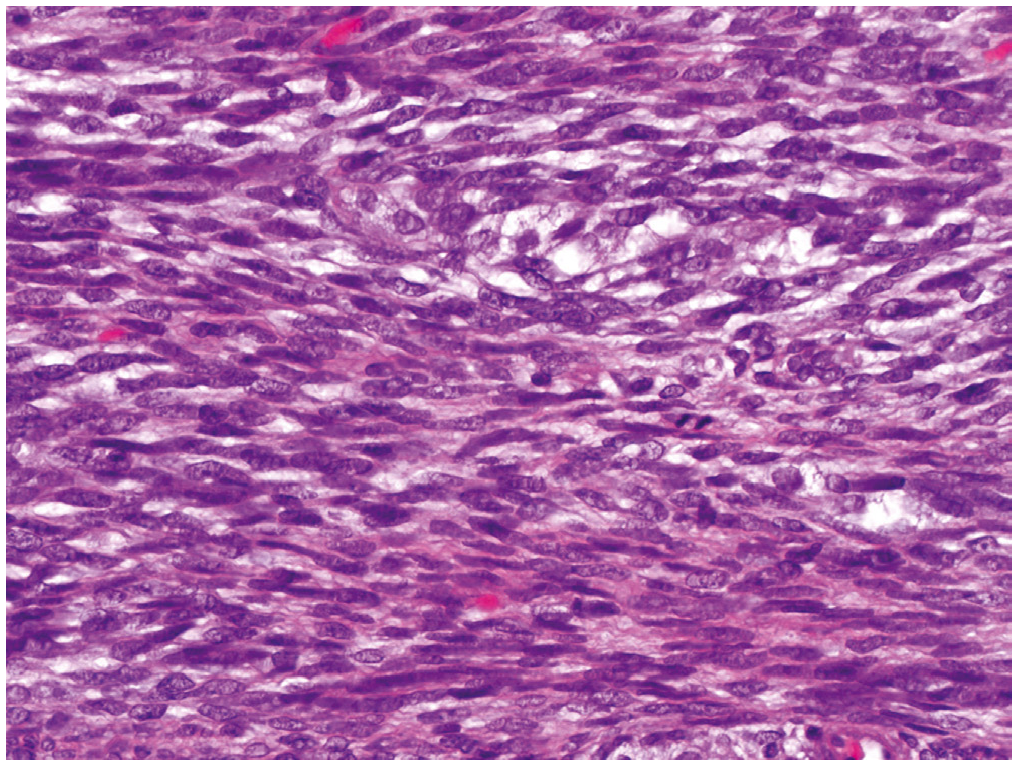
- IHC: Keratin and EMA positive (both epithelial and spindle components) - unusual for a mesenchymal tumor; TLE-1 diffusely positive
- Cytogenetics: t(X;18)(p11.2;q11.2) → SS18::SSX fusion in virtually all cases - pathognomonic
- Firestein & Kelley's Rheumatology
Leiomyosarcoma (Cutaneous / Soft Tissue)
- IHC: α-SMA+, desmin+/-, h-caldesmon+ (distinguishes from other spindle cell tumors)
- Must exclude: cellular dermatofibroma, cellular schwannoma, MPNST, myopericytoma
Malignant Peripheral Nerve Sheath Tumor (MPNST)
- Arises from nerve or in setting of NF1
- IHC: S-100 variable (patchy); loss of SOX10, neurofibromin, and p16
Dermatofibrosarcoma Protuberans (DFSP)
- Storiform spindle cell pattern; infiltrates subcutis in "honeycomb" fashion
- IHC: CD34+ (key marker), SMA-, S100-
- t(17;22) → COL1A1-PDGFB fusion
Inflammatory Myofibroblastic Tumor (IMT)
- Neoplastic myofibroblastic spindle cells + dense polyclonal plasma cell infiltrate
- Favors lungs, mesentery; children and young adults
- IHC/molecular: ALK rearrangement (2p23) in ~50% - ALK IHC is a particularly useful diagnostic tool in small biopsies with a broad spindle cell differential; ROS1 rearrangement in ALK-negative cases
- Absence of ALK expression = greater risk of aggressive behavior - Murray & Nadel's Respiratory Medicine
6. Spindle Cell Lesions in Specific Organs
| Organ | Key Entities |
|---|---|
| Skin | Spitz nevus, pigmented spindle cell nevus, spindle cell SCC, Kaposi sarcoma, dermatofibroma, DFSP, leiomyosarcoma, angiosarcoma, atypical fibroxanthoma, pleomorphic dermal sarcoma |
| Larynx/H&N | Spindle cell carcinoma (SPCC), fibrosarcoma, nodular fasciitis, MPNST |
| GI tract | GIST (spindle cell type), leiomyosarcoma, sarcomatoid carcinoma, Kaposi sarcoma |
| Lung | IMT, solitary fibrous tumor, synovial sarcoma, spindle cell carcinoid |
| Soft tissue | Synovial sarcoma, leiomyosarcoma, MPNST, fibrosarcoma, spindle cell RMS |
| Bone | Periosteal osteosarcoma, BPOP (Nora lesion), clear cell sarcoma |
IHC Panel for Differential Diagnosis of Spindle Cell Lesions
| Marker | Positive in |
|---|---|
| S-100, SOX10, HMB-45, MART-1 | Melanocytic (melanoma, Spitz nevus) |
| CD34 | Spindle cell lipoma, DFSP, solitary fibrous tumor |
| SMA, desmin, h-caldesmon | Smooth muscle (leiomyosarcoma) |
| SMA (without h-caldesmon) | Myofibroblasts (IMT, nodular fasciitis) |
| Keratin, EMA | Spindle cell carcinoma, synovial sarcoma |
| TLE-1 | Synovial sarcoma (diffuse, strong) |
| ALK | IMT |
| ERG, CD31 | Vascular (angiosarcoma, spindle cell hemangioma) |
| STAT6 (nuclear) | Solitary fibrous tumor |
Approach to a Spindle Cell Lesion - Diagnostic Algorithm
- Clinical context - Age, site, size, growth rate, association with NF1, Maffucci syndrome, prior radiation, sun-damage
- H&E morphology - Architecture (storiform, fascicular, herringbone, whorled), cytologic atypia, mitoses, necrosis, Kamino bodies, inflammatory infiltrate
- IHC panel tailored to the differential (keratin, S-100, CD34, SMA, desmin, h-caldesmon, ALK, TLE-1, ERG, HMB-45)
- Molecular/cytogenetics when IHC is inconclusive: FISH for SS18-SSX (synovial sarcoma), IDH1/2 (hemangioma/chondrosarcoma), ALK/ROS1 FISH (IMT), EWSR1 (clear cell sarcoma), t(17;22) (DFSP)
- Correlation with clinical, radiologic, and molecular findings
Mucinous carcinoma hreast
mucinous colloid carcinoma breast histology microscopy mucin pools
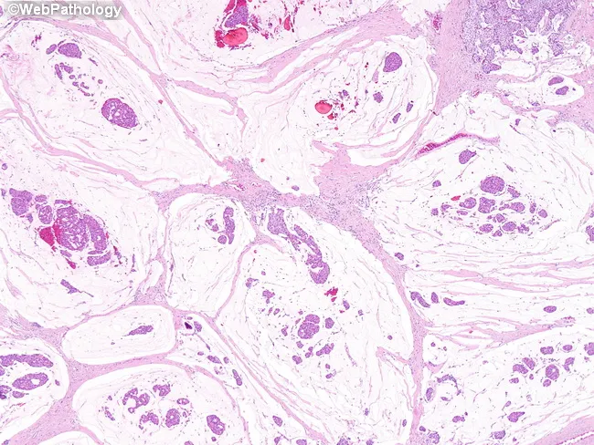
This histopathology image depicts a breast tissue section stained with Hematoxylin and Eosin (H&E) viewed under light microscopy. The primary subject is mucinous (colloid) carcinoma of the breast, an invasive carcinoma variant defined by abundant extracellular mucin. In the field, tumor cells form cohesive clusters floating within large mucin pools; the pools are partially separated by delicate fibrous septa, producing lakes-with-septa architecture. The neoplasm demonstrates multiple architectural patterns, including solid nests, cribriform cords, and trabecular strands embedded in a mucinous matrix. Cellular morphology shows cohesive epithelial cells with round to oval nuclei and vesicular chromatin; mitotic activity is typically low in classical mucinous carcinoma. The mucin-rich stroma imparts a pale, basophilic background with mucin lakes that may appear vacuolated. Invasion into surrounding fibrous stroma may be subtle but detectable at the tumor-stroma interface. Overall, the image highlights the hallmark features: extracellular mucin pools, partial fibrous septation, and tumor proliferation in cribriform, solid, and trabecular patterns, consistent with mucinous breast carcinoma. Clinically, these histology features correlate with a relatively favorable prognosis compared with non-mucinous invasive ductal carcinoma and assist in differential diagnosis from mucin-producing carcinomas of other primaries. This representation is educational for pathology practice, diagnosis, and training. For medical learners.
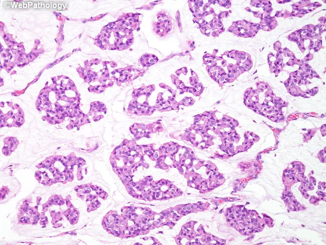
Light microscopy of hematoxylin and eosin stained breast tissue demonstrates a pure mucinous (colloid) carcinoma pattern with abundant extracellular mucin lakes. Tumor cells are arranged in small cohesive clusters and islands that float within pools of mucin, often showing low-grade cytology with round to oval nuclei and minimal pleomorphism. Mitotic figures are infrequent, and desmoplastic stromal reaction is limited, yielding a relatively well-circumscribed rather than aggressively infiltrative appearance. The mucin-rich matrix dominates the histologic landscape, with occasional signet-ring-like cells present but not prominent. Such histology produces a characteristic translucent, gelatinous background on gross examination and a distinctive cellular screed on microscopic slides. Immunophenotype commonly includes estrogen receptor (ER) and progesterone receptor (PR) positivity, with HER2 amplification typically absent, consistent with favorable prognosis. The abundant mucin is thought to hinder vascular invasion and lymphovascular spread, contributing to the excellent prognosis described for pure mucinous breast carcinomas. Clinically, this histology correlates with lower nodal metastasis rates and favorable survival, influencing surgical management and adjuvant therapy decisions. Differential diagnoses include mucinous carcinoma with an invasive ductal component and, less likely, signet-ring cell carcinoma; correlate with imaging, receptor status, and clinical course for definitive classification and treatment planning. This image exemplifies mucinous breast carcinoma.
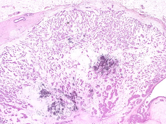
Imaging modality: light microscopy of formalin-fixed, paraffin-embedded breast tissue stained with Hematoxylin and Eosin (H&E). The specimen shows a fibroadenomatous lesion in which malignant mucinous (colloid) carcinoma arises within the epithelial component. The histology demonstrates abundant extracellular mucin pools with floating or discohesive malignant epithelial cells arranged in small clusters and glands. The surrounding stroma resembles a fibroepithelial lesion consistent with a longstanding fibroadenoma, but focal invasion by mucinous carcinoma is evident at the periphery, with cells displaying low to intermediate nuclear grade, mild pleomorphism, and mitotic activity. The mucin pools are typically large and may displace adjacent ducts, with signet ring-like morphology occasionally observed in mucin-producing cells. Immunohistochemical profile (not shown) would typically reveal cytokeratin positivity in tumor cells; myoepithelial layer is diminished at invasive fronts, supporting invasion beyond the fibroadenoma capsule. Clinically, this reflects a rare malignant transformation of a benign fibroepithelial lesion, reported to occur in approximately 0.1% of fibroadenomas, with the majority of malignant cases previously described as in-situ, while invasive mucinous carcinoma arising in a fibroadenoma carries implications for surgical management and prognosis. This image is educational for recognition of carcinoma arising within benign breast lesions, illustrating the need for complete excision and careful histologic assessment of invasion.
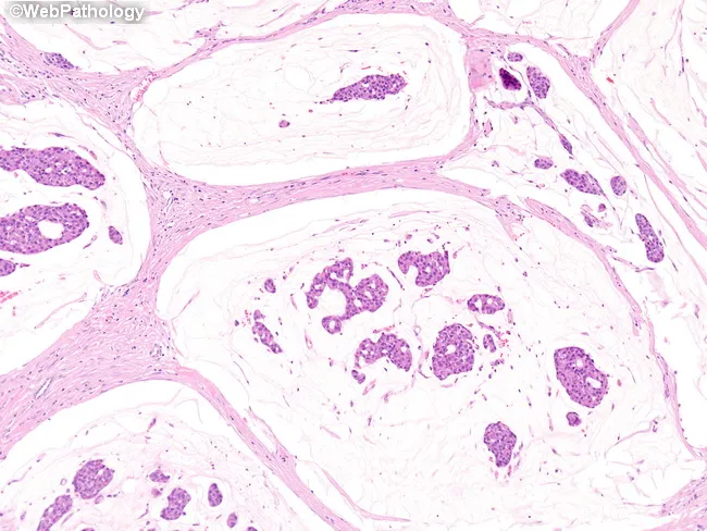
Histopathology, light microscopy of a breast tissue specimen stained with Hematoxylin and Eosin (H&E). The primary subject is an invasive mucinous (colloid) carcinoma of the breast. Diagnostic hallmarks are clusters of tumor cells embedded in abundant extracellular mucin pools, with mucin partially compartmentalized by delicate fibrous septa. Tumor cells arrange in solid, cribriform, and trabecular patterns within the mucin lakes, reflecting glandular differentiation in a myxoid background. The cells typically display round to oval nuclei with moderate cytoplasm, mild pleomorphism, and conspicuous nucleoli variably present; mitotic activity is often low to moderate. Surrounding stroma is fibrous and may show hyalinization; mucin pools appear as pale eosinophilic material separating clusters. These features help distinguish mucinous carcinoma from classic invasive ductal carcinoma and from mucin-producing metastases. Clinically, this histology portends a comparatively favorable prognosis relative to nonmucinous invasive breast cancers, especially when tumor size is limited and nodal involvement is absent. In diagnostic practice, recognizing extracellular mucin with cohesive tumor clusters aids classification, informs surgical planning, and guides adjuvant therapy decisions. Differential considerations include invasive ductal carcinoma with mucinous differentiation and metastatic mucin-secreting tumors. The image provides essential cues for pathology reports, education, and research on mucin biology in breast cancer.
Mucinous Carcinoma of the Breast (Colloid Carcinoma)
Epidemiology
- Accounts for ~2% of all invasive breast cancers
- Presents more commonly in older patients (postmenopausal women)
- May present as a bulky tumor despite its relatively indolent behavior
- Schwartz's Surgery; Mulholland & Greenfield's Surgery
Gross Pathology
- Soft, rubbery, gelatinous consistency on palpation and cut surface
- Cut surface is glistening and gelatinous ("colloid-like")
- Borders are pushing or circumscribed (not infiltrative/spiculated)
- Fibrosis is variable; when abundant, the tumor may feel firm
- Robbins Cotran Kumar; Schwartz's Surgery
Histopathology - The Hallmark
Key histological features:
| Feature | Description |
|---|---|
| Mucin | Extracellular, abundant, pools/lakes |
| Tumor cells | Uniform, low-grade nuclei; vesicular chromatin |
| Architecture | Clusters, nests, cribriform, trabecular cords within mucin |
| Mitoses | Low mitotic activity |
| Borders | Circumscribed/pushing (not spiculated) |
| Desmoplasia | Minimal |
| Associated DCIS | May be present |
Important: Because mucin dominates sections, cancer cells may not be present in every microscopic field. Multiple sections must be examined to confirm the diagnosis. - Schwartz's Surgery
Pure vs. Mixed type:
- Pure mucinous carcinoma: >90% of the tumor is mucinous; best prognosis
- Mixed mucinous carcinoma: mucinous component combined with NST (no special type/ductal NOS); prognosis is closer to NST
Molecular / Receptor Profile
- ER positive: >90% of cases
- PR positive: usually
- HER2: typically negative
- Molecular subtype: Luminal A (low Ki-67, ER+/PR+, HER2-)
- Belongs to the luminal group alongside invasive lobular, tubular, cribriform, and papillary carcinoma
- Robbins Cotran Kumar; Robbins & Kumar Basic Pathology
Clinical Features
| Feature | Details |
|---|---|
| Age | Older/postmenopausal women predominantly |
| Presentation | Palpable breast mass; may be bulky |
| Consistency | Soft, smooth, gelatinous |
| Borders | Well-circumscribed clinically and radiologically |
| Bilateral/multicentricity | Less common than lobular carcinoma |
Imaging
- Mammography: Well-circumscribed, round or lobulated mass - may mimic a benign lesion (fibroadenoma, cyst)
- Ultrasound: Hypoechoic, well-defined mass; posterior acoustic enhancement due to high water content of mucin
- MRI: Hyperintense on T2 (due to mucin water content); variable enhancement; circumscribed borders
Lymph Node Metastasis & Prognosis
| Parameter | Mucinous Carcinoma |
|---|---|
| Lymph node metastases | ~33% of cases (lower than NST) |
| 5-year survival | ~73% |
| 10-year survival | ~59% |
| Prognosis vs NST | Significantly better |
Pure mucinous carcinoma has a favorable prognosis and is one of the rare histological variants that consistently carries a better outlook than invasive carcinoma of no special type (NST). - Bailey & Love's Surgery; Schwartz's Surgery
- Low nuclear grade
- Low mitotic index
- Luminal A biology (ER+, HER2-, low Ki-67)
- Circumscribed growth pattern (less vascular invasion)
- The mucin matrix itself may hinder vascular spread
Treatment
Surgery
- Breast-conserving surgery (BCS/lumpectomy) is the standard for operable cases, followed by radiotherapy
- Sentinel lymph node biopsy (SLNB) for axillary staging (same as other invasive carcinomas)
- Total mastectomy if BCS is not feasible
Adjuvant Systemic Therapy
- Endocrine therapy (tamoxifen or aromatase inhibitor): mainstay given near-universal ER positivity
- Chemotherapy: Given the favorable biology (low grade, Luminal A), many pure mucinous carcinomas have a low Oncotype DX recurrence score, and chemotherapy may be omitted
- HER2-targeted therapy: Not indicated (HER2 negative)
- Radiotherapy: Standard after BCS
Differential Diagnosis
| Entity | Distinguishing Feature |
|---|---|
| Fibroadenoma | No intracellular mucin, no atypia, distinct lobular architecture |
| Mucocele-like lesion | Benign; mucin extravasation without epithelial atypia |
| Invasive carcinoma NST with mucin | <50% mucinous; worse prognosis |
| Metastatic mucinous carcinoma | From colorectum, ovary, appendix - exclude with IHC (CK7, CK20, CDX2, GCDFP-15, mammaglobin) |
| Primary cutaneous mucinous carcinoma | A diagnosis of exclusion - breast/GI primaries must be excluded - Fitzpatrick's Dermatology |
Key Points to Remember
- Colloid carcinoma = mucinous carcinoma - same entity, two names
- Defined by extracellular mucin pools with floating low-grade tumor cell clusters
- Almost always ER+, HER2- (Luminal A)
- Pure type has better prognosis than mixed type
- Lymph node metastases occur in ~1/3 but overall survival is favorable
- Cut sections may be negative for tumor cells - multiple sections are mandatory
- Grossly gelatinous and soft; circumscribed borders on imaging
- Endocrine therapy is the cornerstone of adjuvant systemic treatment