
Superior mediastinum and applied aspects
Reading File
Finding Sources
Finding Sources
Searching PubMed
mediastinum anatomy
Searching the Web
superior mediastinum anatomy contents clinical applied anatomy
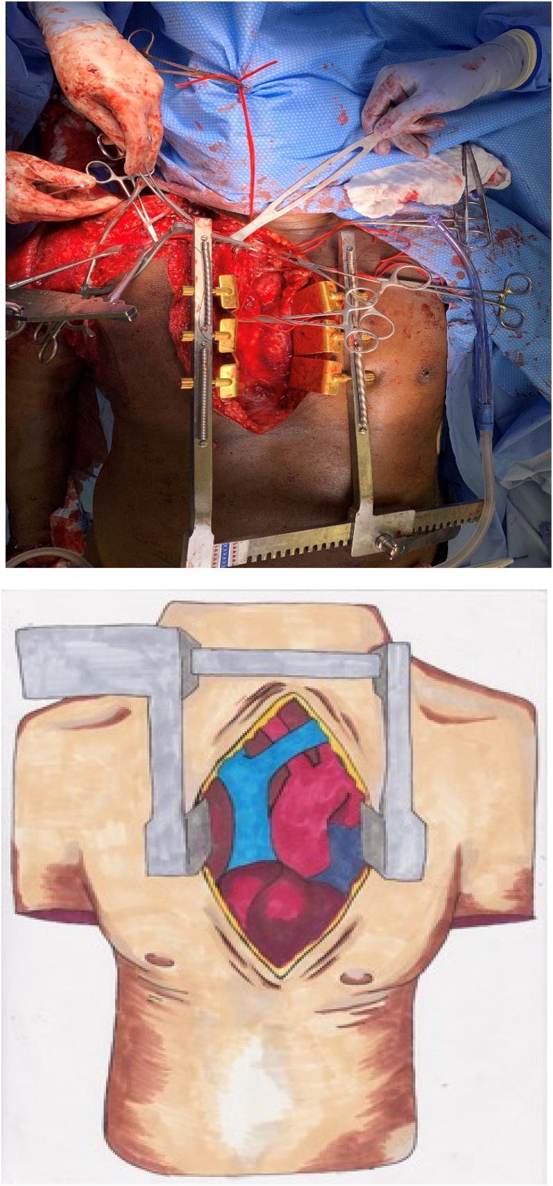
Reading File
Reading File
Reading Web Page
https://www.ncbi.nlm.nih.gov/books/NBK539819
Superior mediastinum
The superior mediastinum is the upper part of the mediastinum. It acts as a passage between the neck and thorax for major blood vessels, nerves, airway, food passage, and lymphatics.
Boundaries
| Boundary | Description |
|---|---|
| Superior | Thoracic inlet, an oblique plane from the jugular notch to the upper border of T1 |
| Inferior | Transverse thoracic plane from sternal angle to the T4-T5 intervertebral disc |
| Anterior | Manubrium sterni |
| Posterior | Bodies of T1-T4 vertebrae |
| Lateral | Mediastinal pleura on each side |
It is continuous with the neck above and the inferior mediastinum below. The sternal angle is an important surface landmark because it marks the plane separating superior from inferior mediastinum. Gray's Anatomy for Students, p. 252.
Contents
The contents can be recalled from anterior to posterior:
- Thymus or its fatty remnant in adults
- Veins
- Right and left brachiocephalic veins
- Upper half of superior vena cava
- Arch of azygos vein entering the SVC
- Left superior intercostal vein
- Arteries
- Arch of aorta
- Its branches, from right to left:
- Brachiocephalic trunk
- Left common carotid artery
- Left subclavian artery
- Nerves
- Right and left phrenic nerves
- Right and left vagus nerves
- Left recurrent laryngeal nerve
- Cardiac nerves and cardiac plexus
- Trachea
- Oesophagus
- Thoracic duct and mediastinal lymph nodes
A useful order is: Thymus - Veins - Arteries - Nerves - Trachea - Oesophagus - Thoracic duct.
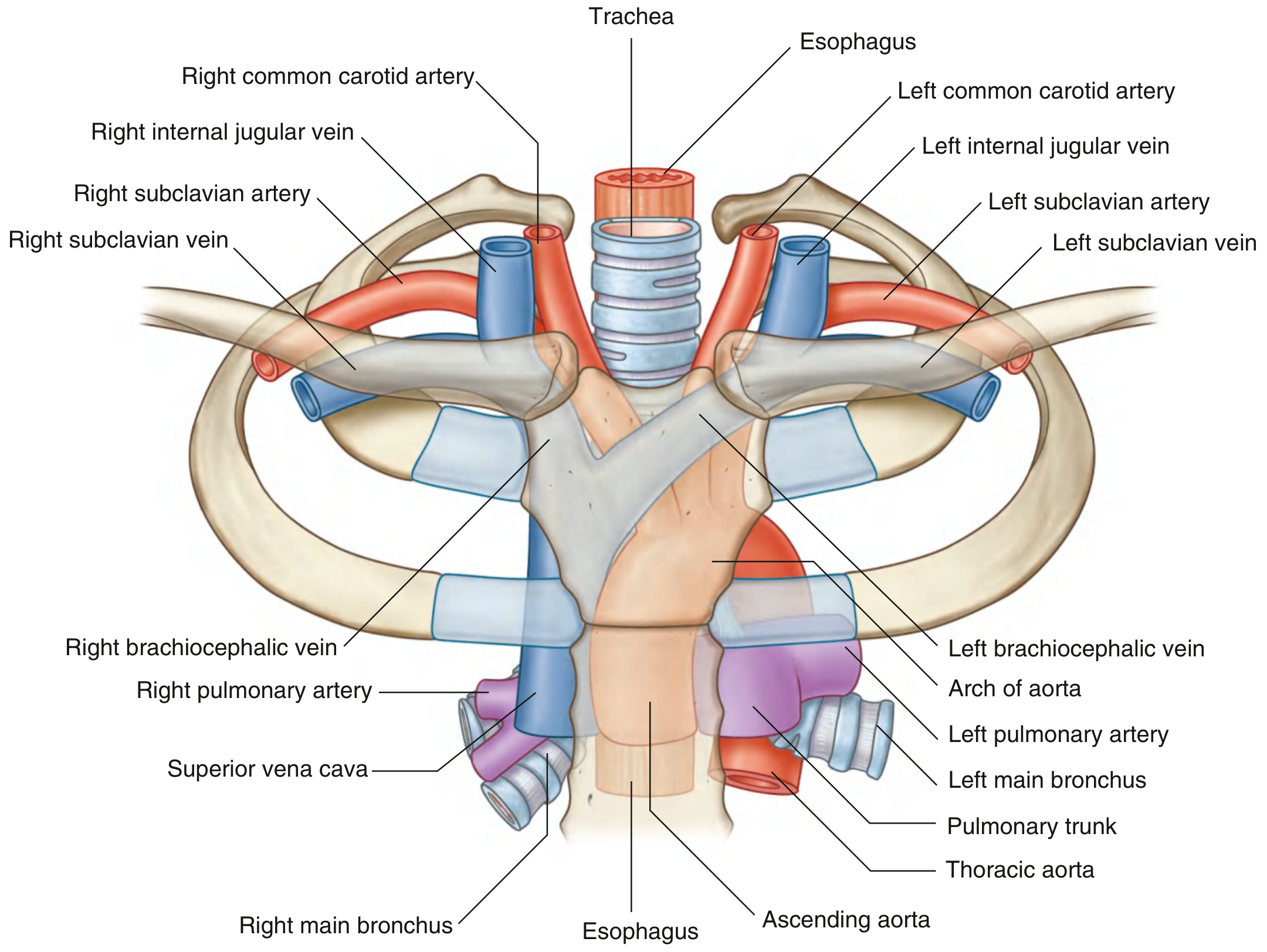
Important relations
- The left brachiocephalic vein crosses obliquely from left to right, anterior to the branches of the aortic arch.
- The arch of aorta passes upward, backward, and to the left over the root of the left lung.
- The left vagus nerve crosses the left side of the arch of aorta.
- The left recurrent laryngeal nerve hooks under the arch of aorta, just posterolateral to the ligamentum arteriosum, then ascends in the tracheo-oesophageal groove.
- The right vagus passes posterior to the root of the right lung, while the left vagus passes posterior to the root of the left lung.
- Both phrenic nerves pass anterior to the root of the lung.
- The trachea lies anterior to the oesophagus.
- The thoracic duct ascends into the superior mediastinum on the left and terminates at the left venous angle.
The anterior-to-posterior arrangement is thymus, brachiocephalic veins/SVC, aortic arch, trachea, oesophagus, and lymphatic trunks. Fischer's Mastery of Surgery, p. 2511.
Applied anatomy
1. Superior mediastinal syndrome
A mass, enlarged lymph nodes, retrosternal goitre, lymphoma, thymoma, or aneurysm may compress structures in this confined space.
| Compressed structure | Clinical feature |
|---|---|
| Superior vena cava | Facial and upper-limb swelling, cyanosis, dilated superficial veins over chest wall, headache |
| Trachea | Dyspnoea, stridor, cough |
| Oesophagus | Dysphagia |
| Recurrent laryngeal nerve | Hoarseness due to ipsilateral vocal-cord paralysis, usually left-sided |
| Phrenic nerve | Raised hemidiaphragm and dyspnoea from diaphragmatic palsy |
| Sympathetic chain, if extension occurs | Horner syndrome |
2. Aortic arch aneurysm
An aneurysm of the arch of aorta may compress:
- Left recurrent laryngeal nerve causing hoarseness, known as Ortner syndrome
- Trachea causing cough, wheeze, or stridor
- Oesophagus causing dysphagia
- Left main bronchus in larger aneurysms
3. Left recurrent laryngeal nerve palsy
The left recurrent laryngeal nerve is especially vulnerable because it loops under the aortic arch. Hoarseness can therefore be a presentation of:
- Aortic arch aneurysm
- Enlarged mediastinal lymph nodes
- Bronchogenic carcinoma
- Left atrial enlargement or other cardiovascular enlargement
4. Retrosternal goitre
An enlarged thyroid may extend through the thoracic inlet into the superior mediastinum and compress the trachea, oesophagus, or great veins. It may present with positional dyspnoea, stridor, dysphagia, or SVC obstruction.
5. Thymic lesions
The thymus lies anteriorly in the superior mediastinum. Thymoma is classically associated with myasthenia gravis. Thymic enlargement or tumour may cause local pressure symptoms.
6. Central venous access
During internal jugular or subclavian venous catheterization, the catheter passes through the brachiocephalic vein into the SVC. Knowledge of the long, oblique course of the left brachiocephalic vein helps explain why left-sided central venous access can be technically more difficult.
7. Mediastinal widening on chest radiograph
Widening of the superior mediastinal shadow may result from:
- Aortic aneurysm or dissection
- Mediastinal haemorrhage after trauma
- Retrosternal goitre
- Lymphadenopathy
- Mediastinal tumour
Because it contains great vessels, airway, and major nerves, trauma or penetrating injury in this region can be rapidly life-threatening, as summarized in the NCBI mediastinum anatomy review.
This is a shared conversation. Sign in to Orris to start your own chat.