
Some Important topics: Leukaemia, anemia, purpura, hodgkin lymphoma, thalassemia, sickle cell anemia, DIC, blood grouping. Everyone please study these topics now tomorrow I have exams on this topics teach ,e from Robbins
Reading File
Now let me search all 8 topics in parallel from Robbins:
Finding Sources
Finding Sources
Finding Sources
Finding Sources
I need to search specifically in Robbins. Let me find the Robbins book first and search for all topics:
Finding Sources
Finding Sources
Finding Sources
Finding Sources
Excellent! Found Robbins books. Now let me search specifically in Robbins for all topics and read the key blocks:
Finding Sources
Now let me read all the key Robbins sections in parallel:
Reading File
Reading File
Reading File
Reading File
Now let me read the remaining key sections - Anemia, Leukemia, Hodgkin Lymphoma:
Reading File
Reading File
Reading File
Now let me read the Hodgkin Lymphoma and AML sections from Robbins, plus blood grouping:
Finding Sources
Reading File
Reading File
Reading File
Now let me read more of the Hodgkin Lymphoma section and then fetch key images:
Reading File
Reading File
Excellent! I have gathered enough content from all major topics. Let me now fetch a couple of the key images (RS cell, sickle cell) and compile the comprehensive notes:
Loading Image
Loading Image
Loading Image
Now let me compile the full exam notes. Here is everything you need from Robbins & Kumar Basic Pathology and Robbins, Cotran & Kumar Pathologic Basis of Disease:
🩸 Hematology Exam Notes - From Robbins Pathology
1. ANEMIA
Definition
Reduction in red cell mass below normal levels, leading to decreased oxygen-carrying capacity.
Classification by Cause
| Mechanism | Examples |
|---|---|
| Blood loss (hemorrhage) | Acute bleed, chronic GI bleed |
| Increased destruction (hemolysis) | Sickle cell, hereditary spherocytosis, malaria |
| Decreased production (marrow failure) | Iron deficiency, thalassemia, aplastic anemia |
Classification by RBC Size
- Microcytic (low MCV): Iron deficiency, thalassemia
- Macrocytic (high MCV): Folate or B12 deficiency; also elevated reticulocyte count
- Normocytic: Hereditary spherocytosis, sickle cell disease (shape changes visible on smear)
Clinical Features
- Acute anemia: Shortness of breath, organ failure, shock
- Chronic anemia: Insidious pallor, fatigue, lassitude
- Extravascular hemolysis: Jaundice + gallstones
- Ineffective erythropoiesis: Iron overload → heart and endocrine failure
- Severe congenital anemia: Growth retardation + bone deformities from marrow hyperplasia
Key Hemolytic Anemias Summary
| Type | Mechanism |
|---|---|
| Hereditary spherocytosis | Autosomal dominant; membrane skeleton mutation; spherocytes removed in spleen |
| Sickle cell anemia | Autosomal recessive; HbS polymer formation → hemolysis + vascular occlusion |
| Thalassemia | Autosomal codominant; reduced α/β globin synthesis → microcytic, hypochromic |
| G6PD deficiency | X-linked; oxidant damage triggers hemolysis |
| Immunohemolytic anemia | Antibodies → opsonization → extravascular hemolysis |
| Malaria | Intracellular parasite; cerebral malaria with falciparum |
Source: Robbins & Kumar Basic Pathology, block4 ~p.433
2. SICKLE CELL ANEMIA
Genetics
- Autosomal recessive - mutation in β-globin gene
- Substitution: Valine replaces glutamic acid at position 6 of β-globin
- Result: HbS instead of HbA; on deoxygenation → HbS polymerizes
Epidemiology
- Most common familial hemolytic anemia
- Prevalent in equatorial Africa, India, Southern Europe, Middle East - areas where falciparum malaria is endemic
- Protective against falciparum malaria (hence high allele frequency)
- US: ~8% of African-descent individuals are HbS carriers; ~1 in 600 have sickle cell anemia
Pathogenesis
- Deoxygenation → HbS polymers form via intermolecular contacts at abnormal valine residue
- Polymers distort RBC → elongated crescentic (sickle) shape
- Initially reversible on reoxygenation, but repeated sickling → calcium influx → loss of K+ and water → membrane damage → irreversibly sickled cells
Three Factors Determining Severity
- Level of other hemoglobins: HbF and HbA inhibit HbS polymerization (heterozygotes rarely sickle; newborns protected until HbF falls ~5-6 months)
- MCHC (mean corpuscular Hb concentration): Higher concentration = more polymerization
- Transit time through microcirculation: Longer transit → more sickling
Peripheral Blood Smear
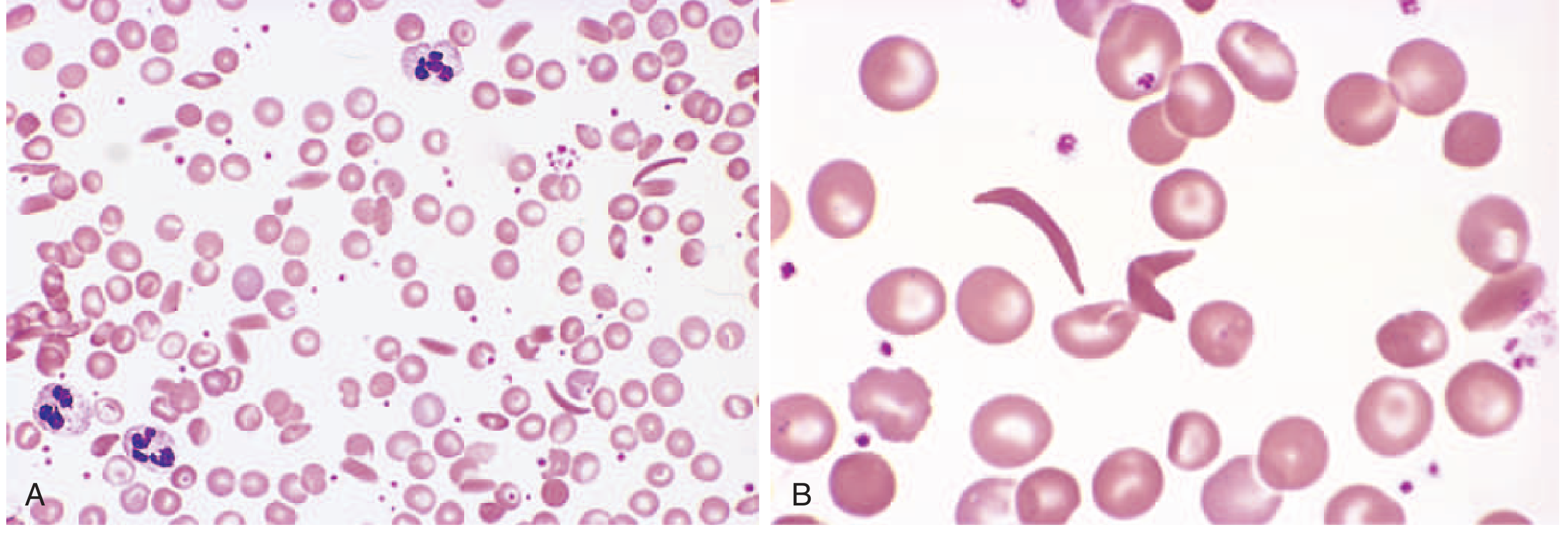
Complications
- Vaso-occlusive crises: Pain crises, tissue infarction, stroke, acute chest syndrome
- Hemolytic anemia with jaundice, pigment gallstones
- Bacterial infections: Functional asplenia (autoinfarction of spleen) → susceptible to encapsulated organisms (Streptococcus pneumoniae, H. influenzae)
- Aplastic crisis: Parvovirus B19 infection
- Splenic sequestration crisis: Acute pooling of blood in spleen
Heterozygotes (Sickle Cell Trait)
- ~40% HbS, 60% HbA - very little in vivo sickling
- Generally asymptomatic but can manifest under extreme hypoxia
Source: Robbins & Kumar Basic Pathology, block4 lines 1695-1764
3. THALASSEMIA
Definition
Inherited disorders with decreased synthesis of α- or β-globin chains, leading to:
- Hemoglobin deficiency
- Intracellular precipitates from excess unpaired normal globin chains → RBC damage + hemolysis
Genetics
- Endemic in Mediterranean, Africa, Asia (malaria belt - similar protective mechanism as HbS)
- β-globin: Single gene on chromosome 11 (point mutations)
- α-globin: Two genes in tandem on chromosome 16 (gene deletions)
β-Thalassemia Classification
| Syndrome | Genotype | Clinical Features |
|---|---|---|
| β-Thal Major (Cooley's anemia) | Homozygous (β⁻/β⁻, β⁺/β⁺) | Severe anemia - requires regular transfusions |
| β-Thal Intermedia | Variable | Moderately severe; transfusions not required |
| β-Thal Minor (trait) | Heterozygous (β⁺/β) | Asymptomatic with mild/absent anemia; red cell abnormalities on smear |
α-Thalassemia Classification
| Syndrome | Gene Deletions | Clinical Features |
|---|---|---|
| Silent carrier | −/α, α/α (1 gene) | Asymptomatic, normal smear |
| α-Thal trait | −/−, α/α or −/α, −/α (2 genes) | Mild anemia, resembles β-thal minor |
| HbH disease | −/−, −/α (3 genes) | Moderately severe; HbH (β4 tetramers) form |
| Hydrops fetalis | −/−, −/− (4 genes, deletion) | Lethal in utero - HbBart's (γ4) - fatal |
Molecular Mutations
- β-thalassemia: Mainly point mutations affecting transcription, splicing, or translation of β-globin mRNA
- α-thalassemia: Mainly gene deletions
Pathophysiology of β-Thal Major
- Deficient β-chain synthesis → excess α chains precipitate in RBC precursors → ineffective erythropoiesis in marrow
- Those cells that leave marrow are destroyed in spleen → hemolysis
- Erythroid marrow hyperplasia → "crew-cut" skull X-ray, "chipmunk facies," bone thinning
- Chronic hemolysis → hepatosplenomegaly (extramedullary hematopoiesis)
- Repeated transfusions → iron overload → hemosiderosis affecting heart, liver, endocrine organs
Source: Robbins & Kumar Basic Pathology, block4 lines 1765-1850
4. LEUKAEMIA
Overview
Leukemias are neoplasms of hematopoietic cells that primarily involve the bone marrow and peripheral blood, as opposed to lymphomas which present as solid masses.
Acute Lymphoblastic Leukemia/Lymphoma (ALL)
- Neoplasms of immature B or T cells (lymphoblasts)
- 85% are B-ALL (childhood acute leukemia)
- T-ALL: Adolescent males, often as thymic mass
- Most common cancer in children (~2500 new cases/year US, peak age ~3 years for B-ALL)
Pathogenesis:
- Chromosomal aberrations in ~90%: most common is hyperploidy (>50 chromosomes)
- Key mutations: NOTCH1 (T-ALL), PAX5, TCF3, ETV6, RUNX1, BCR::ABL1 (B-ALL)
- Philadelphia chromosome [t(9;22)] → BCR::ABL1 fusion → most important adverse prognostic factor
- Mutations disturb "master" regulatory transcription factors → maturation arrest + self-renewal
Clinical Features:
- Fatigue, fever, bleeding (marrow failure → anemia, thrombocytopenia, neutropenia)
- Bone pain (marrow expansion)
- Lymphadenopathy, hepatosplenomegaly
- CNS involvement (headache, nerve palsies)
Immunophenotype markers: TdT positive (terminal deoxynucleotidyl transferase - key marker), CD10 (CALLA), CD19 (B-ALL), CD3 (T-ALL)
Chronic Lymphocytic Leukemia / Small Lymphocytic Lymphoma (CLL/SLL)
- Most common leukemia of adults in Western world
- CLL = >5000 lymphocytes/µL in blood; SLL = predominantly lymph node involvement (4% of NHLs)
- Indolent - increased tumor cell survival (not proliferation) is key
Pathogenesis:
- High levels of BCL2 (anti-apoptotic) - loss of chromosome 13q miRNA regulation
- BTK (Bruton tyrosine kinase) signaling via B-cell receptor is critical
- Immune dysregulation: Hypogammaglobulinemia; paradoxically 15% develop warm autoantibodies
Morphology:
- Small, resting lymphocytes with dark round nuclei, scant cytoplasm
- Proliferation centers = foci of larger dividing cells → pathognomonic for CLL/SLL
- Immunophenotype: CD5+ B cell (key - co-expression of CD5 and CD23 is characteristic)
Acute Myeloid Leukemia (AML)
- Tumor of hematopoietic progenitors with mutations that impede differentiation → immature myeloid blasts accumulate
- Incidence rises with age, peaks after 60 years
- Results in marrow failure → anemia, thrombocytopenia, neutropenia
Key Genetic Subtypes:
| Translocation | Fusion Gene | Prognosis | Feature |
|---|---|---|---|
| t(8;21) | RUNX1::RUNX1T1 | Favorable | Auer rods easily found |
| inv(16) | CBFB::MYH11 | Favorable | Abnormal eosinophilic precursors |
| t(15;17) | PML::RARA | Very favorable | Acute promyelocytic leukemia (APL); Auer rods in bundles; high DIC risk; responds to ATRA |
| t(11q23) | KMT2A rearrangement | Poor | Monocytic differentiation |
Auer rods = crystallized primary granules, pathognomonic for AML
Diagnosis: ≥20% blasts in bone marrow (WHO criteria)
Source: Robbins, Cotran & Kumar PBD, block6 lines 3028-3100 and block7 lines 266-292
5. HODGKIN LYMPHOMA
What Sets HL Apart from NHL
- Distinctive neoplastic Reed-Sternberg (RS) giant cells
- Robust but ineffective host immune response - tumor cells are only a small fraction of the mass
- Arises in a single node or chain and spreads in stepwise contiguous fashion
- Cell of origin: Germinal center B cells (proven by immunoglobulin gene rearrangement studies)
Reed-Sternberg Cell - The Hallmark
- Very large cell: 15-45 µm in diameter
- Enormous multilobate nucleus
- Exceptionally prominent "owl-eye" nucleoli (large acidophilic nucleoli surrounded by clear halo in mirror-image lobes)
- Immunophenotype: CD15⁺, CD30⁺, CD45⁻, B/T-cell marker negative
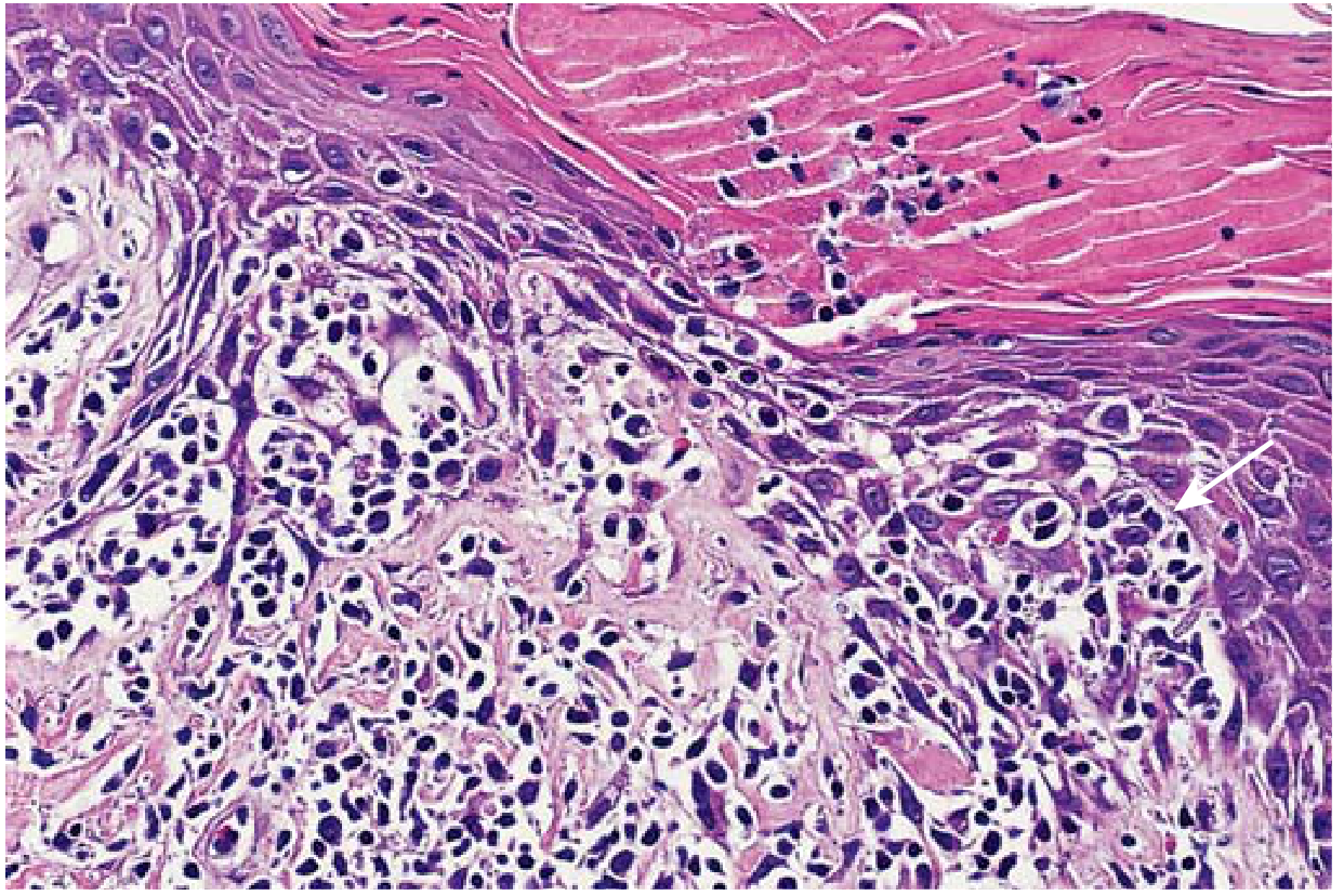
Five Subtypes
| Subtype | Key Features | Epidemiology | Prognosis |
|---|---|---|---|
| Nodular Sclerosis (most common ~70%) | Lacunar cells; collagen bands dividing into nodules | Adolescents/young adults; equal M=F; lower cervical/supraclavicular/mediastinal | Excellent |
| Mixed Cellularity | Classic RS cells; eosinophils, plasma cells, lymphocytes | Older adults, HIV patients; EBV in ~70% | Good |
| Lymphocyte Rich | Abundant lymphocytes, rare RS cells | Good | |
| Lymphocyte Depletion | Many RS cells, few lymphocytes | Elderly, HIV | Worst |
| Nodular Lymphocyte Predominant (NLPHL) | L&H ("popcorn") cells; CD20⁺, CD15⁻, CD30⁻ - different from classic RS | Excellent |
Pathogenesis Highlights
- EBV found in RS cells in ~70% of mixed-cellularity subtype
- RS cells secrete IL-5 (attracts eosinophils), TGF-β (fibrosis), IL-13 (autocrine RS growth)
- Immune escape: RS cells lose MHC class I (loss of β2-microglobulin); express PD-L1 and PD-L2 (checkpoint inhibition) → anti-PD-1 antibodies are highly effective even in refractory disease
Staging (Ann Arbor)
- Stage I: Single node region
- Stage II: Two or more node regions, same side of diaphragm
- Stage III: Both sides of diaphragm
- Stage IV: Disseminated (liver, bone marrow, lung)
- "B" symptoms: Fever, night sweats, weight loss >10% → worse prognosis
Source: Robbins & Kumar Basic Pathology, block4 lines 2728-2850
6. PURPURA
Definition
Small hemorrhages into skin, mucous membranes, or serosal surfaces due to platelet or vascular disorders. Types:
- Petechiae: Pinpoint (<3mm)
- Purpura: 3mm-1cm
- Ecchymoses: >1cm (bruises)
Immune Thrombocytopenic Purpura (ITP)
Two subtypes:
- Chronic ITP - most common; women 20-40 years
- Acute ITP - mostly children after viral infection; self-limited
Pathogenesis (Chronic ITP):
- Antibodies (IgG) against platelet membrane glycoproteins IIb/IIIa or Ib/IX complexes in ~80% of cases
- Spleen = major site of antiplatelet antibody production + site of IgG-coated platelet destruction
- Bone marrow shows increased megakaryocytes (reactive increase)
Clinical Features:
- Insidious onset
- Petechiae, easy bruising, epistaxis, gum bleeding
- Serious intracranial hemorrhage is uncommon
Treatment:
- Immunosuppressive agents
- Splenectomy normalizes platelet count and induces complete remission in >2/3 patients
Thrombotic Thrombocytopenic Purpura (TTP)
- Pentad: Fever + thrombocytopenia + microangiopathic hemolytic anemia + transient neurologic deficits + renal failure
- Widespread platelet-rich thrombi in microcirculation
- Due to deficiency of ADAMTS13 (metalloprotease that cleaves large vWF multimers)
- Very large vWF multimers → platelet aggregation
Heparin-Induced Thrombocytopenia (HIT)
- Develops in 3-5% of patients after 1-2 weeks of unfractionated heparin
- IgG antibodies bind platelet factor 4 in a heparin-dependent manner
- Platelet activation → thrombosis (paradox - both thrombocytopenia AND thrombosis)
- Treatment: Stop heparin; switch to direct thrombin inhibitors
Source: Robbins & Kumar Basic Pathology, block4 lines 3089-3115
7. DISSEMINATED INTRAVASCULAR COAGULATION (DIC)
Definition
Systemic activation of coagulation → microthrombi throughout microcirculation → consumption of platelets and clotting factors + secondary fibrinolysis. Dual problem:
- Microthrombi → tissue hypoxia, microinfarcts
- Bleeding → consumptive coagulopathy (factors and platelets depleted)
DIC Pathophysiology Diagram
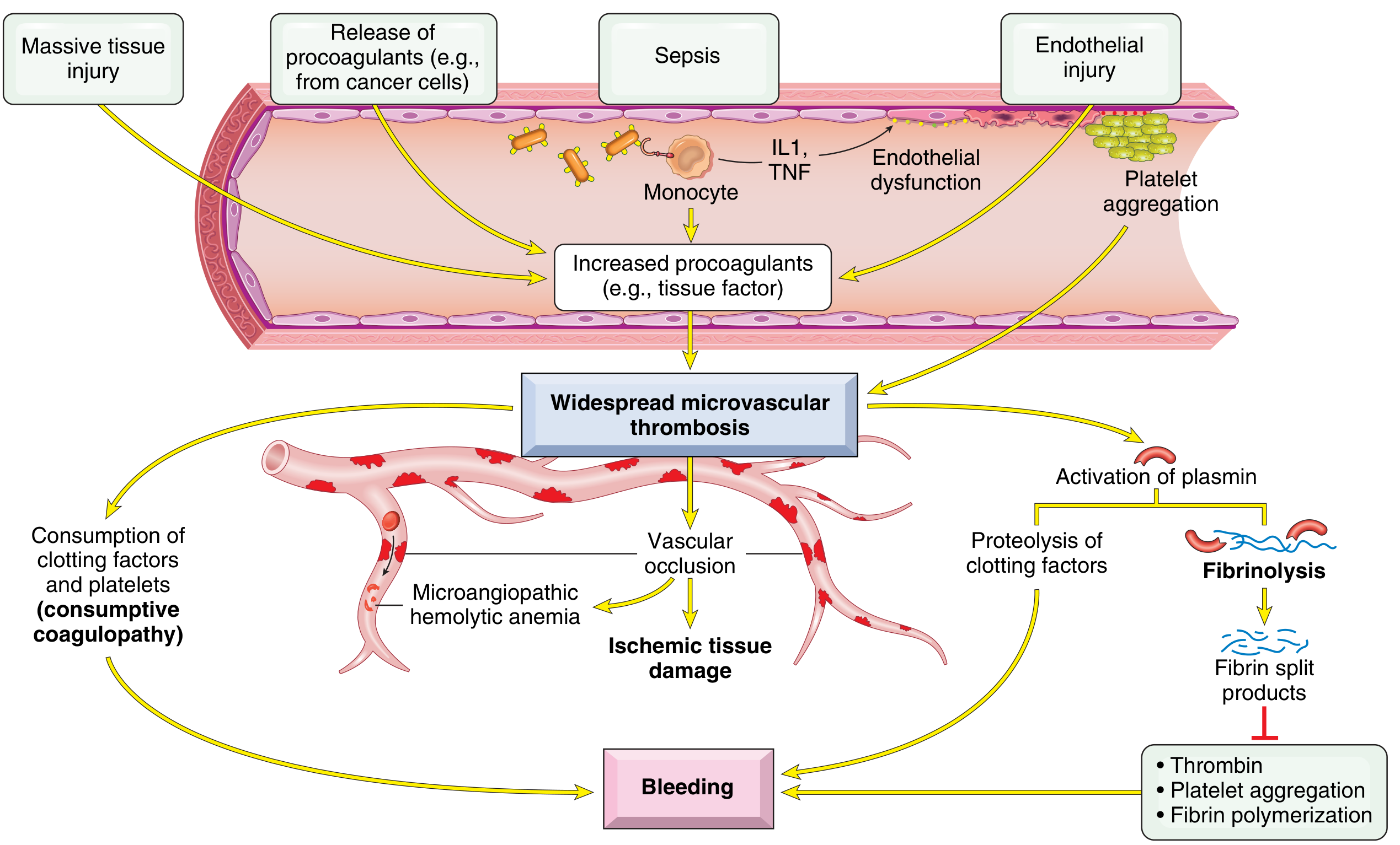
Triggers (Two Main Mechanisms)
-
Release of tissue factor/procoagulants into circulation:
- Obstetric complications (placental release)
- Tumors (especially acute promyelocytic leukemia (APL) and adenocarcinoma)
- Bacterial sepsis: endotoxins stimulate tissue factor expression on monocytes + cytokines (IL-1, TNF) stimulate tissue factor on endothelial cells + decrease thrombomodulin (→ less protein C activation)
-
Widespread endothelial cell injury:
- Antigen-antibody complexes (SLE)
- Temperature extremes (heat stroke, burns)
- Infections (meningococci, rickettsiae)
- Systemic inflammatory response syndrome (SIRS)
Common Clinical Causes
| Category | Examples |
|---|---|
| Obstetric | Abruptio placentae, amniotic fluid embolism, eclampsia, retained dead fetus |
| Infections | Gram-negative sepsis, meningococcemia, Rocky Mountain spotted fever |
| Neoplasms | APL (M3 AML), mucin-secreting adenocarcinomas |
| Massive tissue injury | Trauma, burns, surgery |
| Miscellaneous | Snake venom, transfusion reactions, SLE |
Lab Findings in DIC
| Test | Result |
|---|---|
| Platelet count | ↓ (consumed) |
| PT / aPTT | ↑ (factors consumed) |
| Fibrinogen | ↓ (consumed) |
| D-dimers / FDPs | ↑ (fibrinolysis products) |
| Blood smear | Schistocytes (microangiopathic hemolytic anemia) |
Clinical Presentation
- Bleeding from multiple sites (venipuncture sites, skin, mucous membranes, GI, urinary)
- Thrombotic manifestations: renal failure, stroke, adrenal infarction
- Both simultaneously in many patients
Source: Robbins & Kumar Basic Pathology, block4 lines 3023-3088
8. BLOOD GROUPING (ABO and Rh System)
ABO Blood Group System
| Blood Group | Antigen on RBC | Antibody in Serum | Can Donate To | Can Receive From |
|---|---|---|---|---|
| A | A antigen | Anti-B | A, AB | A, O |
| B | B antigen | Anti-A | B, AB | B, O |
| AB | A and B antigens | None | AB only | Universal recipient |
| O | None | Anti-A and Anti-B | Universal donor | O only |
Key Principles
- ABO antibodies (anti-A, anti-B) are naturally occurring IgM antibodies - no prior sensitization needed
- Formed early in life against environmental antigens that cross-react with A/B antigens
- IgM antibodies fix complement → acute intravascular hemolytic transfusion reaction if mismatched
- ABO system is the most important blood group in transfusion medicine
Rh Blood Group System
- Based on the RhD antigen (most immunogenic)
- Rh positive (Rh⁺): RhD antigen present on RBC surface
- Rh negative (Rh⁻): RhD antigen absent
- ~85% of population is Rh positive
Rh antibodies:
- NOT naturally occurring - require prior sensitization (transfusion or pregnancy)
- IgG antibodies (unlike ABO's IgM) → can cross the placenta
Hemolytic Disease of the Newborn (HDN / Erythroblastosis Fetalis)
- Rh⁻ mother × Rh⁺ father → Rh⁺ baby
- First pregnancy: exposure of Rh⁻ mother to fetal Rh⁺ RBCs at delivery → she forms anti-RhD IgG (sensitization)
- Second Rh⁺ pregnancy: Anti-RhD IgG crosses placenta → destroys fetal RBCs → hemolytic anemia, hydrops fetalis, kernicterus (bilirubin deposition in brain)
- Prevention: Administer Rh immune globulin (RhoGAM) to Rh⁻ mother at 28 weeks and within 72 hours of delivery → passively neutralizes fetal RBCs before maternal sensitization
ABO HDN
- Can occur in first pregnancy (naturally occurring IgG anti-A or anti-B in group O mothers)
- Usually milder than Rh HDN
Blood Type Compatibility in Transfusion
- Always test ABO + Rh before transfusion
- Cross-match: Mix patient serum + donor RBCs to detect unexpected antibodies
- Type and Screen: ABO/Rh typing + antibody screen for less urgent situations
QUICK COMPARISON TABLE - Leukemia Types
| Type | Cell of Origin | Age | Key Markers | Key Mutation | Notes |
|---|---|---|---|---|---|
| B-ALL | Pre-B lymphoblast | Children (~3 yrs) | TdT⁺, CD10⁺, CD19⁺ | BCR::ABL1 (t9;22) | Most common childhood cancer |
| T-ALL | Pre-T lymphoblast | Adolescent males | TdT⁺, CD3⁺ | NOTCH1 | Thymic mass |
| CLL/SLL | Mature B cell | Adults >50 yrs | CD5⁺, CD23⁺, CD20 dim | del 13q, BTK | Most common adult leukemia (West) |
| AML | Myeloid progenitor | Adults >60 yrs | MPO⁺, Auer rods | t(15;17) APL most notable | Auer rods pathognomonic |
| CML | Myeloid stem cell | Adults | BCR::ABL1⁺ | t(9;22) Philadelphia chromosome | Responds to imatinib (Gleevec) |
KEY HIGH-YIELD MNEMONICS
DIC Causes - "STOP Making New Thrombi":
S - Sepsis, Snake bite
T - Trauma, Transfusion reaction
O - Obstetric (abruptio, amniotic fluid embolism)
P - Promyelocytic leukemia (APL/M3)
M - Malignancy
N - Necrosis (burns, surgery)
T - Temperature extremes
Hodgkin vs NHL:
- Hodgkin: RS cells, CD15⁺/CD30⁺, contiguous spread, better prognosis
- NHL: No RS cells, non-contiguous spread, diverse group
Sickle Cell vs Thalassemia:
- Sickle cell: Structural defect (wrong protein)
- Thalassemia: Quantitative defect (less protein)
ITP vs TTP:
- ITP: Immune destruction, splenomegaly absent, treat with steroids/splenectomy
- TTP: Microangiopathic, neurological symptoms, ADAMTS13 deficiency, treat with plasma exchange
ROBBINS MUST-KNOW MORPHOLOGY IMAGES
Sickle cell anemia - peripheral blood shows elongated crescentic/sickle-shaped cells (see image above)
Hodgkin lymphoma - "owl-eye" RS cells with prominent nucleoli in inflammatory background
AML - blasts with Auer rods (needle-like cytoplasmic inclusions)
CLL - sheets of small mature-looking lymphocytes with "smudge cells" on peripheral smear
β-Thal major - severe microcytic hypochromic anemia; nucleated RBCs; target cells
All content sourced directly from:
- Robbins & Kumar Basic Pathology (ISBN 9780323790185)
- Robbins, Cotran & Kumar Pathologic Basis of Disease (ISBN 9780443264528)All 8 topics are covered above. Here is a summary of what was taught and what to focus on tonight before your exam:
Your Exam Prep Summary - All 8 Topics from Robbins
What to focus on (high-yield for each topic):
1. Anemia - Classify by cause (blood loss / hemolysis / decreased production) and by MCV (micro/macro/normo). Know the clinical features of acute vs chronic anemia.
2. Sickle Cell Anemia - Autosomal recessive, Val→Glu substitution at β-globin position 6, HbS polymerizes when deoxygenated. Three modifying factors (HbF, HbA, MCHC). Complications = vaso-occlusion + hemolysis + functional asplenia. Protective against falciparum malaria.
3. Thalassemia - Quantitative defect in globin synthesis. β-thal = point mutations (chr 11); α-thal = deletions (chr 16). Know the 4-tier α-thalassemia spectrum. β-thal major = severe anemia + bone deformities + iron overload from transfusions.
4. Leukaemia - Know ALL (childhood, TdT+, BCR::ABL1 worst prognostic factor), CLL (CD5+ B cell, most common adult leukemia in West, BCL2 overexpression), AML (Auer rods, t(15;17) = APL = very favorable + ATRA sensitive + DIC risk).
5. Hodgkin Lymphoma - Reed-Sternberg cells (owl-eye nucleoli, CD15+/CD30+/CD45-). 5 subtypes: Nodular sclerosis is most common. EBV in ~70% of mixed cellularity. PD-L1 overexpression. Stepwise contiguous spread.
6. Purpura - ITP (IgG anti-platelet antibodies, splenectomy works in >2/3). TTP (pentad: FTMRN - Fever, Thrombocytopenia, Microangiopathic anemia, Renal failure, Neurological; ADAMTS13 deficiency). HIT (anti-PF4 antibody, causes thrombosis paradoxically).
7. DIC - Triggers: tissue factor release (sepsis, APL, obstetric) or endothelial injury. Dual picture: clotting AND bleeding. Labs: ↓platelets, ↓fibrinogen, ↑PT/aPTT, ↑D-dimers, schistocytes on smear.
8. Blood Grouping - ABO: O = universal donor (anti-A + anti-B), AB = universal recipient. ABO antibodies = IgM (naturally occurring). Rh: IgG, not naturally occurring, crosses placenta → HDN. Prevent with RhoGAM.
Good luck on your exam tomorrow! 🩸
Some Important topics: Leukaemia, anemia, purpura, hodgkin lymphoma, thalassemia, sickle cell anemia, DIC, blood grouping. Everyone please study these topics now tomorrow I have exams on this topics teach ,e from Robbins
Reading File
Let me search the Robbins books for all 8 topics simultaneously:
Finding Sources
Finding Sources
Finding Sources
Finding Sources
All section locations confirmed. Now reading all key blocks in parallel:
Reading File
Reading File
Reading File
Reading File
Now reading the remaining sections - sickle cell, purpura, leukemia overview, AML, and blood grouping:
Reading File
Reading File
Reading File
Reading File
Now reading the Hodgkin morphology details, AML, CLL, and DIC clinical/lab sections:
Reading File
Reading File
Reading File
Reading File
Now fetching the key Robbins images - RS cell, sickle cell smear, and DIC diagram:
Loading Image
Loading Image
Loading Image
Here are your complete, exam-ready notes directly from Robbins. Study these carefully tonight!
🩸 HEMATOLOGY EXAM MASTER NOTES
Sourced directly from Robbins & Kumar Basic Pathology + Robbins, Cotran & Kumar Pathologic Basis of Disease
1. ANEMIA
Definition & Classification
Anemia = reduced red cell mass → decreased oxygen-carrying capacity.
Three root causes:
- Blood loss (hemorrhage - acute or chronic)
- Increased destruction (hemolysis - intravascular or extravascular)
- Decreased production (marrow failure - aplastic, megaloblastic, iron deficiency)
Classification by RBC Size (MCV)
| Type | MCV | Common Causes |
|---|---|---|
| Microcytic | Low | Iron deficiency, Thalassemia |
| Macrocytic | High | Folate deficiency, B12 deficiency, high reticulocyte count |
| Normocytic | Normal | Hereditary spherocytosis, sickle cell (shape changes on smear) |
Clinical Features
| Scenario | Presentation |
|---|---|
| Acute anemia (large bleed) | Shortness of breath, organ failure, shock |
| Chronic anemia | Insidious pallor, fatigue, lassitude |
| Extravascular hemolysis | Jaundice, gallstones (pigment stones) |
| Ineffective erythropoiesis | Iron overload → heart and endocrine failure |
| Severe congenital anemias | Growth retardation, bone deformities (marrow hyperplasia) |
Hemolysis - Key Distinction
Extravascular hemolysis (phagocytosis in spleen):
- Hyperbilirubinemia and jaundice
- Splenomegaly
- Pigment gallstones (if chronic)
- Iron is recycled - NO iron deficiency
Intravascular hemolysis (RBC bursts in vessel):
- Hemoglobinemia, hemoglobinuria, hemosiderinuria
- Iron loss → can cause iron deficiency
- Both types: ↓ serum haptoglobin (binds free Hb, then removed)
Summary of Key Hemolytic Anemias (Robbins)
| Disorder | Inheritance | Mechanism |
|---|---|---|
| Hereditary spherocytosis | AD | Membrane skeleton defect → spherocytes → splenic destruction |
| Sickle cell anemia | AR | β-globin mutation → HbS polymerizes when deoxygenated |
| Thalassemia | Codominant | Reduced α/β globin synthesis → microcytic, hypochromic |
| G6PD deficiency | X-linked | Oxidant damage triggers hemolysis |
| Immunohemolytic anemia | Acquired | Antibodies → opsonization → extravascular hemolysis |
| Malaria | Acquired | Intracellular parasite (cerebral malaria = falciparum) |
Robbins & Kumar Basic Pathology, p. 384-385
2. SICKLE CELL ANEMIA
The Basics
- Most common familial hemolytic anemia
- Autosomal recessive (AR)
- Gene: β-globin mutation on chromosome 11
- Mutation: Valine substitutes for Glutamic acid at position 6 of β-globin
- Result: HbS (sickle hemoglobin) instead of HbA
Why It's Prevalent (Epidemiology)
- Protective against Plasmodium falciparum malaria - hence high frequency in:
- Equatorial Africa, India, Southern Europe, Middle East
- USA: ~8% of African-Americans are HbS carriers; ~1 in 600 have sickle cell disease
Pathogenesis - Step by Step
- Deoxygenation → HbS molecules undergo conformational change
- HbS polymerizes via intermolecular contacts at the abnormal valine residue
- Polymers distort RBC into elongated crescentic (sickle) shape
- Initially reversible on reoxygenation
- With repeated sickling: Ca²⁺ influx → K⁺ and water loss → membrane damage → irreversibly sickled cells → prone to hemolysis
Peripheral Blood Smear (Robbins Fig. 10.3)
Robbins Fig. 10.3 - (A) Sickle cell anemia: peripheral blood at low power showing sickled cells, target cells, nucleated RBCs; (B) High power showing classic crescentic sickle cells
Three Factors Determining Sickling Severity
| Factor | Effect |
|---|---|
| 1. Levels of other Hb (HbF, HbA) | HbF and HbA inhibit HbS polymerization; newborns protected until HbF falls (~5-6 months); heterozygotes (40% HbS, 60% HbA) rarely sickle = "sickle cell trait" |
| 2. MCHC (mean corpuscular Hb concentration) | Higher MCHC → more polymerization |
| 3. Transit time through microcirculation | Longer time in low O₂ → more sickling |
Clinical Manifestations
| Problem | Mechanism | Example |
|---|---|---|
| Hemolytic anemia | RBC destruction | Jaundice, pigment gallstones |
| Vaso-occlusive crises | Sickled cells block vessels | Painful crises, stroke, acute chest syndrome, organ infarcts |
| Bacterial infections | Functional asplenia (autoinfarction) | Especially encapsulated organisms: S. pneumoniae, H. influenzae |
| Aplastic crisis | Parvovirus B19 infects erythroid precursors | Sudden drop in Hb |
| Splenic sequestration | Acute pooling of blood in spleen | Life-threatening in children |
Sickle Cell Trait vs. Sickle Cell Disease
- Trait (HbAS): ~40% HbS, ~60% HbA → very little in vivo sickling → asymptomatic (protective against malaria)
- Disease (HbSS): 100% HbS → symptomatic
Robbins & Kumar Basic Pathology, p. 384-392
3. THALASSEMIA
Definition
Inherited disorders of decreased globin chain synthesis (quantitative defect) → Hb deficiency + excess unpaired chains form intracellular precipitates → red cell damage + hemolysis.
Key Points
- Common in Mediterranean, Africa, Asia (malaria endemic regions - same protective logic as HbS)
- β-globin: Single gene on chromosome 11 → mainly point mutations (splicing, transcription, translation defects)
- α-globin: Two genes in tandem on chromosome 16 → mainly gene deletions
β-Thalassemia Classification (Robbins Table 10.3)
| Syndrome | Genotype | Clinical Features |
|---|---|---|
| β-Thal Major (Cooley anemia) | Homozygous (β⁻/β⁻ or β⁺/β⁺) | Severe anemia from 6 months of age; requires regular transfusions |
| β-Thal Intermedia | Variable | Moderately severe; transfusions NOT required |
| β-Thal Minor (trait) | Heterozygous (β⁺/β or β⁻/β) | Asymptomatic or mild; red cell abnormalities on smear |
β⁰ = no β-globin produced; β⁺ = reduced β-globin produced
α-Thalassemia Classification
| Syndrome | Genes Deleted | Clinical Features |
|---|---|---|
| Silent carrier | 1 gene (−/α, α/α) | Asymptomatic; normal smear |
| α-Thal trait | 2 genes (−/−,α/α or −/α,−/α) | Asymptomatic; resembles β-thal minor |
| HbH disease | 3 genes (−/−,−/α) | Moderately severe; HbH = β₄ tetramers form |
| Hydrops fetalis | 4 genes (−/−,−/−) | Lethal in utero; HbBart = γ₄ tetramers |
Pathophysiology of β-Thalassemia Major
Deficient β-chains
↓
Excess α-chains precipitate in RBC precursors
↓
Ineffective erythropoiesis (marrow destroys its own cells)
↓
Severe anemia → EPO ↑↑ → Erythroid marrow hyperplasia
↓
Skeletal changes: "crew-cut" skull X-ray, chipmunk facies, bone thinning
Extramedullary hematopoiesis → hepatosplenomegaly
↓
Repeated transfusions → Iron overload (hemosiderosis)
↓
Heart failure, liver cirrhosis, endocrine failure (diabetes, hypogonadism)
Robbins & Kumar Basic Pathology, p. 392-400
4. LEUKAEMIA
Overview
Leukemias = neoplasms of hematopoietic cells primarily in bone marrow and peripheral blood (vs. lymphomas = solid masses).
All produce marrow failure → anemia (↓ RBC) + thrombocytopenia (↓ platelets) + neutropenia (↓ neutrophils)
A. Acute Lymphoblastic Leukemia / Lymphoma (ALL)
Key Facts
- Neoplasm of immature B or T cells (lymphoblasts)
- 85% are B-ALL (childhood leukemia)
- T-ALL: Adolescent males, thymic mass
- Most common cancer of children; peak B-ALL at age ~3 years
Pathogenesis
- 90% have chromosomal changes; most common = hyperploidy (>50 chromosomes)
- Key mutations:
- T-ALL: NOTCH1 mutations
- B-ALL: PAX5, TCF3, ETV6, RUNX1, BCR::ABL1, KMT2A, PBX1
- Philadelphia chromosome [t(9;22)] → BCR::ABL1 = most important adverse prognostic factor in ALL
Immunophenotype
| Marker | B-ALL | T-ALL |
|---|---|---|
| TdT | ✅ | ✅ |
| CD10 (CALLA) | ✅ | ❌ |
| CD19 | ✅ | ❌ |
| CD3 | ❌ | ✅ |
Clinical Features
- Bone pain, fatigue, fever, bleeding
- Lymphadenopathy, hepatosplenomegaly
- CNS involvement (headache, cranial nerve palsies)
Prognosis
- Children: 85% cure rate with chemotherapy
- Adults/Philadelphia chromosome positive: poor without targeted therapy
B. Chronic Lymphocytic Leukemia / Small Lymphocytic Lymphoma (CLL/SLL)
Key Facts
- Most common leukemia of adults in the Western world
- CLL = peripheral blood lymphocytes >5000/µL; SLL = mostly lymph node involvement
- Less common in Asia
- Indolent - survival measured in years
Pathogenesis
- Increased tumor cell survival (not proliferation) is key
- High BCL2 levels (anti-apoptotic) - from loss of chr 13q miRNAs that suppress BCL2
- BTK (Bruton tyrosine kinase) signaling via B-cell receptor promotes survival
- Immune dysregulation: Hypogammaglobulinemia; paradoxically 15% develop warm autoantibodies
Morphology
- Sheets of small, resting lymphocytes, dark round nuclei, scanty cytoplasm
- Proliferation centers (foci of larger dividing cells) = pathognomonic for CLL/SLL
- Peripheral blood: smudge cells (fragile lymphocytes crushed during smear preparation)
- Immunophenotype: CD5⁺ CD23⁺ B cell (co-expression of CD5 is the key)
C. Acute Myeloid Leukemia (AML)
Key Facts
- Tumor of hematopoietic progenitors; differentiation is blocked → immature myeloid blasts accumulate
- Incidence rises with age, peaks after age 60
- Diagnosis: ≥20% blasts in bone marrow
Key Genetic Subtypes (WHO Classification - Robbins Table 13.10)
| Translocation | Fusion Gene | Prognosis | Notes |
|---|---|---|---|
| t(8;21) | RUNX1::RUNX1T1 | Favorable | Auer rods easily found |
| inv(16) | CBFB::MYH11 | Favorable | Abnormal eosinophilic precursors |
| t(15;17) | PML::RARA | Very favorable | Acute Promyelocytic Leukemia (APL/M3); Auer rods in bundles (faggot cells); high DIC risk; responds to ATRA |
| t(11q23) | KMT2A | Poor | Monocytic differentiation |
| NPM1 mutation | - | Favorable |
Auer rods = needle-like cytoplasmic inclusions of crystallized primary granules = pathognomonic for AML
Robbins, Cotran & Kumar PBD, p. 553-570
5. HODGKIN LYMPHOMA
What Makes HL Unique (Robbins)
- Distinctive neoplastic Reed-Sternberg (RS) cells
- RS cells make up only a small fraction of tumor mass; the rest is reactive inflammatory cells
- Arises in single lymph node/chain → spreads in stepwise contiguous fashion
- Cell of origin: Germinal center B cells (proven by identical Ig gene rearrangements in all RS cells)
The Reed-Sternberg Cell (Robbins Fig. 10.23)
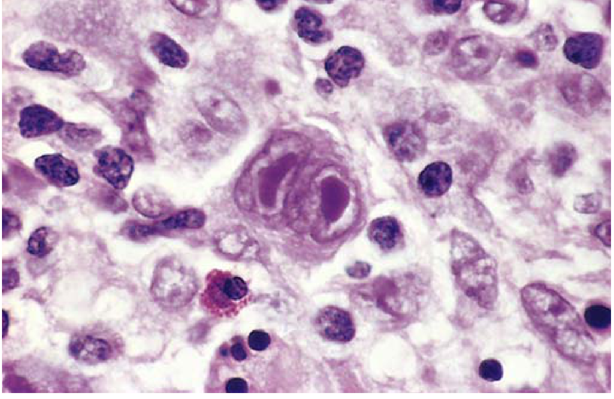
Robbins Fig. 10.23 - Reed-Sternberg cell: large cell (~15-45 µm) with bilobed "owl-eye" nucleus showing prominent eosinophilic nucleoli surrounded by reactive lymphocytes, eosinophils, and macrophages
RS Cell Features (MEMORIZE)
- Size: 15-45 µm (giant)
- Nucleus: Binucleate or bilobed (mirror-image)
- Nucleoli: Large, inclusion-like, acidophilic = "owl-eye" appearance
- Immunophenotype: CD15⁺ CD30⁺ CD45⁻ (negative for B and T cell markers in classic HL)
Five Subtypes of HL
| Subtype | Frequency | Key Features | Age/Sex | Prognosis |
|---|---|---|---|---|
| Nodular Sclerosis | ~70% (most common) | Lacunar cells (RS variant) + collagen bands dividing tissue into nodules | Adolescents/Young adults; M=F; Mediastinal/cervical nodes | Excellent |
| Mixed Cellularity | ~25% | Classic RS cells amid eosinophils, plasma cells, macrophages | >50 yrs; male predominance; EBV in ~70% | Good |
| Lymphocyte Rich | Uncommon | Many lymphocytes, rare RS cells | Good | |
| Lymphocyte Depleted | Rare | Many RS cells, few lymphocytes | Elderly, HIV | Worst |
| Nodular Lymphocyte Predominant (NLPHL) | ~5% | "L&H" (popcorn) cells; CD20⁺, CD15⁻, CD30⁻ | Different biology from classic | Excellent |
Pathogenesis Highlights
- EBV in RS cells in 70% of mixed cellularity subtype (identical integration site in all RS cells)
- RS cells secrete: IL-5 (attracts eosinophils), TGF-β (causes fibrosis), IL-13 (autocrine growth)
- Immune evasion: Loss of MHC class I (β₂-microglobulin loss); overexpression of PD-L1/PD-L2 on chromosome 9 → Anti-PD-1 antibodies are highly effective even in refractory disease
Staging - Ann Arbor
- Stage I: Single node region
- Stage II: Two+ regions, same side of diaphragm
- Stage III: Both sides of diaphragm
- Stage IV: Disseminated (liver, bone marrow, lungs)
- "B" symptoms (fever, night sweats, weight loss >10%) = worse prognosis
Robbins & Kumar Basic Pathology, p. 415-420
6. PURPURA
Definition
Small hemorrhages into skin/mucosa due to platelet or vascular disorders:
- Petechiae: <3 mm (pinpoint)
- Purpura: 3-10 mm
- Ecchymoses: >1 cm (bruises)
A. Immune Thrombocytopenic Purpura (ITP)
Two Forms
| Type | Age | Trigger | Course |
|---|---|---|---|
| Chronic ITP | Women 20-40 yrs | Autoimmune | Persistent; requires treatment |
| Acute ITP | Children | Post-viral infection | Self-limited |
Pathogenesis (Chronic ITP)
- IgG autoantibodies against platelet membrane glycoproteins IIb/IIIa or Ib/IX in ~80% cases
- Spleen = major site of antiplatelet Ab production AND destruction of IgG-coated platelets
- Bone marrow: Increased megakaryocytes (compensatory; reactive to accelerated platelet destruction)
Clinical Features
- Petechiae, easy bruising, epistaxis, gum bleeding
- Serious intracranial hemorrhage = uncommon
- Splenomegaly is absent in uncomplicated chronic ITP
Treatment
- Immunosuppressive agents (steroids first-line)
- Splenectomy → complete remission in >2/3 patients
- IVIG, thrombopoietin receptor agonists
B. Heparin-Induced Thrombocytopenia (HIT)
- Occurs in 3-5% of patients on unfractionated heparin after 1-2 weeks
- IgG antibodies bind platelet factor 4 (PF4) in a heparin-dependent way
- Immune complexes bind platelet Fc receptors → platelet activation → THROMBOSIS (paradox!)
- Both venous and arterial thrombosis → can cause limb loss, death
- Treatment: Stop heparin immediately; use direct thrombin inhibitors
C. Thrombotic Thrombocytopenic Purpura (TTP)
Classic Pentad (FATMR)
- Fever
- Thrombocytopenia
- Microangiopathic hemolytic anemia (schistocytes on smear)
- Renal failure
- Transient neurologic deficits
Pathogenesis
- ADAMTS13 deficiency (metalloprotease that cleaves large von Willebrand factor multimers)
- Uncleaved ultra-large vWF multimers → platelet aggregation → platelet-rich thrombi in microcirculation
- Treatment: Plasma exchange (replaces ADAMTS13, removes antibodies)
TTP vs HUS
| Feature | TTP | HUS |
|---|---|---|
| Neurological symptoms | Prominent | Absent |
| Renal failure | Present | Dominant |
| Age | Adults | Children (often post-E. coli O157:H7) |
Robbins & Kumar Basic Pathology, p. 429-432
7. DISSEMINATED INTRAVASCULAR COAGULATION (DIC)
Definition
Systemic activation of coagulation → microthrombi throughout microcirculation → consumption of platelets and clotting factors → secondary fibrinolysis.
Dual consequence:
- Microthrombosis → ischemia, microinfarcts, microangiopathic hemolytic anemia
- Bleeding → consumptive coagulopathy (factors + platelets used up) + fibrin degradation products inhibit remaining coagulation
Pathophysiology Diagram (Robbins Fig. 10.30)
Robbins Fig. 10.30 - DIC Pathophysiology
Two Triggering Mechanisms
1. Release of Tissue Factor / Procoagulants
- Obstetric complications: Placenta releases thromboplastin (abruptio placentae, amniotic fluid embolism, retained dead fetus, eclampsia)
- Cancer cells: Especially Acute Promyelocytic Leukemia (APL/M3) and adenocarcinoma
- Sepsis: Endotoxins → tissue factor expression on monocytes; IL-1 + TNF from monocytes → tissue factor on endothelial cells + ↓ thrombomodulin (→ less protein C activation)
2. Widespread Endothelial Cell Injury
- Antigen-antibody complexes (SLE)
- Temperature extremes (heat stroke, burns)
- Infections (meningococci, rickettsiae)
- SIRS/sepsis
Major Disorders Causing DIC (Robbins Table 10.10)
| Category | Examples |
|---|---|
| Obstetric | Abruptio placentae, amniotic fluid embolism, retained dead fetus, eclampsia, septic abortion |
| Infections | Gram-negative/positive sepsis, meningococcemia, Rocky Mountain spotted fever, malaria |
| Neoplasms | Acute promyelocytic leukemia, carcinomas of pancreas, prostate, lung |
| Massive tissue injury | Trauma, burns, extensive surgery, brain injury |
| Miscellaneous | Snake venom, transfusion reactions, liver disease, shock, heat stroke |
DIC Lab Findings
| Test | Result | Why |
|---|---|---|
| Platelet count | ↓ | Consumed in thrombi |
| PT | ↑ | Clotting factors consumed |
| aPTT | ↑ | Clotting factors consumed |
| Fibrinogen | ↓ | Consumed + cleaved by plasmin |
| D-dimers / FDPs | ↑ | Secondary fibrinolysis |
| Blood smear | Schistocytes | Microangiopathic hemolytic anemia |
Organs Affected by Microthrombi
- Kidneys (most common): Glomerular fibrin thrombi → bilateral renal cortical necrosis
- Adrenal glands (Waterhouse-Friderichsen syndrome in meningococcemia)
- Brain, heart
Robbins & Kumar Basic Pathology, p. 427-429
8. BLOOD GROUPING
ABO Blood Group System
| Blood Group | Antigen on RBC | Antibody in Serum | Can Donate To | Can Receive From |
|---|---|---|---|---|
| A | A | Anti-B (IgM) | A, AB | A, O |
| B | B | Anti-A (IgM) | B, AB | B, O |
| AB | A and B | None | AB only | All groups (Universal Recipient) |
| O | None | Anti-A + Anti-B (IgM) | All groups (Universal Donor) | O only |
Key Principles of ABO System
- ABO antibodies are naturally occurring IgM - formed early in life against environmental antigens cross-reacting with A/B antigens (no prior transfusion needed)
- IgM antibodies fix complement → acute intravascular hemolytic transfusion reaction if mismatched
- ABO system is the most important in transfusion medicine
- Incompatible ABO transfusion = life-threatening emergency (fever, chills, back pain, hemoglobinuria, renal failure, shock)
Rh Blood Group System
| Rh Positive | Rh Negative | |
|---|---|---|
| Antigen | RhD present on RBC | RhD absent |
| Frequency | ~85% of population | ~15% |
| Antibody type | - | IgG (not naturally occurring; requires sensitization) |
| Antibody formation | - | After exposure to Rh⁺ blood (transfusion or pregnancy) |
Why Rh Matters: Hemolytic Disease of the Newborn (HDN / Erythroblastosis Fetalis)
Rh⁻ mother × Rh⁺ father
↓
First Rh⁺ pregnancy: Fetal RBCs enter maternal circulation at delivery
↓
Mother forms anti-RhD IgG (sensitization - usually occurs at delivery)
↓
SECOND Rh⁺ pregnancy: Anti-RhD IgG crosses placenta
↓
Destroys fetal RBCs → hemolytic anemia → hydrops fetalis
↓
↑ Bilirubin → Kernicterus (bilirubin in brain = brain damage)
Prevention: Rh Immune Globulin (RhoGAM)
- Administered to Rh⁻ mother at 28 weeks gestation and within 72 hours of delivery
- Passively neutralizes fetal Rh⁺ RBCs before maternal sensitization occurs
ABO HDN (Robbins note)
- Can occur in first pregnancy (naturally occurring IgG anti-A/anti-B in group O mothers)
- Usually milder than Rh HDN
Cross-Match Before Transfusion
- ABO and Rh typing of both donor and recipient
- Antibody screen - detect unexpected antibodies
- Crossmatch - mix patient serum + donor RBCs → look for agglutination/hemolysis
Robbins & Kumar Basic Pathology
EXAM HIGH-YIELD COMPARISON TABLES
Leukemia Quick Reference
| Type | Cell Origin | Peak Age | Key Marker | Key Mutation | High-Yield Fact |
|---|---|---|---|---|---|
| B-ALL | Pre-B lymphoblast | Children, ~3 yrs | TdT⁺, CD10⁺, CD19⁺ | BCR::ABL1 = worst prognosis | Most common childhood cancer |
| T-ALL | Pre-T lymphoblast | Adolescent males | TdT⁺, CD3⁺ | NOTCH1 | Presents as thymic (mediastinal) mass |
| CLL/SLL | Mature B cell | >50 yrs | CD5⁺ CD23⁺ | del(13q), BTK | Most common adult leukemia (West); smudge cells |
| AML | Myeloid progenitor | >60 yrs | MPO⁺, Auer rods | t(15;17) = APL subtype | Auer rods pathognomonic; APL = DIC risk |
| CML | Myeloid stem cell | Adults | BCR::ABL1 | t(9;22) Philadelphia chr | Responds to imatinib (Gleevec) |
Hodgkin vs. Non-Hodgkin Lymphoma
| Feature | Hodgkin Lymphoma | Non-Hodgkin Lymphoma |
|---|---|---|
| RS cells | ✅ Present (CD15⁺/CD30⁺) | ❌ Absent |
| Spread | Contiguous, stepwise | Non-contiguous, widespread |
| EBV association | Yes (especially mixed cellularity) | Some types (Burkitt) |
| Treatment | Chemo + radiation | Chemo ± immunotherapy |
| Prognosis | Generally better | Varies widely |
ITP vs TTP vs HIT
| Feature | ITP | TTP | HIT |
|---|---|---|---|
| Platelet count | ↓ | ↓ | ↓ |
| Mechanism | Anti-platelet IgG | ADAMTS13 deficiency | Anti-PF4 IgG |
| Thrombosis | No | Yes (microvascular) | Yes (paradoxical) |
| Neurological symptoms | No | Yes | No |
| Renal failure | No | Yes | Variable |
| Trigger | Autoimmune | Acquired or hereditary | Heparin |
| Treatment | Steroids/Splenectomy | Plasma exchange | Stop heparin |
QUICK MNEMONICS FOR EXAM DAY
DIC causes - "STOP Making New Thrombi":
- S - Sepsis, Snake bite
- T - Trauma, Transfusion reactions
- O - Obstetric (amniotic fluid, abruptio, eclampsia)
- P - Promyelocytic leukemia (APL/M3)
- M - Malignancy (pancreas, lung, prostate)
- N - Necrosis (burns, surgery)
- T - Temperature extremes
TTP Pentad - "FAT RN":
- F - Fever
- A - (micro)Angiopathic hemolytic anemia
- T - Thrombocytopenia
- R - Renal failure
- N - Neurological symptoms
Thalassemia α-gene deletions (1→2→3→4):
- 1 gene = Silent
- 2 genes = Trait (mild)
- 3 genes = HbH disease (moderate)
- 4 genes = Hydrops fetalis (Fatal)
Sickle cell: "VAL replaces GLU at position 6"
- Val→Glu = sickle cell anemia
Reed-Sternberg markers: "CD15 and CD30 POSITIVE; CD45 NEGATIVE"
All notes sourced directly from:
- Robbins & Kumar Basic Pathology (ISBN 9780323790185)
- Robbins, Cotran & Kumar Pathologic Basis of Disease (ISBN 9780443264528)
Good luck on your exam! You've got this. 🩸All 8 topics are fully covered above from Robbins. Here's your quick priority list for tonight's revision:
Tonight's Study Priority Order
1. DIC - Highest yield: know the 2 triggers (tissue factor release vs. endothelial injury), causes table (sepsis, APL, obstetric), the dual picture (clotting + bleeding), and the 6 lab findings. Study the pathophysiology diagram above.
2. Hodgkin Lymphoma - Know the Reed-Sternberg cell (owl-eye nucleoli, CD15⁺/CD30⁺/CD45⁻), the 5 subtypes (nodular sclerosis = most common), EBV association, PD-L1 immune evasion, and Ann Arbor staging.
3. Sickle Cell Anemia - Val→Glu at position 6 of β-globin, the 3 sickling factors, clinical complications (vaso-occlusion, functional asplenia, aplastic crisis), protective against malaria.
4. Thalassemia - The key table: β-thal (chr 11, point mutations; major/intermedia/minor) vs α-thal (chr 16, deletions; 1-2-3-4 gene deletions = silent/trait/HbH/hydrops). Pathophysiology of β-thal major (ineffective erythropoiesis → bone changes → iron overload).
5. Leukaemia - The 3 main types: ALL (children, TdT+, B-ALL vs T-ALL), CLL (CD5+, smudge cells, BCL2), AML (Auer rods, APL subtype t(15;17) = DIC risk + ATRA treatment).
6. Anemia - Classification by cause (hemorrhage/hemolysis/marrow failure) and by MCV (micro/macro/normo). Extravascular vs intravascular hemolysis differences.
7. Purpura - ITP (anti-platelet IgG, splenectomy works), TTP (FAT RN pentad, ADAMTS13 deficiency, plasma exchange), HIT (anti-PF4, paradoxical thrombosis).
8. Blood Grouping - ABO table (O = universal donor, AB = universal recipient), Rh system (IgG, not natural), HDN mechanism, and RhoGAM prevention.
For short notes
Here are your short notes for all 8 topics from Robbins - concise, exam-ready, and easy to memorize fast!
🩸 SHORT NOTES - HEMATOLOGY
(From Robbins Pathology)
1. ANEMIA
Definition: ↓ red cell mass → ↓ O₂ carrying capacity
3 Causes:
- Blood loss (hemorrhage)
- ↑ Destruction (hemolysis)
- ↓ Production (marrow failure)
By MCV:
- Microcytic → Iron deficiency / Thalassemia
- Macrocytic → B12 / Folate deficiency
- Normocytic → Spherocytosis / Sickle cell
Extravascular hemolysis: Jaundice + splenomegaly + gallstones, haptoglobin ↓
Intravascular hemolysis: Hemoglobinuria + hemosiderinuria + iron loss, haptoglobin ↓
2. SICKLE CELL ANEMIA
Gene: β-globin (chr 11) | Inheritance: Autosomal Recessive
Mutation: Val replaces Glu at position 6 of β-globin → HbS
Pathogenesis:
- Deoxygenation → HbS polymerizes → sickle shape
- Reversible initially → repeated sickling → Ca²⁺ influx → irreversible sickled cells → hemolysis
3 Factors modifying severity:
- Other Hb levels (HbF/HbA inhibit polymerization)
- MCHC (↑ = worse)
- Transit time through microcirculation
Complications:
- Vaso-occlusion → pain crises, stroke, acute chest
- Hemolytic anemia + jaundice
- Functional asplenia → susceptible to S. pneumoniae, H. influenzae
- Aplastic crisis → Parvovirus B19
Protects against: Falciparum malaria
Trait (HbAS): 40% HbS, 60% HbA → asymptomatic
3. THALASSEMIA
Definition: ↓ globin synthesis (quantitative) → Hb deficiency + excess chain precipitates
| β-Thalassemia | α-Thalassemia | |
|---|---|---|
| Gene | Chr 11 | Chr 16 |
| Mutation type | Point mutations | Gene deletions |
β-Thalassemia:
- Major (homozygous) → Severe anemia; transfusion-dependent
- Intermedia → Moderate; transfusions not required
- Minor (heterozygous) → Asymptomatic / mild
α-Thalassemia (1→2→3→4 gene deletions):
- 1 gene → Silent carrier
- 2 genes → Thal trait (mild)
- 3 genes → HbH disease (moderate; β₄ tetramers)
- 4 genes → Hydrops fetalis (lethal in utero; HbBart = γ₄)
β-Thal Major complications:
- Erythroid hyperplasia → "Crew-cut" skull X-ray, chipmunk facies
- Extramedullary hematopoiesis → hepatosplenomegaly
- Transfusion iron overload → heart/liver/endocrine failure
4. LEUKAEMIA
ALL (Acute Lymphoblastic Leukemia)
- Most common childhood cancer; B-ALL peak age 3 years
- 85% B-ALL; T-ALL = adolescent males (thymic mass)
- TdT⁺ (key marker)
- B-ALL: CD10⁺ CD19⁺ | T-ALL: CD3⁺
- Philadelphia chromosome t(9;22) → BCR::ABL1 = worst prognosis
- 90% have chromosomal changes; most common = hyperploidy (>50 chr)
CLL/SLL (Chronic Lymphocytic Leukemia)
- Most common adult leukemia (Western world)
- CLL = blood lymphocytes >5000/µL
- Indolent; BCL2 overexpression (chr 13q deletion)
- CD5⁺ CD23⁺ B cell (key immunophenotype)
- Morphology: Smudge cells + proliferation centers (pathognomonic)
- Hypogammaglobulinemia; BTK signaling important
AML (Acute Myeloid Leukemia)
- ≥20% blasts in marrow; peaks >60 yrs
- Auer rods = pathognomonic (crystallized granules)
| Translocation | Subtype | Prognosis | Key Fact |
|---|---|---|---|
| t(8;21) | AML | Favorable | Auer rods present |
| inv(16) | AML | Favorable | Abnormal eosinophils |
| t(15;17) | APL (M3) | Very favorable | DIC risk; ATRA treatment |
| t(11q23) | AML | Poor | Monocytic |
5. HODGKIN LYMPHOMA
Origin: Germinal center B cells
Spread: Stepwise, contiguous (unlike NHL)
Reed-Sternberg Cell (MUST KNOW)
- Size: 15-45 µm
- "Owl-eye" nucleoli (large acidophilic, mirror-image lobes)
- CD15⁺ CD30⁺ CD45⁻
5 Subtypes
| Subtype | % | Key Feature | Age/Notes |
|---|---|---|---|
| Nodular Sclerosis | 70% | Lacunar cells + collagen bands | Young adults; mediastinal; BEST prognosis |
| Mixed Cellularity | 25% | Classic RS cells; eosinophils; EBV 70% | >50 yrs; M>F |
| Lymphocyte Rich | Rare | Many lymphocytes, rare RS | Good |
| Lymphocyte Depleted | Rare | Many RS, few lymphocytes | Elderly/HIV; WORST prognosis |
| NLPHL | 5% | "Popcorn" L&H cells; CD20⁺ CD15⁻ CD30⁻ | Different biology |
EBV: present in RS cells in 70% of mixed cellularity
Immune evasion: ↑ PD-L1/PD-L2 → anti-PD-1 antibodies work
Cytokines from RS: IL-5 (eosinophils), TGF-β (fibrosis), IL-13 (autocrine)
Ann Arbor Staging: I (1 node) → II (2+ nodes, same side) → III (both sides) → IV (disseminated)
B symptoms: Fever + night sweats + weight loss >10% = worse prognosis
6. PURPURA
Petechiae <3 mm | Purpura 3-10 mm | Ecchymoses >1 cm
ITP (Immune Thrombocytopenic Purpura)
- Chronic ITP: Women 20-40 yrs; autoimmune
- Acute ITP: Children after viral infection; self-limited
- IgG against platelet glycoproteins IIb/IIIa or Ib/IX
- Spleen = Ab production site + platelet destruction site
- Marrow: ↑ megakaryocytes
- Symptoms: petechiae, epistaxis, gum bleed, easy bruising
- Splenectomy → complete remission in >2/3 patients
- NO splenomegaly in uncomplicated ITP
HIT (Heparin-Induced Thrombocytopenia)
- 3-5% on unfractionated heparin after 1-2 weeks
- IgG against platelet factor 4 (PF4) + heparin complex
- Paradox: thrombocytopenia + THROMBOSIS
- Treatment: Stop heparin → switch to direct thrombin inhibitors
TTP (Thrombotic Thrombocytopenic Purpura)
- FAT RN pentad: Fever, microAngiopathic hemolytic anemia, Thrombocytopenia, Renal failure, Neurological symptoms
- ADAMTS13 deficiency → ultra-large vWF multimers → platelet thrombi
- Treatment: Plasma exchange
7. DIC (Disseminated Intravascular Coagulation)
Definition: Systemic coagulation activation → microthrombi everywhere → consumption of platelets/factors + secondary fibrinolysis
Dual consequence:
- Thrombosis → ischemia/infarcts/microangiopathic hemolytic anemia
- Bleeding → consumptive coagulopathy (factors + platelets used up)
2 Triggering mechanisms:
- Tissue factor release - obstetric (placenta), APL, cancer, sepsis (endotoxin → monocytes → IL-1/TNF)
- Endothelial injury - SLE, heat/burns, meningococci, rickettsiae
Causes (STOP MNT):
Sepsis | Trauma | Obstetric | Promyelocytic leukemia (APL) | Malignancy | Necrosis | Temperature extremes
Lab findings:
| Test | Result |
|---|---|
| Platelets | ↓ |
| PT / aPTT | ↑ |
| Fibrinogen | ↓ |
| D-dimers | ↑ |
| Smear | Schistocytes |
Organs hit most: Kidneys > Adrenals > Brain > Heart
8. BLOOD GROUPING
ABO System
| Group | Antigen | Antibody | Donate to | Receive from |
|---|---|---|---|---|
| A | A | Anti-B | A, AB | A, O |
| B | B | Anti-A | B, AB | B, O |
| AB | A+B | None | AB only | All (Universal Recipient) |
| O | None | Anti-A + Anti-B | All (Universal Donor) | O only |
- ABO antibodies = naturally occurring IgM
- Mismatch → acute intravascular hemolysis (life-threatening)
Rh System
- RhD antigen: 85% Rh⁺, 15% Rh⁻
- Rh antibodies = IgG (NOT naturally occurring; requires sensitization)
- IgG = crosses placenta → HDN
HDN (Hemolytic Disease of Newborn)
Rh⁻ mother + Rh⁺ fetus
→ 1st pregnancy: sensitization (anti-RhD IgG forms at delivery)
→ 2nd Rh⁺ pregnancy: IgG crosses placenta → fetal RBC destruction
→ Hydrops fetalis / Kernicterus
Prevention: RhoGAM (Rh immune globulin) at 28 weeks + within 72 hrs of delivery
ONE-PAGE QUICK SUMMARY
| Topic | Key Fact 1 | Key Fact 2 | Key Fact 3 |
|---|---|---|---|
| Anemia | 3 causes: loss/hemolysis/↓production | Micro=IDA/thal; Macro=B12/folate | Extravascular: jaundice+splenomegaly |
| Sickle cell | Val→Glu at β-globin pos.6; AR | Deoxygenation → HbS polymerizes | Functional asplenia; protects vs malaria |
| Thalassemia | β=chr 11 point mutations; α=chr 16 deletions | 4 α-gene deletions = hydrops fetalis | β-major: crew-cut skull + iron overload |
| Leukemia | ALL = TdT⁺; most common childhood cancer | CLL = CD5⁺; smudge cells; most common adult | AML = Auer rods; APL = DIC + ATRA |
| Hodgkin | RS cells: owl-eye, CD15⁺ CD30⁺ CD45⁻ | Nodular sclerosis = most common subtype | EBV in 70% mixed cellularity |
| Purpura | ITP = anti-IIb/IIIa IgG; splenectomy works | TTP = ADAMTS13 deficiency; plasma exchange | HIT = anti-PF4; paradoxical thrombosis |
| DIC | Trigger: tissue factor or endothelial injury | Labs: ↓Plat ↑PT/aPTT ↓Fibrinogen ↑D-dimer | APL (M3) = classic DIC-causing cancer |
| Blood group | O = universal donor; AB = universal recipient | ABO = IgM natural; Rh = IgG not natural | RhoGAM prevents HDN in Rh⁻ mothers |
All sourced from Robbins & Kumar Basic Pathology + Robbins, Cotran & Kumar PBD
Best of luck tomorrow! 🩸💪
This is a shared conversation. Sign in to Orris to start your own chat.