
Mimp topic in pathology paper 1
Asking for Preferences
Which exam are you preparing for? · Any specific area focus?
Here are the most important (MIMP) topics in General Pathology - Paper 1 for MBBS university exams:
Most Important Topics - General Pathology (Paper 1)
1. CELL INJURY & ADAPTATION
- Cell injury - causes, mechanisms (ischemic vs toxic)
- Reversible vs irreversible cell injury - morphological changes
- Necrosis - types (coagulative, liquefactive, caseous, fat, fibrinoid, gangrenous) with examples
- Apoptosis - mechanisms, morphology, differences from necrosis (very frequently asked)
- Free radical injury - mechanism, sources, defense
- Cellular adaptations - hypertrophy, hyperplasia, atrophy, metaplasia, dysplasia (with examples)
- Intracellular accumulations - fatty change (steatosis), cholesterol, proteins, pigments
2. INFLAMMATION
- Acute inflammation - vascular & cellular events, mediators (always asked)
- Chemical mediators - histamine, serotonin, kinins, prostaglandins, leukotrienes, complement, cytokines
- Chronic inflammation - types, cells involved (macrophages, epithelioid cells, giant cells)
- Granulomatous inflammation - TB granuloma vs foreign body granuloma (frequently asked)
- Outcomes/sequelae of acute inflammation
- Morphological types - serous, fibrinous, suppurative, hemorrhagic
3. WOUND HEALING & REPAIR
- Healing by primary vs secondary intention
- Factors affecting wound healing - local & systemic (short answer favorite)
- Granulation tissue - composition and significance
- Regeneration vs repair - labile, stable, permanent cells
- Complications of wound healing - keloid vs hypertrophic scar
4. HEMODYNAMIC DISORDERS
- Edema - types and mechanisms (transudate vs exudate) (definite long answer)
- Hyperemia vs congestion - differences, examples
- Thrombosis - Virchow's triad, types, fate of thrombus
- Embolism - types (pulmonary, fat, air, amniotic fluid)
- Infarction - red vs white infarct with examples
- Shock - types, stages, pathogenesis (very important)
- DIC (Disseminated Intravascular Coagulation) - mechanism and features
5. NEOPLASIA (Oncology)
- Benign vs malignant tumors - differences (most asked topic in Paper 1)
- Tumor nomenclature - classification table
- Carcinogenesis - chemical, radiation, viral (oncogenic viruses)
- Oncogenes & tumor suppressor genes - p53, Rb, ras, myc
- Spread of tumors - local invasion, metastasis (routes)
- Paraneoplastic syndromes
- Tumor markers - CEA, AFP, PSA, CA-125, etc.
- Tumor angiogenesis, tumor immunity
- Grading vs staging of tumors
6. IMMUNOPATHOLOGY
- Hypersensitivity reactions - Type I to IV with examples (long answer must)
- Type I: Anaphylaxis, atopy
- Type II: AIHA, Goodpasture's
- Type III: SLE, serum sickness
- Type IV: TB, contact dermatitis
- Autoimmune diseases - SLE (LE cell, anti-dsDNA), Rheumatoid Arthritis
- Amyloidosis - classification (AA, AL), organs involved, staining (Congo red)
- AIDS/HIV - pathogenesis, stages, opportunistic infections
7. GENETIC & DEVELOPMENTAL DISORDERS
- Autosomal dominant - Marfan's, NF
- Autosomal recessive - PKU, cystic fibrosis
- X-linked - Duchenne's, hemophilia
- Chromosomal disorders - Down's (trisomy 21), Turner's (45,X), Klinefelter's (47,XXY)
8. ENVIRONMENTAL PATHOLOGY
- Radiation injury - ionizing radiation effects
- Pneumoconiosis - silicosis, asbestosis, coal worker's (brief)
- Nutritional deficiencies - kwashiorkor vs marasmus, vitamin deficiencies
High-Yield Short Answer / Viva Topics
| Topic | Key Point |
|---|---|
| Coagulative necrosis | Ischemia, architecture preserved |
| Caseous necrosis | TB - "cheese-like" |
| Keloid vs hypertrophic scar | Keloid grows beyond wound margins |
| Congo red staining | Amyloid - apple-green birefringence |
| Reed-Sternberg cell | Hodgkin's lymphoma |
| Psammoma body | Papillary carcinoma thyroid, meningioma |
| LE cell | SLE |
| Virchow's triad | Stasis, endothelial injury, hypercoagulability |
| p53 | "Guardian of the genome" - tumor suppressor |
| Bcl-2 | Anti-apoptotic oncogene |
Exam Strategy Tips
- Long answers (10 marks): Inflammation, Neoplasia (benign vs malignant), Wound healing, Hypersensitivity, Shock
- Short answers (5 marks): Apoptosis, Granuloma, Edema, Amyloidosis, Thrombus fate, Oncogenes
- Diagrams to practice: TB granuloma, Type I-IV hypersensitivity, Virchow's triad
These topics collectively cover ~80% of questions asked in MBBS Paper 1 General Pathology exams across most Indian universities.
CVC lung microscopy diagram
Searching the medical library for CVC lung microscopy images and descriptions.
Searching sources...
Reading File
Finding Sources
Finding Sources
Reading File
Reading File
Found the image. Let me fetch it now.
Loading Image
Here is the microscopy image and a full breakdown for your exam:
CVC Lung (Chronic Venous Congestion) - Microscopy
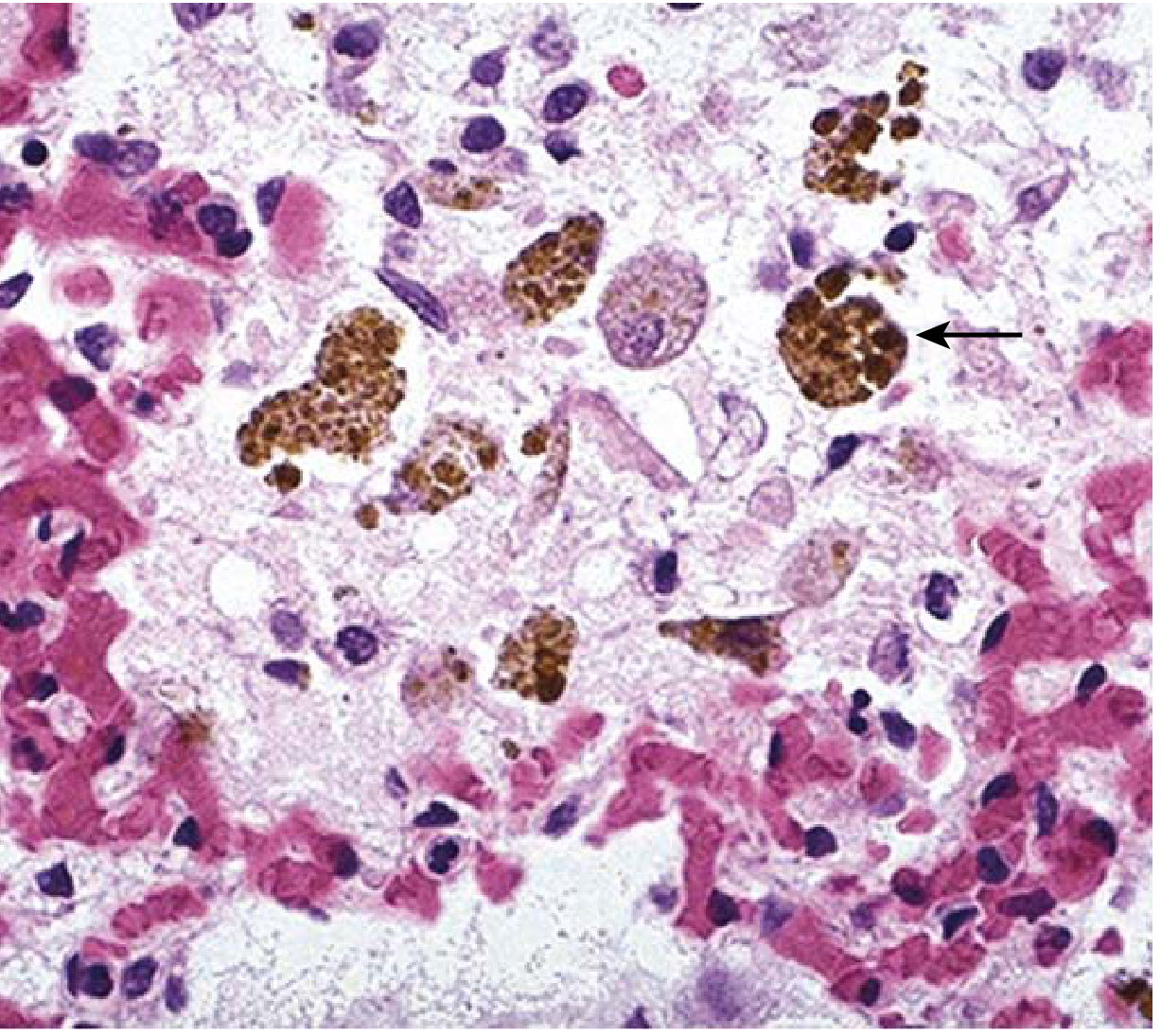
Robbins & Kumar Basic Pathology - eFig. 3.1. Alveolar space with "heart failure cells" - macrophages packed with brown hemosiderin granules (arrow). Pink edema fluid fills the alveoli.
What You See Under the Microscope
| Feature | What It Looks Like |
|---|---|
| Heart failure cells (arrow) | Large macrophages stuffed with coarse brown hemosiderin granules - the most characteristic finding |
| Alveolar spaces | Filled with pink proteinaceous edema fluid |
| Alveolar septa | Thickened and fibrotic (in chronic stage) |
| Capillaries | Congested/engorged with RBCs |
| Stain | H&E; confirm hemosiderin with Perl's Prussian Blue (turns blue) |
Why These Findings Occur
Cause: Left heart failure / mitral stenosis → ↑ pulmonary venous pressure → congestion
- Raised capillary pressure → RBCs leak into alveoli
- Alveolar macrophages phagocytose RBCs → break down hemoglobin
- Hemoglobin → hemosiderin (iron-containing pigment) stored in macrophages
- These macrophages = "Heart Failure Cells" / siderophages
- Chronic congestion → hypoxia → septal fibrosis = "Brown Induration" of lung (gross)
Gross vs Microscopy Summary
| Gross | Microscopy | |
|---|---|---|
| Acute CVC | Wet, heavy, red lungs | Engorged capillaries, alveolar edema |
| Chronic CVC | Brown, firm - "Brown Induration" | Heart failure cells + septal fibrosis |
Key Exam Points
-
Heart failure cells = hemosiderin-laden macrophages in alveoli
-
Stain with Perl's Prussian Blue for iron (positive)
-
Condition = Brown Induration of lung
-
Cause = left heart failure / mitral stenosis
-
The brown color comes from hemosiderin (iron pigment)
-
The "induration" (hardness) comes from septal fibrosis
-
Robbins & Kumar Basic Pathology, p. 77-78
This is a shared conversation. Sign in to Orris to start your own chat.