
subsidence of femoral stem
Reading File
Subsidence of a femoral stem refers to the downward migration (sinking) of a hip prosthesis stem within the femoral canal after total hip arthroplasty (THA). It is a key radiographic and clinical sign in evaluating implant stability. Let me search the textbook library and recent literature simultaneously.
Searching sources...
Finding Sources
Searching PubMed
femoral stem subsidence total hip arthroplasty
Good results. Let me now read the relevant textbook sections in parallel with the most relevant recent meta-analysis.
Reading File
Reading File
Reading Article
PMID: 41989304
Excellent data. Let me fetch the relevant radiograph images and read more of the textbook for cementless stem subsidence classification and impaction grafting context.
Loading Image
Reading File
Finding Sources
Let me now read the Engh classification section and the section about early vs late subsidence to complete the picture.
Reading File
Finding Sources
I now have comprehensive information. Let me fetch one more key image and get the early subsidence / impaction grafting data.
Reading File
I now have all the information needed for a comprehensive answer. Here it is:
Subsidence of the Femoral Stem
Subsidence refers to the downward (axial) migration of the femoral prosthesis stem within the femoral canal following total hip arthroplasty (THA). It is one of the most important radiographic indicators of implant fixation status.
How It Is Measured
Subsidence is quantified by measuring the distance between a fixed point on the stem and a radiographic landmark such as:
- A trochanteric wire or cable
- A bony prominence (lesser or greater trochanter)
Serial radiographs are essential - single films are insufficient because differences in magnification and positioning can mimic or mask migration.
- Campbell's Operative Orthopaedics 15th Ed 2026, p. 325
Subsidence in Cemented Femoral Stems
Subsidence of a cemented stem is one of the cardinal signs of loosening. The full list of loosening signs includes:
- Radiolucency between the superolateral stem (Gruen zone 1) and cement mantle - indicates debonding and possible stem deformation
- Radiolucency between the cement mantle and surrounding bone
- Subsidence of the stem alone, or stem + cement mantle together
- Change into varus position
- Fragmentation of cement (especially Gruen zone 7, superomedial)
- Fracture of the cement mantle (especially Gruen zone 4, near tip)
- Deformation of the stem
- Fracture of the stem
Harris Radiographic Grading of Loosening:
| Grade | Criteria |
|---|---|
| Definite loosening | Migration of the component or cement |
| Probable loosening | Complete radiolucency around cement mantle |
| Possible loosening | Incomplete radiolucency >50% of cement |
Subsidence may not be noticed unless serial films are carefully compared. The stem may subside within the cement (often with cement fracture near the tip), or the entire cement-stem construct may subside together.
Technical factors predisposing to loosening/subsidence:
-
Failure to remove soft cancellous bone from medial femoral neck
-
Inadequate cement mantle thickness
-
Inadequate cement quantity or pressurization
-
Stem motion during cement hardening
-
Malalignment (non-neutral positioning)
-
Cement voids from poor mixing technique
-
Campbell's Operative Orthopaedics 15th Ed 2026, pp. 324-326
Subsidence in Cementless Femoral Stems
Engh Classification of Cementless Fixation
Engh et al. proposed a 3-category radiographic classification:
| Category | Criteria |
|---|---|
| Bone Ingrowth | No subsidence, minimal/no radiopaque lines, spot welds present, possible cortical hypertrophy distally and proximal stress shielding |
| Stable Fibrous Fixation | No progressive migration, but extensive parallel radiopaque lines ~1 mm from stem surface; no cortical hypertrophy |
| Unstable | Progressive subsidence/migration, divergent radiopaque lines (wider at extremities), increased cortical density beneath collar and at stem tip |
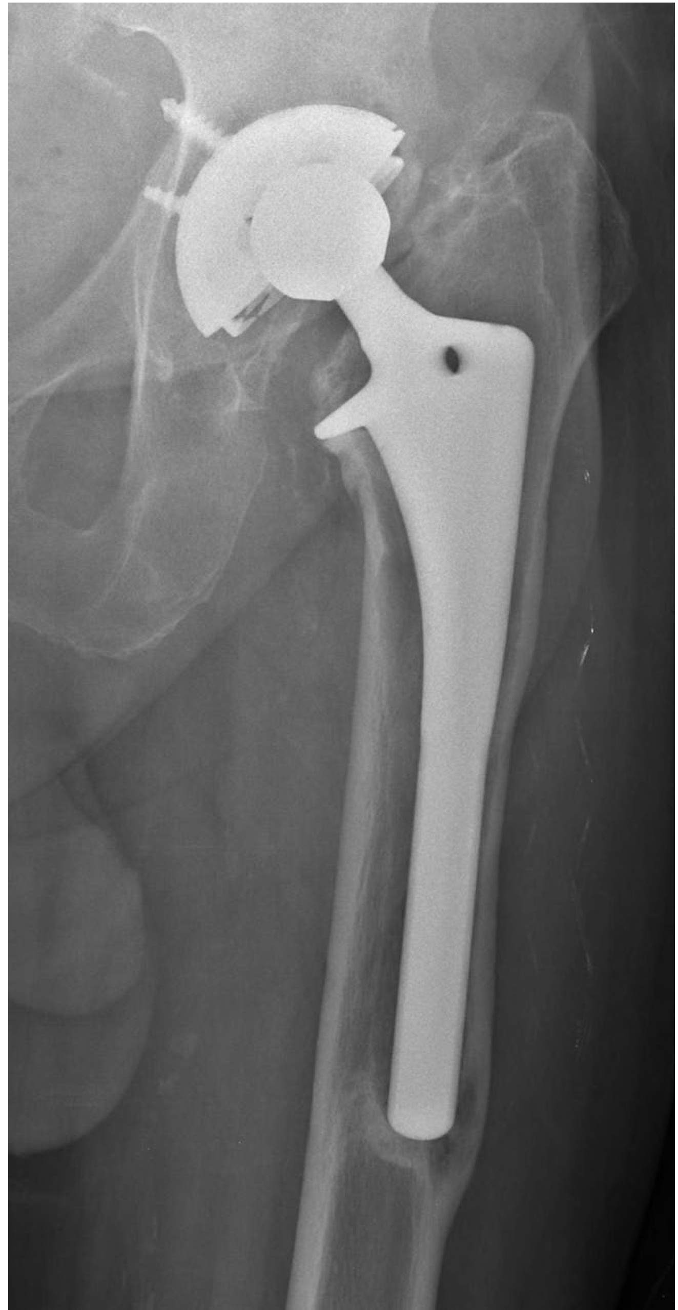
Fig. 4.130 - Unstable cementless stem with subsidence and circumferential radiolucencies (shown below):
Early vs. Late Subsidence - Key Distinction:
- Early subsidence (shortly after surgery): May actually be beneficial - it allows the stem to seek a more stable position within the canal. Bone ingrowth can still occur, and early subsidence is compatible with durable fixation.
- Late subsidence (months to years post-op): Implies unstable fixation. A bony pedestal often develops at the stem tip (Gruen zone 4) - evidence of pistoning.
Other radiographic signs of cementless instability:
-
Progressive bead shedding from porous coating on serial films = micromotion at bone-implant interface
-
Neocortex formation (cortical bone around smooth distal portion of proximally-coated stem) - when seen without subsidence, it is a variant of normal fixation seen with tapered wedge designs
-
Calcar atrophy/rounding - paradoxically, a sign of good proximal osseointegration (stress transfer to proximal stem)
-
Campbell's Operative Orthopaedics 15th Ed 2026, pp. 325-326
Collar vs. Collarless Stems and Subsidence
The role of a collar in preventing subsidence is debated:
-
A collarless stem may allow small amounts of early subsidence, enabling better canal fit and promoting bone ingrowth
-
A collar might resist subsidence but can also prevent optimal seating
-
Campbell's Operative Orthopaedics 15th Ed 2026, p. 283 (Classification of Cementless Stem Designs)
Subsidence in Revision Surgery (Impaction Bone Grafting)
In femoral impaction grafting for revision THA, a small degree of subsidence of the collarless polished tapered stem is expected and acceptable. The graft consolidates as the stem subsides slightly, restoring bone stock.
A 2025 meta-analysis of 2,514 hips specifically examined graft incorporation and stem subsidence in femoral impaction bone grafting, confirming that measured subsidence is a routine part of the technique's outcome assessment.
Recent Evidence (2026 Meta-Analysis)
A landmark 2026 systematic review and meta-analysis (Van der Water et al., Acta Orthopaedica, PMID 41989304) of 73 studies, 2,970 uncemented stems measured by radiostereometric analysis (RSA) found:
| Time point | Pooled subsidence | Pooled retroversion |
|---|---|---|
| 3 months | 0.29 mm (95% CI 0.19-0.39) | 0.51° (CI 0.33-0.70) |
| 2 years | 0.32 mm (CI 0.21-0.43) | 0.70° (CI 0.48-0.93) |
Key findings:
- Most subsidence occurs within the first 3 months, then stabilizes
- Hydroxyapatite (HA)-coated stems showed the least migration (0.26 mm)
- Anterior approach showed significantly more migration (1.04 mm at 3 months) than other approaches
- Migration patterns differ based on stem type, coating, surgical approach, and timing of baseline measurement
Clinical Significance Summary
| Scenario | Interpretation |
|---|---|
| Early, self-limited subsidence (cementless) | Likely benign; stem seeking stable position |
| Progressive subsidence months-years later | Unstable fixation; consider revision |
| Subsidence + pedestal at tip (zone 4) | Pistoning - definite instability |
| Subsidence + cement migration (cemented) | Definite loosening (Harris criteria) |
| Subsidence in impaction grafting revision | Expected; part of normal graft consolidation |
This is a shared conversation. Sign in to Orris to start your own chat.