
Boydes method is age estimation
Reading File
Finding Sources
Searching the Web
Boyd's method age estimation teeth forensic
Finding Sources
Finding Sources
Reading File
Reading File
Reading File
Reading File
Finding Sources
Reading Web Page
https://www.biolscigroup.us/articles/FST-2-105.php
Reading File
Loading Image
Finding Sources
I now have comprehensive information from the forensic medicine textbook. The textbook content covers dental age estimation thoroughly, including the eruption tables and methods. "Boyd's method" is specifically an eruption-based age estimation technique documented in forensic medicine. Let me compile the complete answer using the textbook data.
Boyd's Method of Age Estimation
Boyd's method is a dental age estimation technique used in forensic medicine, applicable primarily to children and adolescents (up to about 14-17 years). It is based on the eruption sequence and timing of permanent teeth.
Basis of the Method
Boyd's method uses the known, predictable sequence of permanent tooth eruption to estimate age. The eruption of teeth follows a fixed biological timetable, making it one of the more reliable indicators of age in younger individuals.
"From birth to fourteen years of age, the degree of formation of root and crown structures, the stage of eruption, and the intermixture of temporary and permanent teeth are useful in age estimation. It is generally accepted that in a child, estimation of age from teeth gives better results than skeleton."
- The Essentials of Forensic Medicine and Toxicology, 36th ed.
Permanent Tooth Eruption Chart (Boyd's Data - Table 4.9)
This is the core data Boyd's method uses:
| Tooth | Eruption Age |
|---|---|
| First molar | 6 to 7 years |
| Central incisor | 6 to 8 years |
| Lateral incisor | 7 to 9 years |
| First bicuspid (premolar) | 9 to 11 years |
| Second bicuspid (premolar) | 10 to 12 years |
| Canine | 11 to 12 years |
| Second molar | 12 to 14 years |
| Third molar (wisdom tooth) | 17 to 25 years |
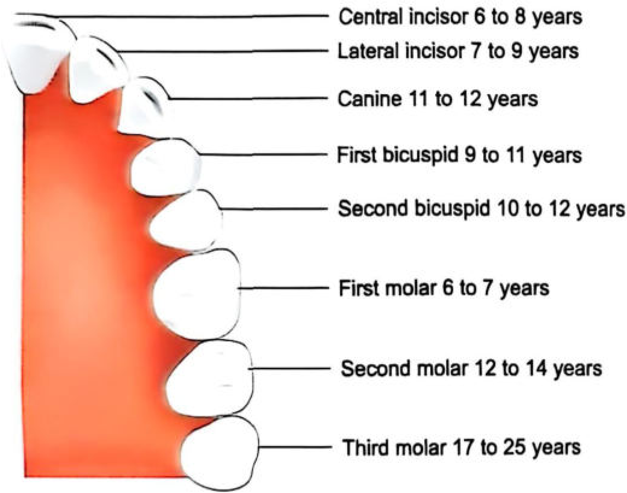
Important Points
-
Age range: Most useful from birth to ~14 years. From 14-20 years, estimation is based on the development stage of the third molar (with accuracy of approximately ±3 years).
-
Third molar rule: If all third molars are fully erupted, the person is above 17 years of age.
-
Sex differences: Tooth eruption tends to occur earlier in females than in males. In females, skeletal age is generally ~1 year ahead of dental age.
-
Method limitations: Ethnic, cultural, hereditary, environmental, endocrine, and nutritional factors all influence eruption timing. Eruption tends to occur earlier in warmer climates and urban areas.
-
Dental X-rays: Orthopantomogram (OPG/dental panoramic tomograph) allows visualization of all teeth in a single X-ray, showing developmental status of unerupted teeth and the degree of root completion in erupted teeth.
Comparison With Other Dental Age Methods
| Method | Age Group | Basis |
|---|---|---|
| Boyd's method | Children/adolescents | Tooth eruption sequence |
| Gustafson's method | Adults (>21 years) | 6 physiological changes (APSRTC - Attrition, Periodontosis, Secondary dentin, Root resorption, Transparency, Cementum apposition) |
| Demirjian's method | Children | Tooth mineralization stages on X-ray |
| Nolla's method | Children | 0-10 graded calcification scale |
Key Rule for Exams
Boyd's method applies up to ~14 years using eruption. Beyond 14 years, dental age estimation transitions to third molar development. Beyond 21 years, Gustafson's method (structural/degenerative changes) takes over.
Source: The Essentials of Forensic Medicine and Toxicology, 36th edition (2026), p. 81-82

photo-1786190328340.jpg
Notes with pictorial representation
analyse_media
Finding Sources
Searching Images
skull sutures fusion age estimation diagram
Reading File
Finding Sources
Finding Sources
Finding Sources
Reading File
Reading File
Reading File
Loading Image
Loading Image
Loading Image
I now have all the content needed. Here are your comprehensive notes on Appearance and Fusion of Ossification Centres (Chapter 18 - Personal Identity, Forensic Medicine):
Appearance and Fusion of Ossification Centres
Age Estimation from Bones - Forensic Medicine
Key General Points
- Development and growth of bones follows a definitive time sequence - useful for age estimation
- Best method: Radiological survey of ossification status
- In young children: X-ray of hand and foot alone can fix age accurately
- Clavicle = first bone to ossify in the body, by membranous ossification at 5th-6th week (IUL)
- All primary ossification centres appear by 3rd month of intrauterine life; others are secondary centres
- At birth: 450 ossification centres present
- Closure of skull sutures gives approximate age of the individual
Skull Suture Closure
The textbook diagram (Fig. 4.43) shows suture closure ages on the skull:
| Suture | Fusion Age |
|---|---|
| Metopic | 2 - 8 years |
| Basi-occiput + Basi-sphenoid (Basal) | Females: 18-22 yrs / Males: 19-24 yrs |
| Sagittal - Posterior 1/3 | 30 - 40 years |
| Sagittal - Anterior 1/3 | 40 - 50 years |
| Sagittal - Middle 1/3 | 50 - 60 years |
| Coronal - Lower half | 40 - 50 years |
| Coronal - Upper half | 50 - 60 years |
| Lambdoid - Upper | 50 - 60 years |
| Lambdoid - Lower | 60 - 70 years |
| Temporal | ~80 years |
Note: Suture closure in skull occurs later in females than in males. Estimation from suture closure is not very reliable - can only give a decade range.
Clavicle - Ossification
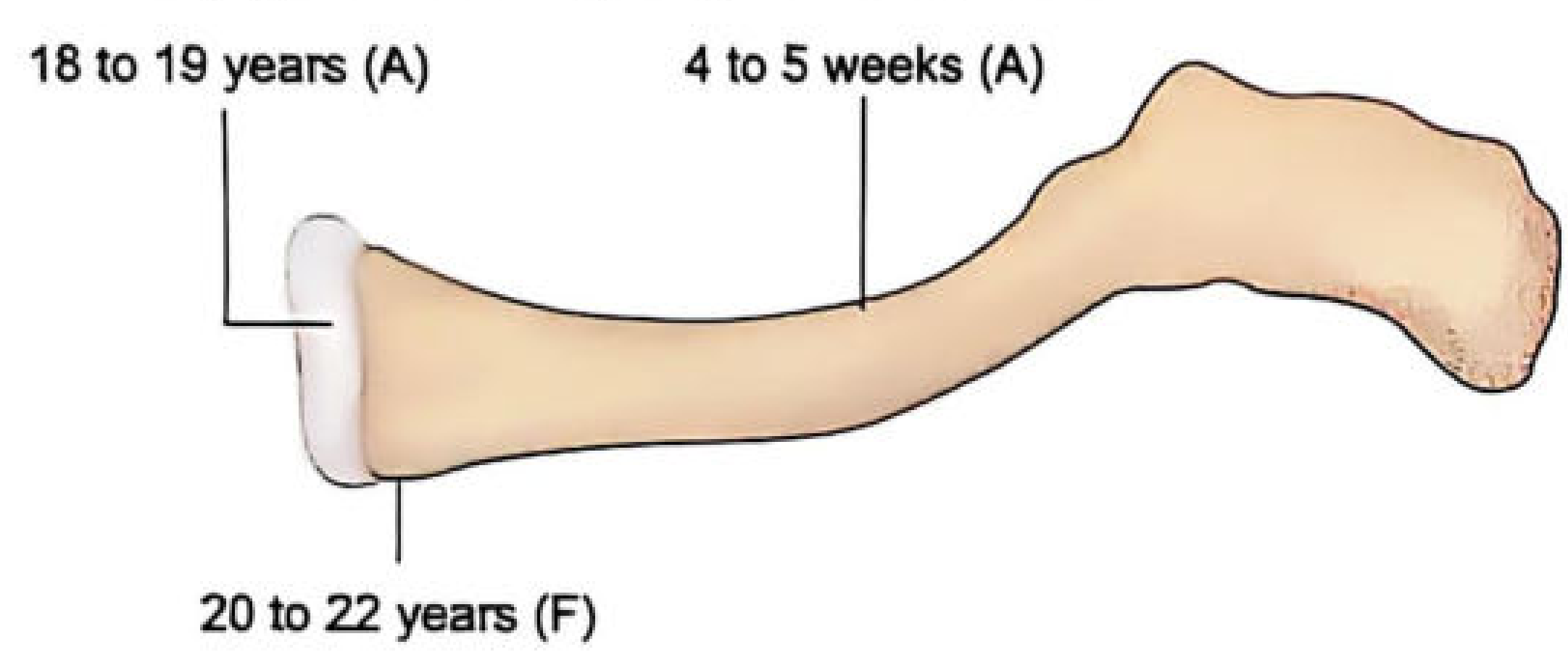
Fig 4.23: The clavicle starts ossifying at 4-5 weeks (A), medial end epiphysis appears at 18-19 years (A), and fuses at 20-22 years (F)
(A) = Appearance, (F) = Fusion
Appearance and Fusion of Various Joints
| Joint | Appearance | Fusion |
|---|---|---|
| Shoulder joint | ||
| Upper end of Humerus | 1 - 6 years | 16 - 18 years |
| Acromial end | 14 years | 17 - 18 years |
| Coracoid process | 10 years | 16 years |
| Elbow joint | ||
| Lower end of Humerus | 2 - 8 years | 14 - 17 years |
| Upper end of Radius | 6 years | 14 - 17 years |
| Upper end of Ulna | 10 years | 14 - 17 years |
| Wrist joint | 2 - 11 years | 16 - 18 years |
| Pelvis | ||
| Iliac crest | 14 years | 18 years |
| Ischial tuberosity | 16 years | 19 yrs (F) / 21 yrs (M) |
| Femur | 1 - 11 years | 15 - 17 years |
Sternum - Fusion of Centres
- Xiphi sternum fuses with body: 40 years
- Manubrium sternum: Above 50 years
- Thyroid and Hyoid bone ossify between 35 - 45 years
- Sacrum: Fuses from below upwards, completed at 20 - 25 years
Hand Bones - Ossification
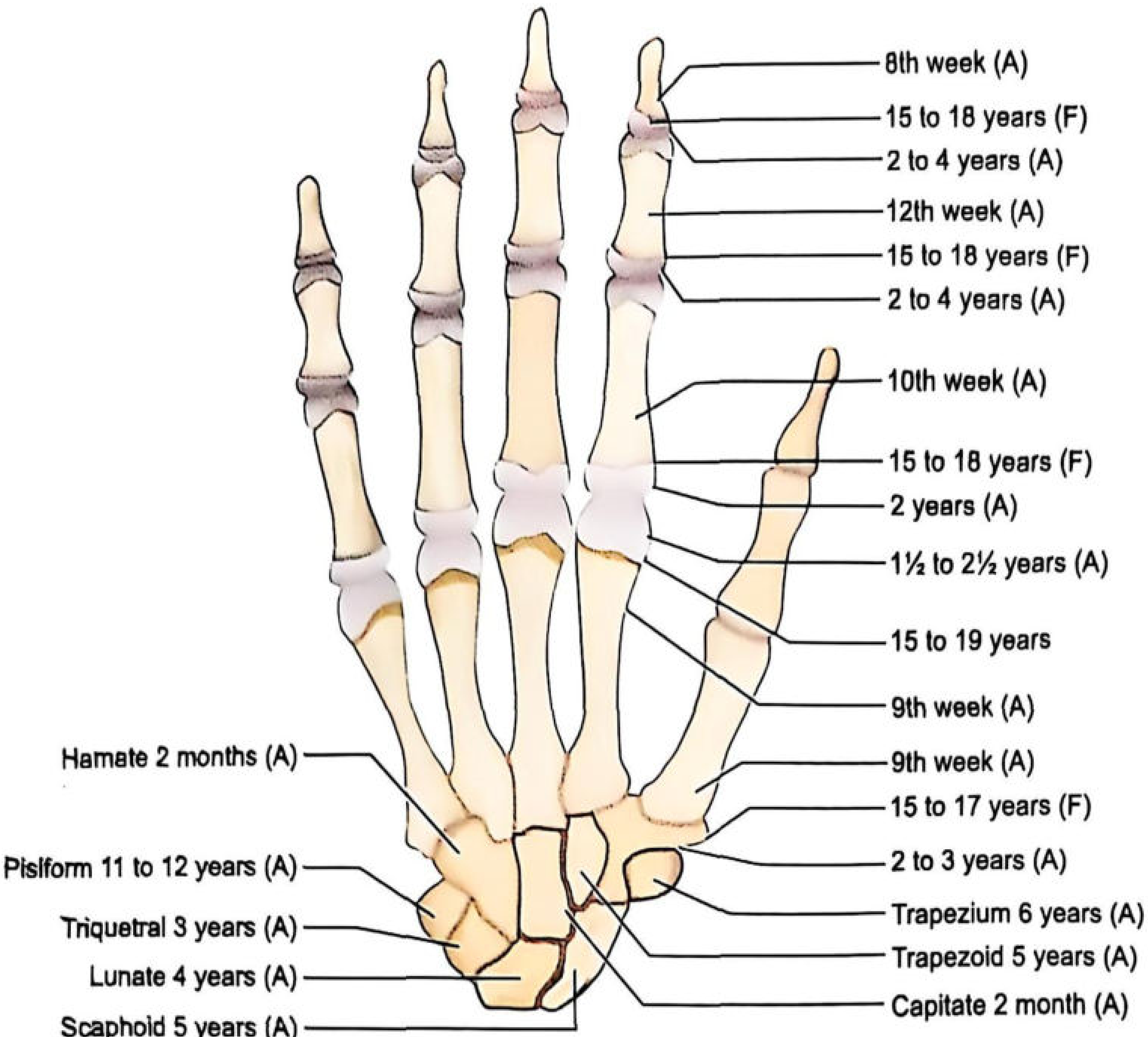
Fig 4.28: Carpal bones - Capitate 2 months, Hamate 2 months, Triquetral 3 yrs, Lunate 4 yrs, Scaphoid 5 yrs, Trapezoid 5 yrs, Trapezium 6 yrs, Pisiform 11-12 yrs. Finger epiphyses appear 1½ - 4 yrs, fuse 15-19 yrs.
Rule: Between 2-6 years, number of carpal bones visible on X-ray ≈ age in years
Foot Bones - Ossification
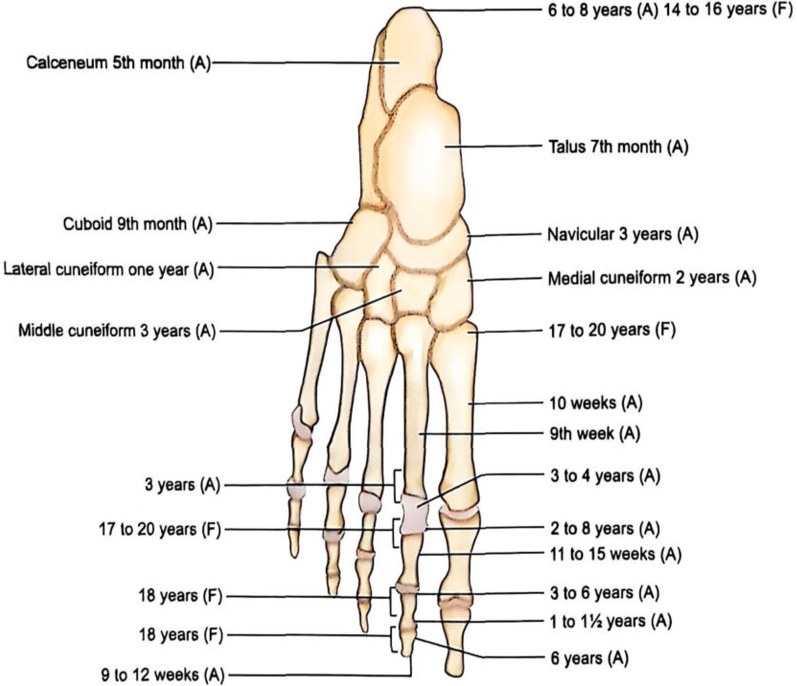
Fig 4.29: Calcaneum 5th month, Talus 7th month, Cuboid 9th month, Navicular 3 yrs, Medial cuneiform 2 yrs, Lateral cuneiform 1 yr, Middle cuneiform 3 yrs. Epiphyses fuse 17-20 yrs.
Important Sex Differences
| Feature | Males | Females |
|---|---|---|
| Epiphyseal union | Later | Earlier (by ~1 year) |
| Skull suture closure | Earlier | Later |
| Ossification in tropics | ~2 years earlier than temperate zones in both sexes |
Radiology - X-rays to Take at Different Ages
| Age | X-ray Site |
|---|---|
| 6 - 12 years | Elbow + Wrist joint |
| 13 - 16 years | Elbow + Pelvis |
| After 17 years | Pelvis + Sternum |
Source: The Essentials of Forensic Medicine and Toxicology, 36th ed. (2026), Ch. 18 - Personal Identity, pp. 271+
This is a shared conversation. Sign in to Orris to start your own chat.