
Fibreoptic intubation
I'll start by reading the medical library skill, then search for comprehensive information on fibreoptic intubation.
Reading File
Now let me search the library in parallel for fibreoptic intubation content.
Finding Sources
Finding Sources
Searching PubMed
"fibreoptic intubation" OR "fiberoptic intubation" awake
Good, I found relevant sections. Now let me read both textbook sections in parallel.
Reading File
Reading File
Finding Sources
Now let me read more of the Cummings Otolaryngology section for the detailed technique and key steps, and also search for the anesthesia textbook content.
Reading File
Finding Sources
Excellent - rich content retrieved. Let me also read the continuation of the Cummings technique section and fetch the relevant image.
Reading File
Finding Sources
I now have comprehensive information. Let me fetch the relevant image from the textbook and compile the full answer.
Loading Image
I now have all the information needed for a comprehensive response. Here is the full overview:
Fibreoptic Intubation (FOI)
Fibreoptic intubation (FOI) is the passage of a flexible fibreoptic bronchoscope/endoscope loaded with an endotracheal tube (ETT) through the upper airway under direct vision, allowing controlled placement of the tube into the trachea. It is the gold-standard technique for managing the known or anticipated difficult airway, particularly when performed awake.
Historical Background
Flexible fibreoptic technology was first used for nasotracheal intubation in 1967 on a patient with Still's disease using a choledochoscope. In 1972 it was used in a patient with severe rheumatoid arthritis via a fibreoptic bronchoscope. Stiles et al. subsequently reported the first series of 100 fibreoptic endotracheal intubations. Although not originally designed for intubation, the technique was rapidly adopted by anaesthesiologists for managing the difficult airway. (Cummings Otolaryngology, p. 124)
Indications
I. Difficult Intubation
- Known or anticipated difficult intubation (e.g. trismus, mandibular injuries, cervical spine rigidity, obstructing mass in the oropharynx/upper aerodigestive tract, severe rheumatoid arthritis, ankylosing spondylitis)
- Unanticipated failed intubation
II. Compromised Airway
- Upper airway compromise (supraglottic/glottic mass, supraglottic oedema, marked obesity)
- Lower airway compromise (tracheal compression)
III. Conscious Patient Intubation Preferred
- High risk of aspiration
- Movement of neck not desirable (cervical spine instability)
- Known difficult mask ventilation
- Morbid obesity
- Self-positioning needed
IV. High Risk of Dental Damage
(Cummings Otolaryngology, Box 6.1, p. 124)
Contraindications
- Fixed stenotic lesions at all levels that will not allow ETT passage without dilatation
- Significant bleeding obscuring visualisation
- Uncooperative patient unable to comply during examination
(Cummings Otolaryngology, p. 125)
Equipment
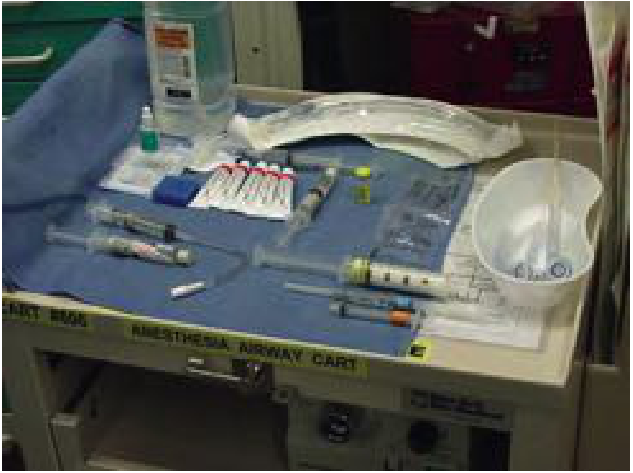
A fully equipped videobronchoscopy cart should be available in any ICU, emergency department, critical care ward, and operating room. The cart should include:
- Flexible fibreoptic bronchoscope connected to a video monitor
- Endotracheal tubes (standard and long microlaryngoscopy tubes)
- Nasopharyngeal airway / nasal trumpets (graduated sizes)
- Syringes, topical anaesthetic agents (lidocaine 2% and 4%)
- Nasal decongestants (oxymetazoline 0.05% or phenylephrine 1%)
- Lubricating gel
- Resuscitation and monitoring equipment (pulse oximetry, ECG, capnography)
- High-flow oxygen delivery (nasal cannula)
Preparation of the Patient
1. Psychological Preparation
Success depends heavily on a relaxed, cooperative patient. The process begins with a reassuring pre-procedure visit. The patient is informed about the procedure and their active participation - maintaining head position, deep breathing, clearing secretions - is requested. (Cummings Otolaryngology, p. 125)
2. Pharmacological Preparation
The three components are premedication, limited sedation, and topical/local anaesthesia:
| Agent | Role | Dosing Notes |
|---|---|---|
| Glycopyrrolate | Antisialagogue (reduces secretions, essential for good FOI) - drug of choice; does not cross BBB, causes less tachycardia | Given ~30 min before procedure |
| Midazolam (IV) | Mild sedation, anxiolysis | Observe 3 min before repeat dosing |
| Fentanyl | Analgesia and conscious sedation | Combined with midazolam |
| Remifentanil infusion | Conscious sedation (alternative) | Titrated carefully |
Key principle: A patient who is awake and mildly uncomfortable is preferred over one who is unresponsive, poorly protecting their airway, or confused.
3. Topical/Local Anaesthesia
Goal: minimise pharyngeal, laryngeal, and tracheobronchial reflexes and patient discomfort. Plasma levels rise more rapidly from mucosal application than from injection - careful dosing is essential.
For oropharyngeal anaesthesia:
- Benzocaine 20% spray to the mouth - gargle and swallow
- 5 mL of 2% plain lidocaine inhaled via nebuliser (if time allows)
For glottic/subglottic anaesthesia:
- Transcricothyroid injection: 2 mL of 2% plain lidocaine through the cricothyroid space - patient instructed to cough
- Or bilateral/unilateral superior laryngeal nerve block with 2% lidocaine
For the nasal route:
- Oxymetazoline 0.05% or phenylephrine 1% spray (vasoconstrictors) - several sprays
- Aerosolised 4% lidocaine nasal spray or cotton pledgets soaked in 4% lidocaine passed between inferior turbinates and septum
- Applied at least 5 minutes before any instrumentation
(Cummings Otolaryngology, pp. 125-126)
Box 6.2: Keys to Successful Awake Fibreoptic Intubation
Pharmacological preparation:
- Antisialagogue (glycopyrrolate)
- Sedation (judicious) - IV midazolam ± fentanyl or remifentanil
- Topical/local anaesthesia:
- Oral intubation: oropharynx, then laryngotracheal
- Nasal intubation: nasal mucosa, then laryngotracheal
- Monitoring and supplemental oxygen throughout
(Cummings Otolaryngology, Box 6.2, p. 126)
Technique
FOI can be performed transorally or transnasally. The transnasal route is generally preferred by otolaryngologists (and many anaesthesiologists), as the scope typically points directly at the vocal cords on entering the oropharynx. There is no strong evidence for superiority of one route over the other.
Transnasal Technique (Step-by-Step)
-
Position: Head of bed elevated as much as tolerated (near upright). Operator faces the patient, with the video monitor in view. Cart positioned on the patient's left at the head of the bed; operator stands on the patient's right.
-
ETT preparation: Placed in warm saline to soften it; cuff fully deflated.
-
Nasal dilatation: Nasal passages progressively dilated with increasingly larger lubricated nasal trumpets (generous lidocaine gel).
-
ETT insertion: ETT introduced into the nostril, directed inferiorly toward the nasopharynx. If resistance is met at the bend toward the oropharynx, pull back, rotate 90° to the right or left, and reintroduce.
-
Fibreoptic passage: The fibreoptic bronchoscope is passed through the ETT lumen. Video monitor used for real-time visualisation. The glottic inlet is identified - in most patients it is visible directly.
-
Scope advancement into trachea: The scope is advanced through the glottis into the trachea. The tracheal rings and carina serve as confirmation of correct placement.
-
ETT railroading: With the scope held stationary in the trachea, the ETT is advanced over the scope ("railroaded") into the trachea.
-
Scope removal and confirmation: The scope is withdrawn while holding the ETT in position. Position confirmed by capnography and direct vision through the scope before removal.
-
Cuff inflation and ventilation: ETT cuff inflated, ventilation commenced.
(Cummings Otolaryngology, p. 126-127)
Oral Approach
In the oral approach, an intubating oral airway (e.g., Ovassapian or Williams airway intubator) is often used to guide the scope past the tongue and toward the glottis, and to protect the scope from being bitten.
Advantages of FOI
- Maintains airway patency throughout - the patient continues to breathe spontaneously during awake FOI
- Allows visual confirmation of tube placement in real time
- Can be used in patients with cervical spine instability - no neck extension required
- Avoids the haemodynamic responses of standard laryngoscopy
- Preferred in emergency or elective settings where conventional laryngoscopy is predicted to fail
- Particularly useful in: trismus, mandibular injuries, cervical spine rigidity, obstructing masses in the oral cavity or upper aerodigestive tract (Bailey and Love's Surgery, p. 820)
Complications
- Failure to intubate - due to obscured vision (secretions, bleeding, oedema)
- Epistaxis (transnasal route) - minimised by nasal decongestants and gentle dilatation
- Laryngospasm - inadequate topical anaesthesia
- Hypoxaemia during prolonged attempts - managed by supplemental O₂ via nasal cannula
- Lidocaine toxicity - from excessive topical application
- Patient non-cooperation - prevented by adequate psychological and pharmacological preparation
Special Considerations
- Emergency setting: In a "cannot ventilate, cannot intubate" scenario, surgical airway (cricothyroidotomy, tracheostomy, transtracheal jet ventilation) takes precedence. FOI is primarily an elective/semi-elective technique for anticipated difficulty.
- Significant bleeding: Is a contraindication to FOI - blood obscures the fibreoptic view. A rigid bronchoscope or surgical airway may be required.
- Paediatrics: Smaller-diameter scopes (paediatric bronchoscopes) are used; the LMA can be used as a conduit for fibreoptic-guided intubation in children with difficult airways.
- Unsedated FOI: Used when the airway/physical condition is so compromised that no sedative or opioid can safely be given before the procedure.
Sources
- Cummings Otolaryngology Head and Neck Surgery, Chapter 6: Awake Fiberoptic Intubation, pp. 124-127
- Bailey and Love's Short Practice of Surgery, 28th Edition: Fibreoptic Endotracheal Intubation, p. 820
This is a shared conversation. Sign in to Orris to start your own chat.