
Malunion of fore arm
Reading File
Now let me search the medical library for content on forearm malunion.
Finding Sources
Finding Sources
Excellent - found content in Campbell's Operative Orthopaedics and Rockwood & Green's. Let me read the key sections.
Reading File
Finding Sources
Reading File
Reading File
Reading File
Reading File
Reading File
Reading File
Reading File
Finding Sources
Reading File
Reading File
Reading File
Loading Image
I now have comprehensive information. Let me also quickly fetch the Monteggia malunion image and look at the key section on angulation thresholds and pediatric considerations.
Reading File
Loading Image
I now have comprehensive content. Let me compile the full response.
Malunion of the Forearm
Definition
Malunion of the forearm refers to healing of one or both bones of the forearm (radius and/or ulna) in a position of unacceptable angulation, rotation, or shortening after fracture. Because the radius and ulna function as a linked unit, even subtle malalignment of either bone can significantly disrupt the complex biomechanics of forearm rotation.
Functional Anatomy Relevant to Malunion
The forearm is not simply two parallel bones - it is a bicondylar joint system where the radius rotates around a fixed ulna. Key anatomical features that must be restored:
- Radial bow: The radius has a characteristic lateral bow peaking at ~60% of the distance between the bicipital tuberosity and the ulnar aspect of the distal articular surface. The maximum distance of this bow averages 15.3 mm. Even small deviations from normal bow magnitude or position reduce forearm rotation.
- Interosseous space: Angular deformity of either bone narrows the interosseous space, mechanically blocking pronation/supination.
- DRUJ geometry: Malunion alters the relationship between the ulnar head and the sigmoid notch, causing DRUJ instability and eventual arthritis (especially after >20 years).
Types of Deformity
| Type | Mechanism | Effect |
|---|---|---|
| Angulation | Failure to correct volar/dorsal or radial/ulnar tilt | Narrows interosseous space; reduces rotation |
| Malrotation | Rotational malalignment at fracture site | Proximal fragment typically supinated due to biceps/supinator insertions |
| Loss of radial bow | Flattening of normal curvature | Reduced rotation and grip strength |
| Shortening | Comminution, bone loss | Alters DRUJ mechanics |
| Combined deformity | High-energy fractures | Worst outcomes |
Biomechanical Thresholds (Cadaveric Studies)
- 10° angulation: Insignificant reduction in forearm rotation
- 20° angulation: Clinically significant loss of pronation and supination
- 15° total deformity: Forearm motion reduced by >27% (except in distal-third fractures, which tolerate more angulation)
- Rotational deformity compounds angular deformity, and the location of angulation along the forearm matters more than the direction of deformity
(Campbell's Operative Orthopaedics 15th Ed, p. 3712; Rockwood & Green's 10th Ed, p. 1772)
Clinical Features
- Loss of forearm rotation (pronation/supination) - the dominant complaint
- Reduced grip and pinch strength
- DRUJ instability (pain, clicking, weakness on load-bearing)
- Cosmetic deformity
- Long-term: PRUJ/DRUJ arthritis, heterotopic ossification, interosseous membrane contracture
Assessment
Clinical:
- Measure full arc of pronation-supination and compare with contralateral side
- Assess elbow flexion/extension
- Stress test proximal and distal radioulnar joints (palmar/dorsal)
Radiographic:
- Full-length AP and lateral radiographs of both forearms (pronation and supination views)
- Assess radial bow, bone lengths, DRUJ/PRUJ alignment
- CT scan with both forearms in maximal pronation and supination: essential for quantifying rotational deformity - cross-sectional cuts proximal and distal to the fracture are compared to the uninjured side
Indications for Surgery
Surgery is indicated based on functional limitation and physical demands, not purely radiographic appearance:
- Loss of forearm rotation (most common)
- DRUJ instability
- Unacceptable cosmetic deformity
Note: Restoring bony alignment does NOT always improve deficits caused by soft-tissue injury or prolonged immobilization - these factors must be considered in counseling.
Surgical Treatment
Timing
- Best results within the first 12 months of injury
- Trousdale & Linscheid (27 patients, 15-year follow-up):
- Treated <12 months: average 79° gain in rotation (range 20-160°)
- Treated >12 months: average 30° gain (range 25-95°)
- Complication rate: 48% when treated >1 year vs 15% when treated earlier
Principle of Corrective Osteotomy (Technique 63.29 - Trousdale & Linscheid, Modified)
- Full preoperative documentation: ROM, radiographs (bilateral), CT for rotation
- Identify whether one or both bones require osteotomy
- Single bone malunion: osteotomy of that bone only
- Both bones malaligned: osteotomize the more severely deformed bone first
- If radius osteotomy allows smooth passive rotation, the ulna may not need correction
- If both equally malaligned: ulna first to establish correct length and alignment, then radius conforms
- Approach:
- Radius: 10-15 cm anterior Henry approach centered over malunion
- Ulna: 10-15 cm longitudinal subcutaneous approach between ECU and FCU
- Osteotomy at malunion site, correct deformity in all planes
- Compression plating (3.5 mm DCP/LC-DCP) + bone grafting (iliac crest)
- Postoperative immobilization: above-elbow splint in supination
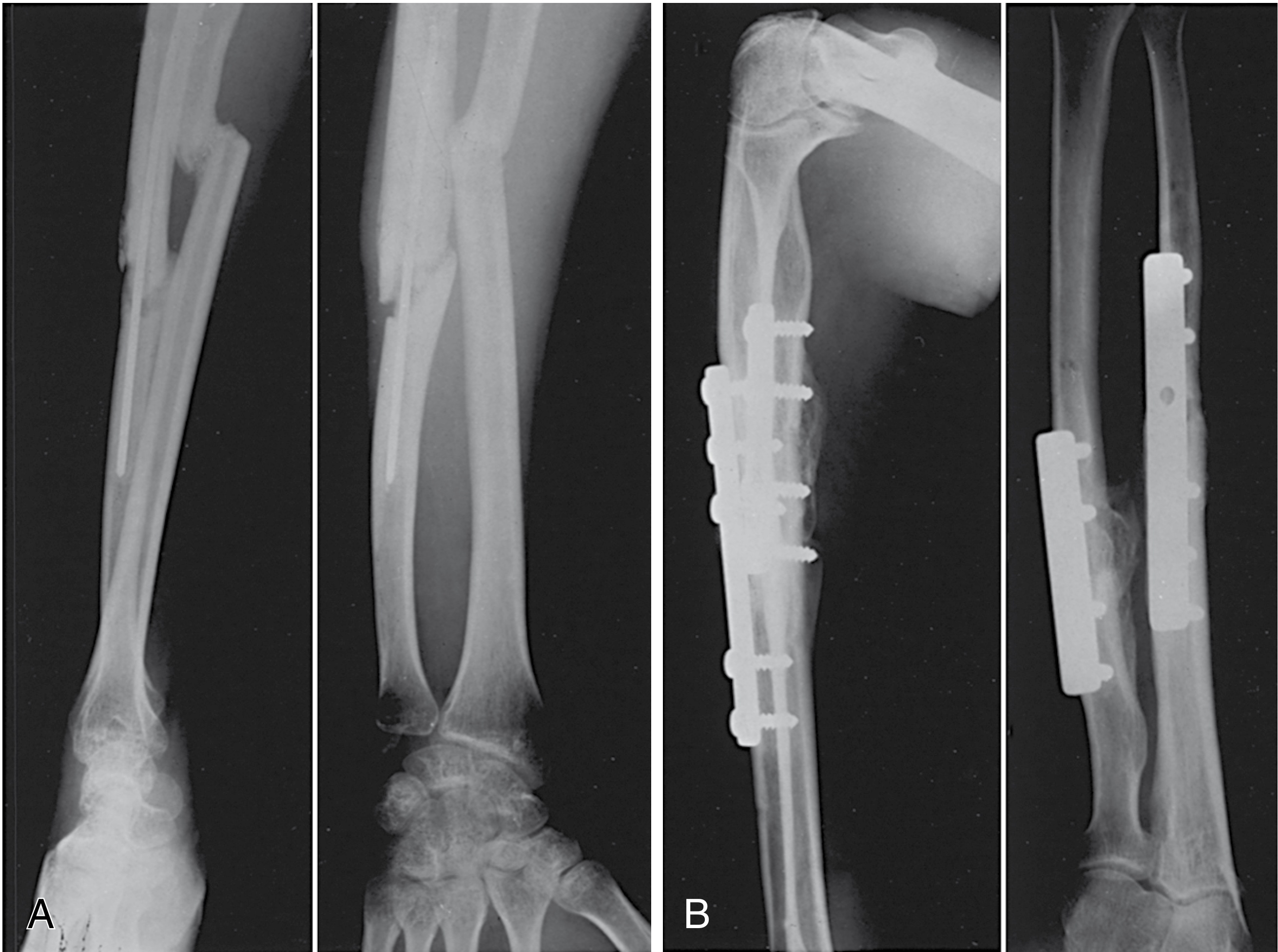
DRUJ Instability with Malunion (Technique for Trousdale & Linscheid)
When forearm malunion coexists with DRUJ instability, corrective osteotomy of the involved bone plus stabilization of the DRUJ is performed. Six patients treated in this manner achieved a stable wrist.
Special Situations
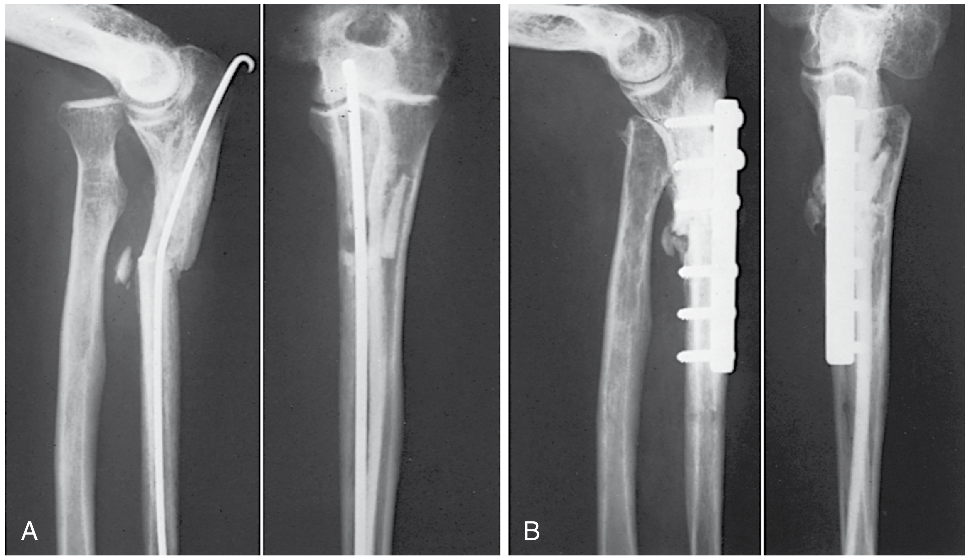
Monteggia Malunion (Proximal Ulna + Radial Head Dislocation)
When the ulna heals in malalignment with a persistent dislocation of the radial head:
- Osteotomy of ulna + compression plating
- Radial head: may require excision if fragmented (used as bone graft for ulna)
- Results in excellent elbow and forearm motion when performed correctly
Olecranon Malunion
- Osteotomy and realignment are generally not recommended (almost always increases disability)
- Excision of the malunited fragment improves function
- On lateral radiograph at 90° flexion: at least 0.3 cm of olecranon must project posterior to the humeral axis to prevent anterior subluxation
Radial Neck Malunion (Technique 63.26)
- Expose through posterolateral approach
- Osteotomy ~1.5 cm from articular surface
- Fix with Herbert screws or minifragment screws
- Bone graft from lateral epicondyle
- Postoperative: immobilize in midflexion and mid-supination x 2 weeks
Radioulnar Synostosis
- Jupiter & Ring Classification:
- A: Synostosis at or distal to bicipital tuberosity
- B: Involves radial head and proximal radioulnar joint
- C: Extends across elbow to distal humerus
- Treatment: resection of synostosis (with or without interposition material); early resection (6-12 months) preferred for better motion recovery
- Alternative: 1-cm resection of proximal radial shaft (Kamineni technique) when synostosis is too extensive or involves the articular surface
Ulnar Shaft Alone
- Isolated ulnar malunion requiring surgery is rare
- Principles mirror those for radial shaft malunion
Distal Radius Malunion
- Common source of residual disability
- Symptomatic malunions in younger/active patients warrant corrective osteotomy
- Risk of malunion highest in patients in their 4th and 5th decades
- Early corrective osteotomy should be considered in active patients
Pediatric Considerations
- Children have significant remodeling potential (especially <10 years old, fractures near the physis)
- When surgery is needed: options include open osteotomy and plating OR drill osteoclasis (Blackburn technique)
- Drill osteoclasis advantages: no plate removal needed, eliminates refracture through screw holes
- Technique: stab incision, multiple 3.2 mm drill holes at malunion, manual osteoclasis, long-arm cast x 3-6 weeks
- 3D simulation and patient-specific instruments are increasingly used for complex pediatric deformities
- The physis must be preserved in any corrective procedure
Complications of Corrective Surgery
- Delayed union / nonunion
- Infection (3-6% in forearm surgery generally)
- Persistent loss of motion
- Radial nerve paresthesias (posterior interosseous nerve at risk during Henry approach)
- Wrist pain / DRUJ instability
- Heterotopic ossification along interosseous membrane
- Subluxation of ulnar head
- Refracture after plate removal
- Radioulnar synostosis
(Campbell's Operative Orthopaedics 15th Ed 2026, pp. 3711-3714; Rockwood & Green's Fractures in Adults 10th Ed 2025, pp. 1813-1818)
Postoperative Rehabilitation
- Above-elbow splint in supination initially
- Cast/splint changed at 2 weeks; maintained until 6 weeks
- K-wires removed at 6 weeks (if used); ROM exercises begun
- Kamineni protocol (for radioulnar synostosis resection): alternating static pronation-supination splinting with progressive weaning over 3 months
Key Takeaways
| Point | Detail |
|---|---|
| Primary deficit | Loss of forearm rotation (pronation/supination) |
| Threshold for significant loss | ≥20° angulation |
| Best surgical timing | Within 12 months of injury |
| Correction principle | Osteotomy + compression plating + bone graft |
| Approach | Henry (radius), subcutaneous (ulna) |
| Sequence if both bones | More deformed bone first; if equal, ulna first |
| Complication rate (late surgery) | ~48% when corrected >1 year post-injury |
This is a shared conversation. Sign in to Orris to start your own chat.