
Give detailed mbbs notes on the physiology of ovulation and menstruation
Reading File
Reading File
Finding Sources
Finding Sources
Searching PubMed
"ovulation"[MeSH Terms] AND "menstrual cycle"[MeSH Terms]
Let me read the key textbook sections.
Reading File
Reading File
Searching Images
menstrual cycle hormone levels ovarian and endometrial changes
Reading File
Reading File
query_images_db
Physiology of Ovulation and Menstruation (MBBS Notes)
1. Overview
- The menstrual cycle is a regular, cyclic change in the ovary (ovarian cycle) and uterus (uterine cycle). It is controlled by the hypothalamic-pituitary-ovarian (HPO) axis.
- The average cycle is 28 days, with a normal range of about 21 to 35 days. Day 1 is the first day of menstrual bleeding (Ganong's Review of Medical Physiology, 26e, p. 403).
- There are three linked cycles:
- Ovarian: follicular phase, ovulation, luteal phase.
- Uterine/endometrial: menstrual, proliferative, secretory phases.
- Hormonal: GnRH, FSH, LH, estrogen, progesterone, inhibin.
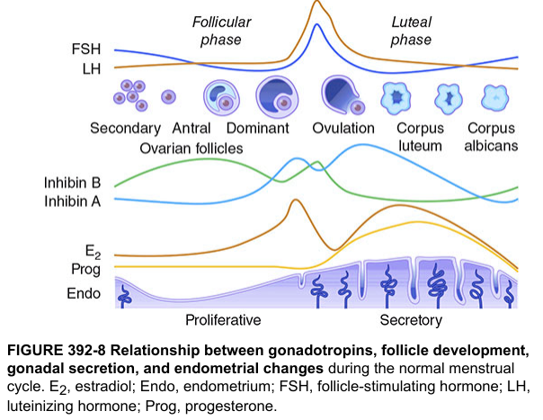
2. Hormones involved
| Hormone | Source | Main action in the cycle |
|---|---|---|
| GnRH | Hypothalamic arcuate/preoptic neurons (pulsatile; kisspeptin-driven) | Stimulates FSH and LH release |
| FSH | Anterior pituitary gonadotrophs | Recruits follicles, induces granulosa aromatase, induces LH receptors on granulosa cells |
| LH | Anterior pituitary gonadotrophs | Theca cell androgen synthesis, ovulation, luteinization, corpus luteum maintenance |
| Estradiol | Granulosa cells (androgens from theca are aromatized) | Endometrial proliferation, cervical mucus changes, negative then positive feedback |
| Progesterone | Corpus luteum (small amount from the preovulatory follicle) | Secretory endometrium, thick cervical mucus, rise in basal body temperature |
| Inhibin B / A | Granulosa cells / corpus luteum | Selective negative feedback on FSH |
| Activin | Granulosa cells | Contributes to the FSH surge |
Two-cell, two-gonadotropin theory:
- LH acts on theca interna cells. These make androgens (androstenedione, testosterone).
- Androgens diffuse into the granulosa cells. FSH induces aromatase there, which converts androgens to estradiol.
- The textbook states that granulosa cells are the primary source of circulating estrogen, but theca cell androgens are necessary for its production (Ganong, p. 403).
3. The ovarian cycle
3.1 Follicle reserve
- The fetal ovary has more than 7 million primordial follicles. About 2 million remain at birth, and fewer than 300,000 remain at puberty.
- Only about 500 oocytes ovulate in a reproductive lifetime. The rest undergo atresia by apoptosis.
- Oocytes are arrested in prophase of meiosis I from fetal life until just before ovulation (Ganong, p. 404).
3.2 Follicular phase (days 1 to 14)
- Recruitment: A rise in FSH at the end of the previous luteal phase recruits a cohort of antral follicles. This happens because estradiol, progesterone and inhibin have fallen after corpus luteum regression.
- Selection: Around day 6 to 7, one follicle becomes dominant. It is the one with the most FSH receptors and the best estradiol production. The others undergo atresia. The exact mechanism is uncertain, but it is related to the follicle's ability to secrete estrogen (Ganong, p. 403).
- Dominance: Rising estradiol and inhibin B from the dominant follicle suppress FSH by negative feedback. Smaller follicles lack enough FSH and regress. FSH also induces LH receptors on granulosa cells.
- Follicle stages: primordial, primary, secondary (preantral), tertiary (antral/graafian). The antrum fills with follicular fluid.
3.3 Ovulation (about day 14)
Trigger: the positive feedback mechanism (LH surge)
- Estradiol above about 200 pg/mL, sustained for about 48 hours, switches from negative to positive feedback.
- The primary trigger is a rise of estradiol to very high threshold levels just before the surge. It has two effects (Medical Physiology, Boron, Ch. 55):
- It sensitizes pituitary gonadotrophs to GnRH.
- It induces a GnRH surge through hypothalamic (preoptic/kisspeptin) neurons.
- Rising activins and low-level progesterone from the preovulatory follicle augment the effect. Progesterone is not the primary trigger.
- LH surge timing: It peaks about 12 hours after onset and lasts about 48 hours. The peak is about 3-fold the baseline. A smaller FSH surge accompanies it.
- Ovulation occurs about 24 to 36 hours after the LH surge begins, and about 10 to 12 hours after the LH peak. Urinary LH detection predicts ovulation (The Developing Human; Tietz).
Events triggered by the LH surge:
- Resumption of meiosis I. The oocyte completes it and forms a secondary oocyte plus the first polar body.
- The secondary oocyte starts meiosis II and arrests in metaphase II. It completes meiosis II only after sperm penetration (Ganong, p. 404).
- Cumulus expansion. Hyaluronic acid synthesis is increased.
- Increased prostaglandins (PGE2, PGF2α) and proteolytic enzymes (plasmin, collagenase, MMPs). These weaken the follicular wall and the stigma.
- Local inflammatory-type response with increased vascular permeability.
- Follicular rupture and release of the oocyte with its cumulus and corona radiata cells.
- Luteinization. Granulosa and theca cells change to luteal cells. The steroid output shifts from estradiol to progesterone (Boron).
Clinical signs of ovulation:
- Mittelschmerz: Fleeting lower abdominal pain. Minor bleeding into the peritoneum from the ruptured follicle causes peritoneal irritation (Ganong).
- Clear, watery, stretchy cervical mucus with ferning and spinnbarkeit.
- Biphasic basal body temperature. It rises by about 0.3 to 0.5 °C after ovulation because of progesterone's thermogenic action.
- Mid-cycle LH peak. Mid-luteal progesterone is above about 3 ng/mL.
- Ultrasound shows follicle collapse and free pelvic fluid.
Ovum pickup: The fimbriae sweep the oocyte into the uterine tube. The oocyte stays viable for about 12 to 24 hours.
3.4 Luteal phase (days 15 to 28)
- The ruptured follicle fills with blood, forming the corpus hemorrhagicum. Luteal cells then proliferate and become yellow and lipid-rich, forming the corpus luteum. VEGF-driven angiogenesis is essential for this (Ganong).
- The corpus luteum secretes progesterone (peak about day 21 to 22, mid-luteal), estradiol (second smaller peak) and inhibin A.
- Its lifespan is a fixed 14 days, under LH support.
- If pregnancy does not occur: The corpus luteum degenerates at about day 24, roughly 4 days before menses. It is replaced by scar tissue, the corpus albicans (Ganong). Falling estradiol, progesterone and inhibin release the pituitary from negative feedback, so FSH rises again and a new cohort is recruited.
- If pregnancy occurs: hCG from the trophoblast rescues the corpus luteum (the same receptors as LH). It persists until the placenta takes over steroid production at about 8 to 10 weeks.
4. The uterine (endometrial) cycle
Endometrial layers and blood supply:
- Stratum functionalis (superficial two-thirds): shed in menstruation. It is supplied by long, coiled spiral arteries.
- Stratum basalis (deep layer): retained, and the source of regeneration. It is supplied by short, straight basal arteries (Ganong, p. 405).
| Phase | Days | Ovarian phase | Dominant hormone | Endometrial changes |
|---|---|---|---|---|
| Menstrual | 1 to 5 | Early follicular | Falling E2 and P | Shedding of the functionalis |
| Proliferative | 5 to 14 | Follicular | Estrogen | Rapid thickening. Glands elongate but are straight and non-secretory. Stroma proliferates. Spiral arteries grow. Progesterone receptors are induced. |
| Secretory | 15 to 28 | Luteal | Progesterone (with E2) | Glands become coiled and tortuous, with glycogen vacuoles and secretions. Stroma is edematous. Spiral arteries coil more. Maximum receptivity for implantation about days 20 to 24 (the "window of implantation"). Late secretory: predecidual change. |
Endometrial thickness is about 2 mm just after menses, about 11 mm in the late proliferative phase, and about 14 mm in the mid-secretory phase.
Estrogen and progesterone have the following effects on the other tract structures:
- Cervical mucus: Under estrogen it is copious, watery and elastic, with a ferning pattern. Sperm can penetrate it. Under progesterone it is scant, thick and non-elastic, with no ferning (Costanzo Physiology, 7e, p. 478).
- Uterine tube: Estrogen increases ciliary activity and contractility. Progesterone increases secretion and decreases contractility.
- Vagina: Estrogen causes epithelial proliferation. Progesterone causes differentiation, with desquamation.
- Myometrium: Estrogen increases contractility. Progesterone decreases it.
- Breast: Estrogen drives duct growth. Progesterone drives alveolar and secretory development.
5. Physiology of menstruation
Definition: Cyclic shedding of the functional endometrium with bleeding. It follows regression of the corpus luteum and the withdrawal of estrogen and progesterone.
Normal parameters (standard clinical values):
- Duration is 3 to 7 days (average 4 to 5).
- Blood loss is about 30 to 80 mL, and over 80 mL is menorrhagia.
- The discharge consists of blood, endometrial tissue, mucus and vaginal epithelial cells. It is generally non-clotting because of fibrinolysins in the endometrium.
Mechanism (steroid withdrawal):
- The corpus luteum regresses (luteolysis) about 4 days before menses. Estrogen and progesterone fall abruptly.
- Loss of progesterone destabilizes lysosomal membranes in endometrial cells. Hydrolytic enzymes are released.
- Local prostaglandin synthesis rises sharply (PGF2α and PGE2, via COX-2 and phospholipase A2).
- PGF2α causes intense vasoconstriction of the spiral arteries. This leads to endometrial ischemia, hypoxia and necrosis of the functionalis. PGF2α also causes myometrial contractions, which explain the pain of dysmenorrhea.
- Matrix metalloproteinases (MMPs) break down the extracellular matrix. Tissue breaks down and the vessels rupture.
- Cyclic vasodilatation follows the ischemia, and blood escapes into the uterine cavity. Bleeding stops through the following mechanisms:
- Myometrial contraction.
- Constriction of the spiral arteries.
- Platelet and fibrin plugs.
- Rising estrogen from the new follicular cohort, which restores the epithelium.
- Repair starts from the stratum basalis within about 48 hours, even while menstruation continues. Estrogen from the new follicles drives the re-epithelialization.
Hormonal summary at menses: Estradiol and progesterone are at their lowest. FSH is rising (the intercycle FSH rise). LH is low.
6. Feedback control (HPO axis) summary
| Phase | Feedback | Result |
|---|---|---|
| Early follicular | Low E2, P and inhibin; FSH is permitted to rise | Follicle recruitment |
| Mid to late follicular | E2 and inhibin B: negative feedback | FSH falls, one dominant follicle remains |
| Pre-ovulatory | E2 above threshold for about 48 h: positive feedback, helped by activin and low P | LH surge, ovulation |
| Luteal | P, E2 and inhibin A: negative feedback (progesterone increases opioid inhibition of GnRH pulses) | Low FSH and LH, no new follicles develop |
| Late luteal | Corpus luteum regresses and steroids fall | FSH rises, next cycle begins |
GnRH pulse frequency: It is faster in the follicular phase, which favors LH. It is slower in the luteal phase under progesterone, which favors FSH.
7. Factors that modify the cycle (brief)
- Low body fat, intense exercise or stress reduces GnRH pulses and can cause hypothalamic amenorrhea.
- Hyperprolactinemia suppresses GnRH.
- Perimenopause: Follicle depletion leads to falling inhibin B and rising FSH. Cycles become irregular.
- Hormonal contraceptives suppress the LH surge, so ovulation does not occur.
8. Clinical correlations (viva and short-note points)
- Anovulatory cycles: Seen at menarche, perimenopause and in PCOS. Estrogen acts unopposed with no progesterone, which gives a thick proliferative endometrium. This can lead to heavy irregular bleeding or endometrial hyperplasia.
- Primary amenorrhea: No menses by age 15 (or 3 years after thelarche).
- Dysmenorrhea: Caused by excess PGF2α. NSAIDs (prostaglandin synthase inhibitors) are effective.
- Menorrhagia: More than 80 mL blood loss per cycle.
- Luteal phase defect: Inadequate progesterone leads to poor endometrial receptivity.
- Ovulation induction: hCG mimics the LH surge (Medical Physiology, Boron).
9. Quick revision table
| Feature | Detail |
|---|---|
| Cycle length | 28 days (21 to 35) |
| Ovulation | Day 14 (about 36 h after LH surge onset) |
| Oocyte arrest at ovulation | Metaphase II |
| Luteal phase duration | Fixed, about 14 days |
| Corpus luteum regression | About day 24 |
| Peak progesterone | Mid-luteal (about day 21) |
| Mediator of endometrial ischemia | PGF2α-induced spiral artery vasoconstriction |
| Layer shed | Stratum functionalis |
| Layer retained | Stratum basalis |
Recent evidence check
I searched PubMed for systematic reviews and meta-analyses from the last 2 years on this topic. The results were on ovulation triggers in assisted reproduction (PMID 41370423), QT interval across ovulatory cycles (PMID 40378165) and foot biomechanics across the cycle (PMID 41861605). None changes the core physiology above.
Sources: Ganong's Review of Medical Physiology 26e, pp. 403 to 405 (ovarian and uterine cycle). Costanzo Physiology 7e, p. 478 (menstrual cycle and cervical mucus). Medical Physiology (Boron), Ch. 55 (LH surge and feedback). The Developing Human (ovulation timing). Points on prostaglandins, spiral artery vasoconstriction and the blood-loss values come from standard teaching. They were not in the textbook passages I read, because the Ganong menstruation section was truncated in retrieval. Please check them against your prescribed text.
This is a shared conversation. Sign in to Orris to start your own chat.