Chest x ray padna nhi aata hai mujhe samjha do
chest X-ray reading systematic approach hindi
Chest X-Ray Padhna Kaise Seekhein?
Sabse Pehle - 3 Basic Cheezein Samjho
| Color | Kya dikhta hai |
|---|---|
| Kala (Black) | Hawa/Air (lungs) |
| Dark grey | Fat |
| Grey | Soft tissue, muscles |
| Light grey/white | Paani/fluid, blood |
| Bilkul safed (White) | Haddi (bones), dhatu |
Step-by-Step System: "RIP + ABCDE"
Step 1 - Pehle Film ki Quality Check karo (RIP)
- Dekho: Spine ke center se dono clavicles ki distance equal honi chahiye
- Agar unequal hai - film rotate hai, findings galat lag sakti hain
- Peeche se 9 ribs count karo spine se
- 9 ya zyada ribs dikhni chahiye - tabhi adequate inspiration hai
- Kam ribs = lungs compressed dikhenge - galat reading hogi
- Spine ki haddiyaan dhundhi dhundhi dikhni chahiye lungs ke peeche
- Zyada dark = overexposed (vessels nahi dikhenge)
- Zyada white = underexposed (details nahi dikhenge)
Step 2 - Lateral View Dekho
- Spine - koi abnormality?
- Diaphragm - normal shape?
- Anterior clear space (sternumके aage) - dark/clear hona chahiye
- Retro-cardiac space (heart ke peeche) - koi density?
Step 3 - Bones & Soft Tissue
- Ribs, clavicles, sternum, vertebrae mein koi fracture, lytic lesion to nahi?
- Soft tissue mein abnormal swelling?
Step 4 - Mediastinum (Seene ka beech ka hissa)
- Normal width: 8 cm se kam (adults mein)
- Agar wide mediastinum hai - sochna:
- Aortic dissection
- Lymphoma
- Thymus/thyroid tumor
- Pericardial tamponade
- Carina level par: mediastinum chest width ka 25% se zyada nahi hona chahiye
Step 5 - Heart (Cardiac Silhouette)
- Cardiomegaly rule (PA view): Heart ka diameter > chest diameter ka 50% = bada dil (cardiomegaly)
- Left side mein extra "hump" = enlarged pulmonary artery
- Heart ke around kali line = pneumopericardium
- Normal: COPD mein heart thin (narrow) dikhta hai - ye normal hai
Step 6 - Diaphragm
- Right diaphragm hamesha left se 2-20 mm upar hoti hai (liver ke wajah se)
- Diaphragm ke neeche hawa (air) = EMERGENCY - bowel perforation!
- Elevation ho sakti hai: atelectasis, paralysis, effusion mein
Step 7 - Lungs (Sabse Important!)
- 70% normal logon mein left hilum right se thoda upar hoti hai
- Right hilum kabhi bhi left se upar nahi hoti normally
- Hilum bada ho = infection (TB), sarcoidosis, ya cancer
- Dono lungs ko ping-pong tarike se compare karo (left-right-left-right)
- Koi white patch, haziness, ya blackness asymmetric?
- Lung ke bahar koi white crescent = pleural effusion (paani)
- Lung ke bahar kali line = pneumothorax (hawa bahar aa gayi)
Common Findings Quick Guide
| Finding | Dikhega kya |
|---|---|
| Pneumonia | White patchy opacity ek jagah |
| Pleural effusion | Lower zone mein white blunting |
| Pneumothorax | Lung border ke bahar kali jagah |
| Cardiomegaly | Heart >50% chest width |
| Pulmonary edema | Dono lungs mein "butterfly" white haziness |
| TB | Upper zones mein white infiltrates/cavities |
| Pneumoperitoneum | Diaphragm ke neeche hawa |
Yaad Rakhne ka Shortcut: "RSVP-MCD-L"
| Letter | Kya check karein |
|---|---|
| R | Rotation |
| S | Soft tissue & bones |
| V | Validity (inspiration, penetration) |
| P | Pleura |
| M | Mediastinum |
| C | Cardiac (heart) |
| D | Diaphragm |
| L | Lungs (hilum + parenchyma) |
Kya image me samjha sakte ho
A detailed educational chest X-ray anatomy diagram for medical students. Show a frontal PA chest X-ray with clear labeled annotations in both English and Hindi. Include labels pointing to: Trachea (Shwasnaali), Carina, Right lung (Dahina Fefda), Left lung (Baaya Fefda), Heart shadow with cardiothoracic ratio line showing 50% rule, Right hilum, Left hilum (slightly higher), Right diaphragm (higher than left), Left diaphragm, Costophrenic angles (sharp and clear), Aortic knuckle, Mediastinum width indicator (less than 8cm), Ribs numbered 1-9 on posterior side. Use clean white background with realistic grayscale X-ray appearance. Add colored arrows for each label. Include a small legend box showing 5 densities: black=air, dark grey=fat, grey=soft tissue, light grey=fluid, white=bone. Make it look like a professional medical teaching poster.
A medical education poster showing 6 common chest X-ray abnormalities side by side in a grid layout. Each panel shows a chest X-ray illustration with the abnormality highlighted in color: 1. Top-left: Pneumonia - white patchy opacity in right lower lobe, label "Pneumonia - Safed patch" 2. Top-middle: Pleural Effusion - white blunting of left costophrenic angle, label "Pleural Effusion - Paani" 3. Top-right: Pneumothorax - visible lung edge with dark space outside right lung, label "Pneumothorax - Hawa bahar" 4. Bottom-left: Cardiomegaly - enlarged heart shadow more than 50% of chest width, with measurement lines, label "Cardiomegaly - Bada dil" 5. Bottom-middle: Pulmonary Edema - bilateral butterfly shaped white haziness, label "Pulmonary Edema - Dono lungs mein paani" 6. Bottom-right: Pneumoperitoneum - air under diaphragm as black crescent, label "Pneumoperitoneum - EMERGENCY" Clean white background, professional medical illustration style, grayscale X-ray with colored highlights, clear bold text labels.
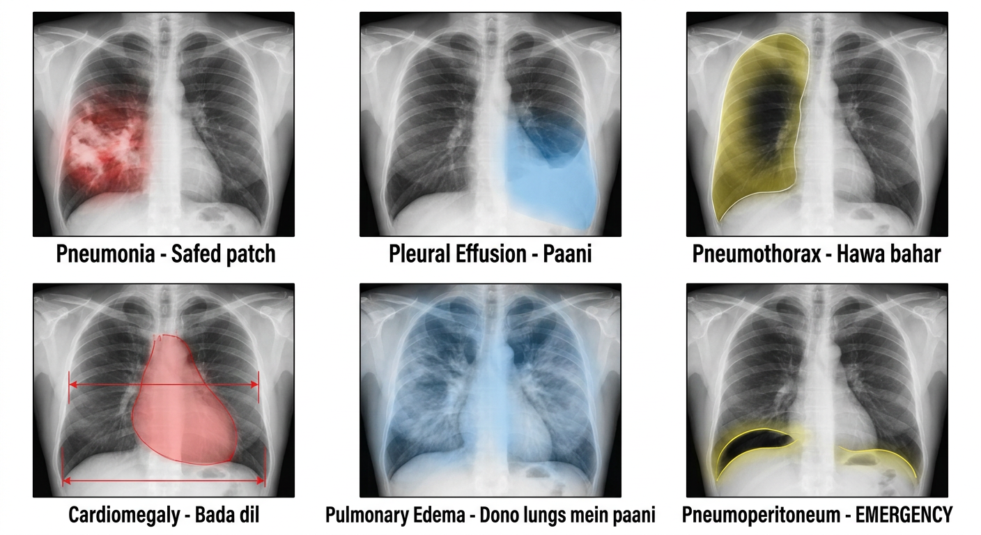
Images ka Summary:
- Sabhi important structures labeled hain
- Cardiothoracic ratio (50% rule) dikhaya gaya hai
- 5 densities ka legend (black se white tak)
- Right diaphragm left se upar dikhti hai
| # | Abnormality | Kya dikhega |
|---|---|---|
| 1 | Pneumonia | Safed patchy opacity |
| 2 | Pleural Effusion | Corner (costophrenic angle) blunt ho jaata hai |
| 3 | Pneumothorax | Lung ke bahar kali jagah, lung border visible |
| 4 | Cardiomegaly | Heart >50% chest width |
| 5 | Pulmonary Edema | Dono taraf butterfly shape mein safed |
| 6 | Pneumoperitoneum | Diaphragm ke neeche hawa - EMERGENCY |