
analyse_media
Reading File
Now let me search the medical library for relevant information on this finding while also doing a web search in parallel.
Finding Sources
Finding Sources
Searching the Web
giant cell tumor bone distal femur epiphysis soap bubble lytic lesion young adult radiology
Now let me read the key textbook sections on GCT for a comprehensive answer.
Reading File
Reading File
I have enough information for a comprehensive answer. Let me fetch the textbook X-ray image for comparison.
Loading Image
Here is a full radiological interpretation and clinical analysis:
X-Ray Interpretation - Left Knee (AP + Lateral)
Patient: Sanjeevulu, 30-year-old Male | Left knee | Mediciti Hospital, Ghanpur
Radiological Findings
Location:
- Large, eccentric lytic lesion involving the medial femoral condyle of the distal left femur
- The lesion is epiphyseal-metaphyseal in distribution, extending to the subchondral bone (abutting the articular surface)
- The physeal plates appear closed - consistent with a skeletally mature individual
Lesion Characteristics:
- Well-defined geographic lysis with a narrow zone of transition
- Classic "soap-bubble" appearance - internal bony septations and trabeculations visible on both AP and lateral views
- No periosteal reaction (no Codman triangle, no sun-burst pattern)
- No mineralized tumor matrix (no chondroid calcification, no osteoid)
- The lateral cortex appears thinned and potentially expanded but not overtly destroyed
- No significant soft tissue mass visible
- Joint space appears relatively preserved
Most Likely Diagnosis: Giant Cell Tumor (GCT) of Bone
This X-ray has a near-textbook appearance of GCT. The key features that point to this diagnosis are:
| Feature | Present in this X-ray | GCT Hallmark |
|---|---|---|
| Age 20-40 years | Yes (30 yr) | Yes |
| Closed physis | Yes | Required |
| Epiphyseal location | Yes | Required |
| Extends to subchondral bone | Yes | Classic |
| Eccentric lytic lesion | Yes | Classic |
| Soap-bubble trabeculation | Yes | Classic |
| No periosteal reaction | Yes | Classic |
| No matrix calcification | Yes | Classic |
| Distal femur location | Yes | Most common site |
As Robbins Pathology states: "Giant cell tumors develop within the epiphysis and may extend into the metaphysis. The majority occur near the knee in the distal femur or proximal tibia." - Robbins, Cotran & Kumar Pathologic Basis of Disease
The Rheumatology textbook further notes: "Giant cell tumor of bone is a benign but locally aggressive neoplasm that usually affects young adults... about two thirds of patients are between ages 20 and 40 years. Giant cell tumors occur most commonly in the distal femur, proximal tibia, and distal radius. These locations account for about 65% of cases." - Rheumatology, 2-Volume Set (Elsevier)
For comparison, here is a classic textbook GCT of the distal femur:
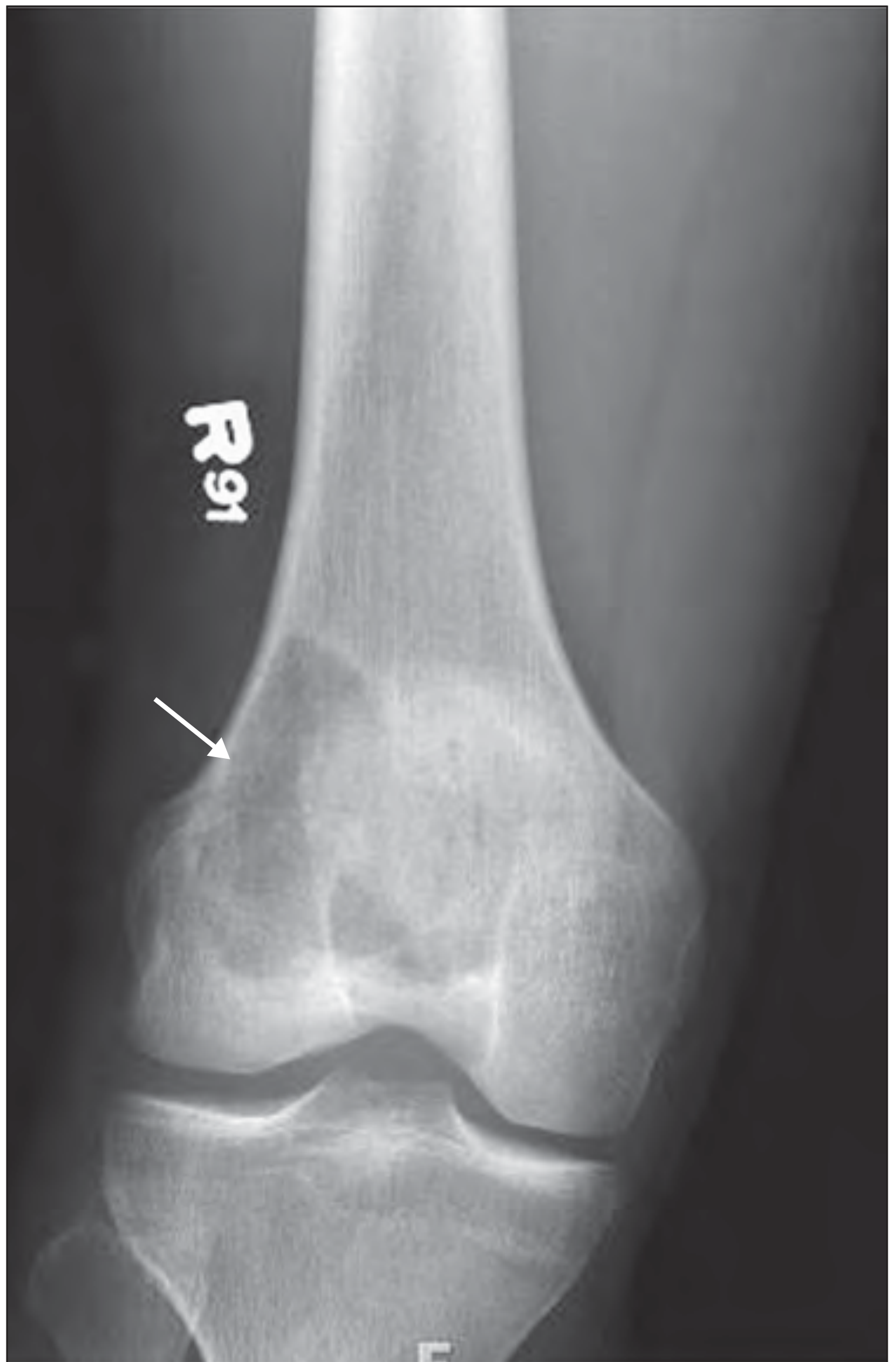
Pathophysiology (Brief)
The neoplastic cells in GCT are primitive osteoblast precursors that overexpress RANKL, driving uncontrolled osteoclast proliferation and bone destruction. The giant cells themselves (osteoclast-type, up to 100+ nuclei) are non-neoplastic - they are the effectors, not the drivers. Mutations in the H3-3A gene (histone 3.3) are the molecular hallmark.
Differential Diagnosis
| Diagnosis | Why Considered | Why Less Likely Here |
|---|---|---|
| Aneurysmal Bone Cyst (ABC) | Soap-bubble, lytic, young adult | More metaphyseal; physis often open; this is epiphyseal with closed physis |
| Chondroblastoma | Epiphyseal lytic, young patient | Occurs before physeal closure (age 10-20); often has chondroid matrix calcification |
| Clear Cell Chondrosarcoma | Epiphyseal, distal femur | Very rare; usually has some calcification; older age |
| Brown Tumor (Hyperparathyroidism) | Similar histology | Polyostotic; check PTH/Ca²+ levels |
| Telangiectatic Osteosarcoma | Lytic, destructive | Periosteal reaction, soft tissue mass, aggressive margins |
Recommended Next Steps
-
MRI of the left knee (most important) - to assess:
- Soft tissue extension beyond cortex
- Involvement of neurovascular structures
- Presence of secondary ABC change (fluid-fluid levels on T2)
- True extent before surgery planning
-
CT chest - baseline staging (GCT can metastasize to lung in ~4% of cases, even when histologically benign)
-
Biopsy - preferably a core needle or incisional biopsy for histological confirmation (the classic appearance of osteoclast-type giant cells admixed with mononuclear stromal cells)
-
Labs - serum calcium, PTH, ALP to exclude brown tumor of hyperparathyroidism
Treatment Overview
- Extended curettage + high-speed burring - the standard surgical approach, followed by packing with bone cement (PMMA) or bone graft
- Local recurrence rate is 40-60% after curettage alone
- Denosumab (RANKL inhibitor) - used preoperatively to shrink the tumor, or as primary treatment when surgery would cause unacceptable morbidity (as per [Robbins Pathology])
- Wide resection + reconstruction may be needed for extensive lesions with joint compromise
- Adjuvants (liquid nitrogen, phenol, hydrogen peroxide) reduce recurrence risk post-curettage
Important caveat: This interpretation is based on radiographic appearance only. Definitive diagnosis requires histopathological confirmation after biopsy. Clinical correlation and MRI are strongly advised before surgical planning.
This is a shared conversation. Sign in to Orris to start your own chat.