
Inverted papilloma
inverted papilloma nasal sinonasal histology
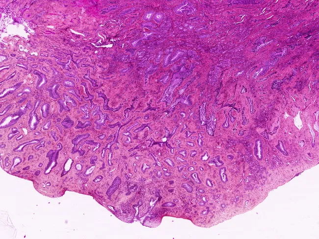
Light microscopy of a hematoxylin-eosin stained nasal polyp from the left nares of a 48-year-old man. Site: nasal cavity with predominant posterior septal involvement. Histology reveals a proliferation of back-to-back glandular spaces lined by ciliated, pseudostratified respiratory epithelium with a continuous basal cell layer. Glands are well differentiated and variably sized, forming tubular to cystic structures embedded in a fibromyxoid stroma. There is no cytologic atypia, mitotic activity, or destructive invasion. Immunohistochemistry shows p63 positivity in basal cells and absence of smooth muscle actin (SMA) labeling in myoepithelial cells, supporting a benign respiratory epithelial adenomatoid hamartoma (REAH). The overall pattern is characteristic of a hamartomatous lesion rather than a true adenocarcinoma, though some discuss its neoplastic relation to low-grade sinonasal adenocarcinoma. Differential diagnosis includes low-grade sinonasal adenocarcinoma, inverted papilloma, and inflammatory polyps; architectural stability and lack of atypia favor REAH. Clinical correlation: this lesion commonly presents with nasal obstruction or polyp formation; treatment is surgical excision with favorable prognosis and low recurrence. Significance: accurate recognition prevents overtreatment, informs prognosis, and guides ENT management. Terminology: REAH, respiratory epithelial adenomatoid hamartoma, nasal glandular hamartoma. This description emphasizes differential diagnosis, immunohistochemical profile, and clinical decision-making in sinonasal glandular lesions.
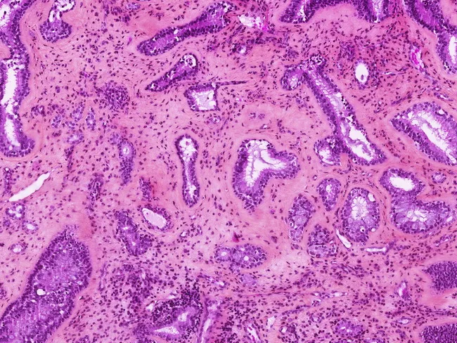
This is a hematoxylin and eosin stained histology image of a sinonasal mucosal lesion evaluated by light microscopy. The specimen is a biopsy/tissue fragment from nasal/paranasal sinus mucosa. The section reveals a network of irregular, gland-forming structures embedded in a fibromyxoid stroma. Glandular lumina vary in size and shape, lined by cuboidal to columnar epithelium with modest basophilic nuclei and scant cytoplasm. Mucin-containing cells or secretory material may be present within luminal spaces. There is a notable absence of overt cytologic atypia or high-grade mitotic activity in this field; no obvious stromal invasion is evident in the captured cortex. Inflammatory cells are scattered, and the architecture shows focal cribriform or tubular patterns consistent with glandular differentiation along Schneiderian mucosa. The histologic differential diagnosis includes intestinal-type adenocarcinoma and other salivary-type adenocarcinomas, as well as inverted papilloma (Schneiderian papilloma) when architectural features and epithelial redundancy are considered. Diagnostic significance rests on distinguishing ITAC from benign/inflammatory sinonasal lesions; immunohistochemistry and clinical context (exposure history, imaging) aid in differential. This image is relevant for surgical pathology, educational histology, and radiologic-pathologic correlation in ENT oncology workups and pathology teaching files. Clinically, it supports multidisciplinary assessment and informs surgical planning and follow-up management for patient care.
inverted papilloma sinonasal
Inverted Papilloma (Sinonasal)
Definition and Classification
| Type | Growth Pattern | HPV | Malignant Risk |
|---|---|---|---|
| Inverted (most common) | Endophytic - epithelium grows inward into stroma | Controversial (22-26% positivity) | 5-15% |
| Exophytic | Exophytic fronds with fibrovascular cores | Low-risk HPV 6/11 (dominant) | Exceptional |
| Oncocytic | Both inverted + exophytic | Not demonstrated | 4-17% |
Epidemiology
- Sex: Male predominance (M:F = 2-3:1)
- Age: Peak in the 5th-6th decades
- Bilateral involvement: Exceptional
Site of Origin
- Lateral nasal wall and maxillary sinus (medial wall, especially near the fontanelles) - most common
- Ethmoid region: ~48% in some series
- Frontal sinus: ~1.6-15% (rare primary site)
- Sphenoid sinus: Rarely involved primarily
- Multiple sites simultaneously in ~30% of cases, making identification of the precise origin challenging
Histology
- Epithelium: multilayered squamous or ciliated columnar cells mixed with mucocytes and transmigrating neutrophils
- Stromal invasion is absent in benign IP
- May occasionally coexist with sinonasal hamartomas
- Activating EGFR mutations are characteristic and preserved even in malignant transformation
- KRAS mutations are never found (in contrast to oncocytic papilloma, which always has KRAS mutations)
Etiology and Risk Factors
| Factor | Evidence |
|---|---|
| Organic solvent exposure | Significant dose-response relationship |
| HPV | Controversial - overall positivity ~22-26%; high-risk HPV subtypes found in 56% of dysplasia and 55% of carcinoma ex-IP in older studies, but transcriptionally active HPV not detected in most recent series |
| Smoking | Not linked to IP development, but confers 12-fold higher risk of malignant transformation |
| Alcohol | No association demonstrated |
Clinical Features
- Unilateral nasal obstruction (most common presenting complaint)
- Watery rhinorrhea
- Unilateral rhinosinusitis (headache, facial pain from sinus obstruction)
- Epiphora, proptosis, diplopia - with orbital involvement; raise suspicion for malignant transformation
Imaging
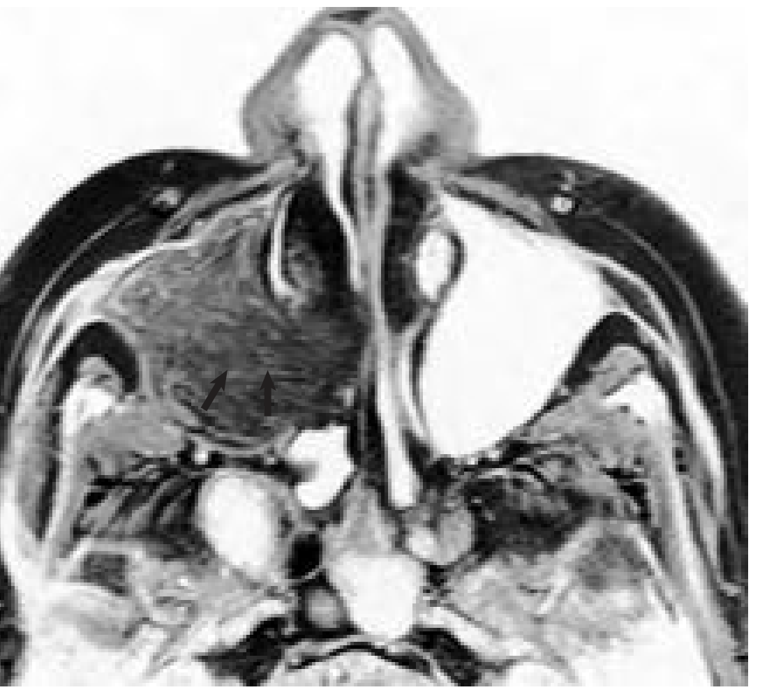
- Better differentiates tumor from retained secretions and inflammatory mucosal changes
- Cerebriform-columnar pattern on MRI (alternating parallel folds of hypercellular epithelium and less cellular stroma) is highly predictive of IP
- Identifies site of origin via bony spur, focal hyperostosis, or osteitic changes
- Loss of the cerebriform-columnar pattern + infiltrative growth suggests malignant transformation
Malignant Transformation
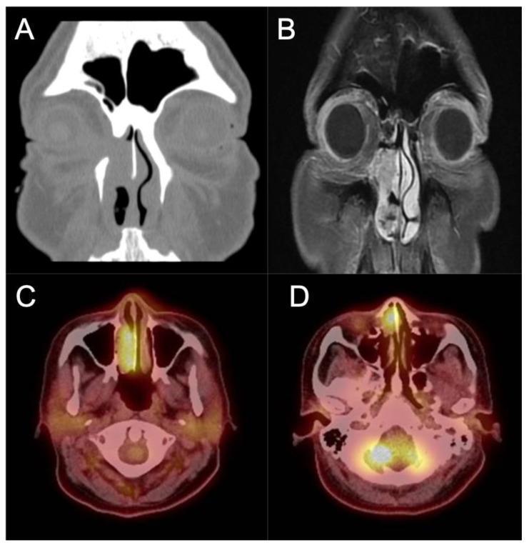
- Occurs in 5-15% of cases
- Synchronous (found at same time as IP) more frequent than metachronous
- Majority are squamous cell carcinomas; rare: sinonasal undifferentiated carcinoma, mucoepidermoid carcinoma, verrucous carcinoma
- Smoking is the strongest modifiable risk factor (12-fold increased risk)
Staging (Krouse Classification)
| Stage | Description |
|---|---|
| T1 | Tumor confined to nasal cavity |
| T2 | Tumor in nasal cavity + ethmoid sinuses/medial maxillary wall |
| T3 | Tumor involves lateral, inferior, superior, anterior, or posterior maxillary walls; sphenoid or frontal sinuses |
| T4 | All tumors with extra-sinonasal extension or malignancy |
Treatment
- No facial incision
- Negligible facial swelling
- Shorter inpatient stay
- Less postoperative pain/paraesthesia
- Endoscopy reduces recurrence risk by one-third to one-half vs. external approaches
- Identify the site of origin - the pedicle/attachment point is the single most important factor
- Subperiosteal dissection of the involved mucosa
- Drilling of the underlying bone at the attachment site to remove microscopic tumor digitations within bone
- Extent of surgery is dictated by site of origin, not by total tumor bulk
- Concomitant malignancy in critical areas
- Site of origin on the anterior wall/lateral recess of the frontal sinus, or massive frontal sinus mucosal involvement
- Orbital involvement (rare, usually in recurrent cases)
Recurrence
- Predominantly due to residual disease at the attachment site rather than true biologic recurrence
- HPV status and recurrence: a 2025 meta-analysis (Alqudrah et al., PMID 39739414) examined the link between HPV and recurrence
- A 2026 systematic review (Aguilera et al., PMID 41593213) examined pheno-endotyping and predictive markers of recurrence and malignancy - the most current evidence suggests the field is moving toward molecular stratification of recurrence risk