
Skin tumours surgery
- Benign lesions - cysts, lipomas, nevi, seborrhoeic keratoses
- Skin cancer surgery - basal cell carcinoma, squamous cell carcinoma, melanoma
- Operative principles - biopsy choices, excision margins, primary closure, grafts and flaps
- Mohs micrographic surgery
- Sentinel lymph-node biopsy and nodal surgery
- Reconstruction and postoperative care
- Complications and follow-up surveillance
Explain all
-
Assessment and diagnosis
History, full skin and lymph-node examination, dermoscopy, photographic documentation, biopsy selection, histopathology, staging, and multidisciplinary referral. -
Benign and premalignant lesions
Excision of epidermoid cysts, lipomas, nevi, and treatment of actinic keratosis, Bowen disease, and keratoacanthoma where indicated. -
Basal cell carcinoma (BCC)
Usually managed by surgical excision or Mohs surgery. Risk category and location determine margins and whether margin-controlled surgery is preferred. -
Cutaneous squamous cell carcinoma (cSCC)
Requires risk assessment for local recurrence, perineural invasion, and lymph-node spread. Treatment is complete excision, often with wider margins for high-risk disease. -
Melanoma
Requires a correctly planned diagnostic excision biopsy, then definitive wide local excision based on Breslow thickness. Some patients need sentinel lymph-node biopsy and staging. -
Other malignant skin tumours
Merkel cell carcinoma, dermatofibrosarcoma protuberans, adnexal tumours, and cutaneous sarcomas generally require specialist multidisciplinary care. -
Operative techniques and reconstruction
Elliptical excision, curettage, shave procedures, primary closure, skin grafts, local flaps, and Mohs surgery. -
Nodal surgery and systemic coordination
Sentinel-node biopsy, therapeutic nodal dissection in selected circumstances, imaging, radiotherapy, and systemic treatment are coordinated with oncology. -
Complications, pathology review, and surveillance
Bleeding, infection, wound dehiscence, scar problems, incomplete excision, recurrence, new primary tumours, and structured follow-up.
Which topic should I explain first in full exam-note detail?
- Benign lesions - cysts, lipomas, nevi, seborrhoeic keratoses
- Skin cancer surgery - basal cell carcinoma, squamous cell carcinoma, melanoma
- Operative principles - biopsy choices, excision margins, primary closure, grafts and flaps
- Mohs micrographic surgery
- Sentinel lymph-node biopsy and nodal surgery
- Reconstruction and postoperative care
- Complications and follow-up surveillance
-
Assessment and diagnosis
History, full skin and lymph-node examination, dermoscopy, photographic documentation, biopsy selection, histopathology, staging, and multidisciplinary referral. -
Benign and premalignant lesions
Excision of epidermoid cysts, lipomas, nevi, and treatment of actinic keratosis, Bowen disease, and keratoacanthoma where indicated. -
Basal cell carcinoma (BCC)
Usually managed by surgical excision or Mohs surgery. Risk category and location determine margins and whether margin-controlled surgery is preferred. -
Cutaneous squamous cell carcinoma (cSCC)
Requires risk assessment for local recurrence, perineural invasion, and lymph-node spread. Treatment is complete excision, often with wider margins for high-risk disease. -
Melanoma
Requires a correctly planned diagnostic excision biopsy, then definitive wide local excision based on Breslow thickness. Some patients need sentinel lymph-node biopsy and staging. -
Other malignant skin tumours
Merkel cell carcinoma, dermatofibrosarcoma protuberans, adnexal tumours, and cutaneous sarcomas generally require specialist multidisciplinary care. -
Operative techniques and reconstruction
Elliptical excision, curettage, shave procedures, primary closure, skin grafts, local flaps, and Mohs surgery. -
Nodal surgery and systemic coordination
Sentinel-node biopsy, therapeutic nodal dissection in selected circumstances, imaging, radiotherapy, and systemic treatment are coordinated with oncology. -
Complications, pathology review, and surveillance
Bleeding, infection, wound dehiscence, scar problems, incomplete excision, recurrence, new primary tumours, and structured follow-up.
Benign tumor
benign skin lesions AND excision
benign skin lesion excision guidance epidermoid cyst lipoma nevus official
Benign skin tumours: surgical overview
1. Preoperative assessment
- Duration and rate of growth
- Pain, itching, discharge, ulceration, trauma, infection, bleeding
- Previous excision or recurrence
- Personal or family history of skin cancer
- Anticoagulant use, diabetes, immunosuppression, allergy, keloid tendency
- Lesion site, size, depth, mobility, relation to nerves and vessels
- Regional lymph nodes if there is diagnostic concern
- Recent rapid enlargement
- Pigment change, asymmetry, irregular border, multiple colours
- Persistent ulceration or spontaneous bleeding
- Induration, fixation, nerve symptoms
- Recurrent lesion after adequate excision
- A deep, firm, enlarging soft-tissue mass
Common benign lesions and their surgery
| Lesion | Typical clinical features | Usual management |
|---|---|---|
| Epidermoid cyst | Slow-growing, firm mobile dermal nodule, often with a central punctum | Observe if asymptomatic. Definitive treatment is complete excision of cyst and wall |
| Pilar cyst | Smooth scalp nodule, usually no punctum | Excision, often easily delivered through a small incision |
| Lipoma | Soft, lobulated, mobile subcutaneous mass | Reassure or excise if painful, enlarging, symptomatic, or uncertain diagnosis |
| Melanocytic nevus (mole) | Symmetrical stable pigmented papule or macule | Observation if clinically typical. Excisional biopsy if changing or suspicious; cosmetic excision can be considered |
| Skin tag | Soft, pedunculated lesion in flexures | Snip excision, electrosurgery, or cryotherapy if symptomatic |
| Dermatofibroma | Firm papule, commonly legs, often dimples on pinching | Usually observe. Excision only if symptomatic or diagnosis is uncertain |
| Seborrhoeic keratosis | “Stuck-on,” waxy, pigmented plaque | Curettage, shave removal, or cryotherapy if irritated or diagnosis unclear |
| Pyogenic granuloma | Rapidly growing red friable nodule that bleeds easily | Curettage or shave excision with destruction of the base; send tissue for pathology |
| Neurofibroma | Soft papule or nodule, sometimes multiple | Observation or excision for symptoms, function, or cosmetic reasons |
| Hemangioma / vascular lesion | Red-blue vascular lesion | Observation, laser, sclerotherapy, or selected excision depending on type and location |
2. Epidermoid cyst
Indications for excision
- Recurrent inflammation or infection
- Pain, pressure symptoms, foul discharge
- Repeated rupture
- Cosmetic concern
- Uncertain diagnosis
Surgical principle
Technique, in principle
- Local anaesthesia.
- Elliptical or small incision, including the punctum or any scar tethered to the cyst.
- Gentle dissection and removal of the cyst intact where possible.
- Haemostasis, irrigation if needed, and closure.
- Send specimen for histology if atypical, recurrent, solid, ulcerated, or clinically uncertain.
Infected or inflamed cyst
- A fluctuant abscess may require incision and drainage.
- Antibiotics are considered when there is surrounding cellulitis or systemic infection, not for every inflamed cyst.
- Delayed definitive excision is often easier once acute inflammation has settled.
3. Lipoma
When to investigate or refer
- Deep to fascia
- Firm or fixed
- Rapidly enlarging
- Painful without a clear reason
- Recurrent after removal
- Large, especially more than about 5 cm
- Associated with neurologic symptoms
Excision
- Incision is placed along skin-tension lines where possible.
- The lipoma is dissected out, generally by marginal excision.
- The specimen should be sent for pathology when diagnosis is not completely certain.
4. Melanocytic nevus (mole)
Indications for removal
- Suspicion of melanoma
- Persistent irritation from shaving or clothing
- Recurrent trauma or bleeding
- Atypical appearance or documented change
- Cosmetic reason after clinical assessment
Important surgical point
5. Dermatofibroma
- Stable, asymptomatic lesions can be left alone.
- Excision may be chosen for pain, repeated trauma, diagnostic doubt, or cosmetic concern.
- Because it extends into the dermis, full-thickness excision gives better diagnostic tissue and more complete removal than superficial shaving.
- Scarring can be more noticeable than the original lesion, so the benefit should be discussed.
6. Pyogenic granuloma
Management
- Curettage or shave excision
- Electrocautery, chemical cautery, or other destruction of the base to reduce bleeding and recurrence
- Histopathology is advisable because amelanotic melanoma and other lesions can occasionally resemble it
7. General operative principles
Incision and excision
- Plan the scar along relaxed skin-tension lines when possible.
- For an elliptical excision, the long axis is usually aligned with these lines to allow primary closure.
- Handle tissue gently and achieve careful haemostasis.
- Avoid excessive cautery, which can delay healing and worsen scarring.
- Close in layers for deeper defects and reduce tension.
Specimen handling
- Diagnosis is uncertain
- The lesion is pigmented, ulcerated, firm, recurrent, rapidly growing, or unusual
- A malignancy cannot confidently be excluded
- The appearance differs from a routine benign lesion
Reconstruction
- Undermining and layered closure
- Local skin flap
- Full-thickness or split-thickness skin graft
- Specialist plastic-surgical reconstruction, especially on the face, eyelid, ear, lip, hand, genitalia, or lower leg
8. Postoperative care and complications
Usual advice
- Keep the wound clean and dry according to the clinician’s instructions.
- Watch for increasing pain, redness, warmth, swelling, pus, fever, bleeding, or wound separation.
- Avoid tension, stretching, and friction at the wound site.
- Use sun protection after healing to limit scar pigmentation.
Complications
- Bleeding or haematoma
- Infection
- Wound dehiscence
- Hypertrophic scar or keloid
- Nerve injury, numbness, altered sensation
- Recurrence, especially after incomplete cyst-wall or nevus removal
- Unexpected malignant diagnosis on histology
Explain acc to standard book
benign skin lesions AND treatment
Schwartz Principles of Surgery benign skin tumors epidermal cyst lipoma dermatofibroma
Benign tumours of skin - standard textbook style
Definition
General principles of management
- Confirm the diagnosis clinically, with dermoscopy for pigmented lesions where available.
- Do not destroy an uncertain lesion by cautery, cryotherapy, laser, or superficial shaving without obtaining histology.
- Send the specimen for histopathology when diagnosis is doubtful, the lesion is recurrent, rapidly enlarging, ulcerated, bleeding, unusually pigmented, fixed, or otherwise suspicious.
- Excise completely if removal is indicated.
- Plan the incision in a relaxed skin-tension line and use layered closure if the lesion is deep or the closure is under tension.
- Suspected melanoma, squamous-cell carcinoma, or other malignancy requires appropriate diagnostic excision or specialist referral, not cosmetic destruction.
Classification
A. Epidermal and adnexal lesions
- Epidermoid cyst
- Pilar or trichilemmal cyst
- Seborrhoeic keratosis
- Syringoma
- Sebaceous hyperplasia
- Hidrocystoma
B. Melanocytic lesions
- Acquired melanocytic nevus
- Congenital melanocytic nevus
- Spitz nevus
- Blue nevus
C. Fibrous and histiocytic lesions
- Dermatofibroma
- Keloid
- Neurofibroma
D. Adipose and soft-tissue lesions
- Lipoma
- Angiolipoma
E. Vascular lesions
- Cherry angioma
- Pyogenic granuloma, also called lobular capillary hemangioma
- Hemangioma
F. Other common lesions
- Acrochordon or skin tag
- Verruca
- Xanthelasma
1. Epidermoid cyst
Definition and pathology
Clinical features
- Commonest cutaneous cyst
- Usually occurs on face, neck, upper trunk, or scrotum
- Slow-growing, round, mobile dermal or subcutaneous swelling
- Skin-coloured or yellowish
- Characteristic central punctum
- May discharge foul-smelling, cheesy keratin
- Infection or rupture causes pain, erythema, tenderness, and abscess formation
Treatment
- Observation if small and asymptomatic.
- Definitive treatment: complete excision of the cyst together with its wall.
- The punctum and attached scar should be included in the excision.
- Incomplete removal of the lining causes recurrence.
- Elective excision is best performed when the cyst is not inflamed, that is, in the “cold” stage.
- In an abscess, perform incision and drainage. Antibiotics are used when there is surrounding cellulitis or systemic infection. Definitive excision can follow after inflammation settles.
2. Pilar or trichilemmal cyst
Features
- Arises from the outer root sheath of the hair follicle.
- Commonly occurs on the scalp.
- Often multiple and can be familial.
- Usually smooth, firm, mobile, and lacks a central punctum.
- Contents are more solid and calcification may occur.
Treatment
- Surgical excision is curative.
- These cysts are often more easily enucleated through a small incision than epidermoid cysts.
3. Lipoma
Definition
Clinical features
- Soft, doughy, lobulated, and freely mobile subcutaneous swelling
- Usually painless
- Common on trunk, shoulder, neck, forearm, and thigh
- Usually small, but may grow large
- Overlying skin is normal
Differential diagnosis
- Epidermoid cyst
- Angiolipoma
- Neurofibroma
- Enlarged lymph node
- Soft-tissue sarcoma or atypical lipomatous tumour
Treatment
- Observation if typical and asymptomatic.
- Excision for pain, pressure symptoms, cosmetic concern, enlargement, or uncertain diagnosis.
- A deep, fixed, painful, recurrent, rapidly growing, or large mass should be investigated, often with ultrasonography or MRI, and referred appropriately before excision.
- Marginal excision is usually adequate for a typical superficial lipoma.
4. Melanocytic nevus
Definition
Clinical features
- Usually a symmetrical, stable, uniformly coloured macule, papule, or nodule
- Colour varies from skin coloured to tan, brown, blue, or black
- Acquired nevi usually arise in childhood or early adult life
When to excise
- Cosmetic concern
- Recurrent trauma or irritation
- Recent change in size, colour, surface, or symptoms
- Irregular pigmentation or asymmetry
- Bleeding or ulceration
- Concern for melanoma
Treatment principles
5. Seborrhoeic keratosis
Features
- Benign epidermal proliferation in middle-aged and older people
- Waxy, verrucous, sharply demarcated plaque
- Classically has a “stuck-on” appearance
- May be tan, brown, grey, or black
- Usually occurs on trunk, face, and scalp
Treatment
- Cryotherapy with liquid nitrogen
- Curettage, with or without cautery
- Shave excision
6. Dermatofibroma
Definition and features
- Commonly occurs on the legs
- Firm, small, brown or reddish-brown papule/nodule
- May itch or be tender
- Characteristic dimple sign: lateral compression produces central dimpling
Treatment
- Reassure if stable and asymptomatic.
- Excision is indicated for symptoms, diagnostic doubt, repeated trauma, or cosmetic concern.
- Because the lesion is dermal, complete excision is preferable to superficial shaving when removal is needed.
- Consider dermatofibrosarcoma protuberans in a large, enlarging, atypical, or recurrent lesion.
7. Pyogenic granuloma
Definition
Clinical features
- Rapidly growing red, friable papule or pedunculated nodule
- Bleeds easily after trivial trauma
- Common on fingers, face, oral mucosa, and sites of trauma
- May occur in pregnancy on the gingiva, called granuloma gravidarum
Treatment
- Shave excision followed by curettage and electrodesiccation of the base
- Alternatively, full-thickness surgical excision
- Histopathology is important because amelanotic melanoma can resemble a pyogenic granuloma
- Residual lesion at the base can lead to recurrence
8. Acrochordon or skin tag
Features
- Soft, pedunculated, skin-coloured fibroepithelial polyp
- Occurs in neck, axilla, groin, eyelids, and other flexures
- More common in obesity, pregnancy, and insulin resistance
Treatment
- Snip excision
- Electrocautery
- Cryotherapy
9. Neurofibroma
Features
- Benign peripheral-nerve sheath tumour
- Soft, skin-coloured, sessile or pedunculated nodule
- May show the “buttonhole” sign, in which it invaginates on pressure
- Multiple neurofibromas should raise concern for neurofibromatosis type 1
Treatment
- Observation for asymptomatic isolated lesions
- Excision for pain, functional impairment, diagnostic uncertainty, or cosmetic reasons
- Multiple lesions need assessment for associated genetic syndrome and systemic manifestations.
10. Cherry angioma
Features
- Common benign capillary proliferation in adults
- Bright red to purple, small dome-shaped papules
- Often multiple on the trunk
Treatment
- Electrocautery
- Laser
- Shave excision
Short surgical answer: principles of excision of a benign skin tumour
- Confirm benign clinical diagnosis and obtain consent.
- Mark lesion and incision along relaxed skin-tension lines.
- Prepare and infiltrate local anaesthetic.
- Make an elliptical incision if complete skin excision is required.
- Dissect carefully around the lesion and remove it intact where possible.
- Ensure haemostasis.
- Send specimen for histopathology where indicated.
- Close without tension, usually with deep absorbable sutures and superficial skin sutures if required.
- Apply dressing, provide wound-care instructions, and arrange review for histology and suture removal.
Indications for histopathology
- Uncertain diagnosis
- Pigmented or changing lesion
- Ulceration, spontaneous bleeding, or rapid growth
- Recurrent lesion
- Atypical clinical appearance
- Any lesion where malignancy has not been confidently excluded
Explain malignant
cutaneous melanoma AND surgery
NCI PDQ skin cancer treatment basal cell squamous cell melanoma surgery
clinical comparison basal cell carcinoma squamous cell carcinoma melanoma skin cancer
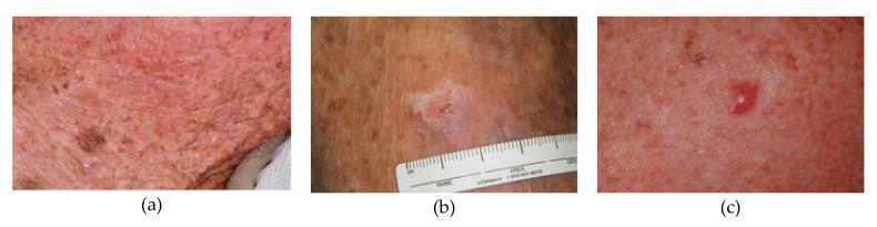
This clinical photograph set presents a side-by-side comparison of three distinct types of skin cancer lesions labeled (a), (b), and (c). Image (a) demonstrates malignant melanoma, characterized by an irregularly pigmented, dark brown to black macule situated on a background of sun-damaged, erythematous skin with a rough texture and multiple small papules. Image (b) shows a squamous cell carcinoma (SCC), presenting as a raised, scaly, flesh-colored plaque with an irregular border and central crusting; a clinical ruler is included for scale. Image (c) displays a basal cell carcinoma (BCC), visible as a small, well-circumscribed, pearly erythematous nodule with possible central ulceration. This comparison chart is designed for medical education to assist in the clinical differentiation of primary skin malignancies based on morphology, color, and surface characteristics. Key concepts illustrated include lesion asymmetry, border irregularity, and characteristic tissue changes associated with different histopathological subtypes of skin cancer.
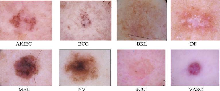
This clinical comparison chart presents eight dermoscopic images of various skin lesions, serving as an educational reference for dermatological classification. Each image represents a category from the ISIC 2019 dataset: Actinic Keratoses and Intraepithelial Carcinoma (AKIEC), Basal Cell Carcinoma (BCC), Benign Keratosis-like Lesions (BKL), Dermatofibroma (DF), Melanoma (MEL), Melanocytic Nevi (NV), Squamous Cell Carcinoma (SCC), and Vascular Lesions (VASC). The visual comparison highlights diagnostic morphological features, including the asymmetrical pigment distribution and irregular borders in MEL and NV, the translucent or pinkish hue with small dark spots in BCC, the central white area characteristic of DF, and the distinct reddish vascular structure in the VASC category. This figure is designed for medical students and clinicians to distinguish between malignant and benign skin tumors based on color variegation, textural patterns, and structural organization. It serves as a visual benchmark for training diagnostic algorithms and improving clinical accuracy in identifying high-level features associated with skin cancer.
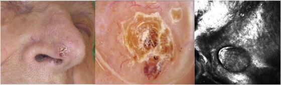
This composite figure illustrates the clinical, dermoscopic, and reflectance confocal microscopy (RCM) findings of a basosquamous carcinoma (BSC) on the nose. Image A (Clinical Photograph) shows an erythematous, squamous nodule located on the nasal tip, characterized by a central hyperkeratotic crust and irregular borders. Image B (Dermoscopy) reveals diagnostic features suggestive of both basal cell and squamous cell lineages: a central white-to-yellow keratin mass, focal blood spots, and peripheral arborizing telangiectasia against a whitish structureless background. Image C (RCM) displays the microscopic architecture at the dermo-epidermal junction, showing dark peritumoral clefts and basaloid islands with peripheral palisading, alongside larger polygonal cells representing atypical keratinocytes. This multimodal imaging comparison highlights the overlapping features of BCC and SCC that define BSC, providing high-resolution visual evidence for the diagnosis of this aggressive non-melanoma skin cancer subtype.
Malignant tumours of skin
- Basal cell carcinoma (BCC)
- Cutaneous squamous cell carcinoma (cSCC)
- Malignant melanoma
- Less common tumours: Merkel cell carcinoma, dermatofibrosarcoma protuberans, sebaceous carcinoma, angiosarcoma, cutaneous lymphoma, and Kaposi sarcoma.
General surgical principles
Assessment
- Duration, enlargement, pain, itch, bleeding, ulceration, or non-healing wound
- Sun or ultraviolet exposure, radiotherapy, arsenic exposure, chronic wound/scar
- Immunosuppression, especially organ transplant
- Previous skin cancer and family history of melanoma
- Change in a pigmented lesion
- Entire skin surface, lesion size, border, pigmentation, ulceration, and fixation
- Regional lymph nodes
- Cranial nerve function or altered sensation if head/neck SCC is suspected, because pain, paraesthesia, or anaesthesia can indicate perineural invasion.
Diagnosis
- Use dermoscopy for suspicious pigmented lesions.
- Confirm diagnosis by biopsy and histopathology.
- A likely melanoma should usually undergo complete excisional biopsy with a narrow margin, allowing Breslow thickness and ulceration to be assessed.
- For a large lesion or a site where total diagnostic excision is impractical, an incisional or punch biopsy should target the thickest or most atypical area.
- Do not treat a suspicious lesion with cryotherapy, laser, cautery, or superficial destruction before histological diagnosis.
Definitive surgery
- Complete tumour clearance with histologically negative margins.
- Preservation of function and acceptable reconstruction.
- Accurate staging.
- Nodal staging or treatment where appropriate.
- Surveillance for local recurrence, nodal disease, and new primary tumours.
1. Basal cell carcinoma
Definition
Risk factors
- Chronic ultraviolet exposure and fair skin
- Increasing age
- Immunosuppression
- Previous BCC
- Ionising radiation or arsenic exposure
- Genetic syndromes, for example basal cell nevus syndrome
Clinical types
- Nodular BCC: pearly papule or nodule, rolled edge, telangiectasia, possible central ulcer
- Superficial BCC: well-defined erythematous scaly patch, often trunk
- Pigmented BCC
- Morpheaform or infiltrative BCC: scar-like, ill-defined, more aggressive
- Basosquamous carcinoma: has aggressive behaviour and requires careful management
Treatment
Low-risk BCC
- Standard surgical excision with histological margin assessment
- Curettage and electrodesiccation in selected superficial, low-risk lesions
- Cryotherapy, topical imiquimod, topical 5-fluorouracil, or photodynamic therapy for selected superficial disease
High-risk BCC
- Central face, eyelid, nose, lip, ear, genital area, hands, or feet
- Recurrent tumour
- Large size
- Ill-defined border
- Aggressive histology, such as infiltrative or morpheaform type
- Perineural invasion
- Immunosuppression
Advanced disease
2. Cutaneous squamous cell carcinoma
Definition
Risk factors
- Cumulative ultraviolet exposure
- Actinic keratosis and Bowen disease
- Fair skin and older age
- Immunosuppression, particularly transplant recipients
- Chronic wounds, ulcers, burns, sinuses, and scars, called Marjolin ulcer
- Ionising radiation, arsenic, HPV in selected anogenital lesions
- Chronic inflammation
Clinical features
- Hyperkeratotic, scaly, indurated papule, plaque, or nodule
- Crusting, ulceration, or bleeding
- May be rapidly growing and painful
- Occurs mainly on bald scalp, face, neck, dorsal hands, forearms, and legs
Precursor lesions
Actinic keratosis
Bowen disease
Risk stratification
- Diameter more than 2 cm
- Thickness more than 2 mm or invasion beyond subcutaneous fat
- Poor differentiation
- Desmoplastic histology
- Perineural or lymphovascular invasion
- Ear, lip, central face, genital, hand, or foot location
- Recurrence
- Immunosuppression
Surgical treatment
Low-risk cSCC
- Complete surgical excision with histological margin assessment.
- Curettage and electrodesiccation may be used only in carefully selected low-risk lesions, not in hair-bearing or high-risk sites.
High-risk cSCC
- Mohs micrographic surgery or excision with comprehensive margin control.
- Consider imaging if there is concern for deep invasion, bone involvement, named-nerve involvement, or nodal metastasis.
- Perform ultrasound-guided needle sampling or biopsy of suspicious regional lymph nodes.
Nodal disease
3. Malignant melanoma
Definition
Risk factors
- Intermittent intense ultraviolet exposure and severe sunburn
- Fair skin, freckles, light eyes/hair
- Numerous or atypical nevi
- Personal or family history of melanoma
- Giant congenital melanocytic nevus
- Immunosuppression
- Genetic susceptibility
Warning signs: ABCDE
- A - Asymmetry
- B - Border irregularity
- C - Colour variation
- D - Diameter, especially enlargement or a lesion larger than 6 mm
- E - Evolution, any change in size, shape, colour, surface, bleeding, or symptoms
Diagnosis
- Breslow thickness
- Ulceration
- Mitotic activity
- Histological subtype
- Peripheral and deep margins
- Microsatellitosis, if present
Definitive treatment: wide local excision
| Melanoma thickness | Recommended clinical excision margin |
|---|---|
| Melanoma in situ | 0.5 cm |
| Less than or equal to 1 mm | 1 cm |
| More than 1 to 2 mm | 1 to 2 cm |
| More than 2 mm | 2 cm |
Sentinel lymph-node biopsy
- Discussed for clinically node-negative melanomas 0.8 to 1.0 mm thick with adverse factors
- Recommended or strongly considered for clinically node-negative melanomas more than 1 mm thick
Advanced melanoma
4. Merkel cell carcinoma
Features
Management
- Wide local excision or margin-controlled surgery
- Sentinel lymph-node biopsy is generally recommended for clinically node-negative disease
- Adjuvant radiotherapy is frequently used because of high local and regional recurrence risk
- Immunotherapy is important for advanced or metastatic disease
5. Other malignant skin tumours
Dermatofibrosarcoma protuberans
- Slow-growing, locally aggressive dermal sarcoma
- Often on trunk or proximal limbs
- Needs wide excision with clear margins or Mohs surgery
- Local recurrence is a concern
Sebaceous carcinoma
- Often arises in eyelid meibomian glands but can occur elsewhere
- May mimic recurrent chalazion or chronic blepharoconjunctivitis
- Requires complete excision with margin control and specialist assessment
Angiosarcoma
- Aggressive vascular malignancy, often on scalp or face of elderly people
- Requires multidisciplinary management with surgery where possible, radiotherapy, and systemic therapy
Cutaneous lymphoma and Kaposi sarcoma
- Management is usually nonsurgical and depends on disease subtype and systemic involvement.
Follow-up and prevention
- Lifelong skin self-examination
- Prompt review of any new, changing, bleeding, ulcerated, or non-healing lesion
- Sun-protective clothing, shade, and broad-spectrum sunscreen
- Examination of regional lymph nodes where relevant
- Regular clinical follow-up based on tumour type, stage, recurrence risk, and immunosuppression status
Exam summary
- BCC: locally destructive, metastasis is rare. Treat mainly by excision or Mohs surgery.
- cSCC: has risk of nodal metastasis. Excise completely; use Mohs surgery for high-risk tumours; assess nodes.
- Melanoma: diagnose by excisional biopsy; prognosis depends mainly on Breslow thickness; treat by wide local excision with thickness-based margins and consider sentinel-node biopsy.
- Merkel cell carcinoma: rare and aggressive; treat with surgery, sentinel-node staging, and usually radiotherapy.
Create a comparison table of BCC, SCC, and melanoma
cutaneous melanoma AND carcinoma
NCI skin cancer treatment basal cell squamous cell melanoma clinical features diagnosis surgery
Comparison of BCC, SCC, and melanoma
| Feature | Basal cell carcinoma (BCC) | Cutaneous squamous cell carcinoma (SCC) | Malignant melanoma |
|---|---|---|---|
| Cell of origin | Basal keratinocytes of epidermis/hair follicle | Squamous keratinocytes of epidermis | Melanocytes |
| Relative frequency | Most common skin cancer | Second most common skin cancer | Less common, but responsible for most skin-cancer deaths |
| Major risk factors | Chronic UV exposure, fair skin, age, immunosuppression, prior BCC, radiation, genetic syndromes | Chronic UV exposure, actinic keratosis, Bowen disease, chronic scars/ulcers, immunosuppression, arsenic, HPV in selected sites | Intermittent intense UV exposure and sunburn, fair skin, numerous/dysplastic nevi, family or personal history, immunosuppression |
| Common sites | Sun-exposed face, nose, eyelids, ears, scalp, neck, upper trunk | Bald scalp, face, ears, lips, neck, dorsal hands, forearms, shins; may occur in chronic wounds | Anywhere. Often trunk/head-neck in men; legs in women; can occur on nail unit, acral skin, mucosa, or eye |
| Typical appearance | Pearly or translucent papule/nodule; rolled edge; telangiectasia; central ulceration in advanced lesion | Scaly, hyperkeratotic, indurated papule, plaque, or nodule; crusting, ulceration, bleeding | Asymmetric pigmented macule, papule, plaque, or nodule with irregular border and variable colour; may be amelanotic |
| Useful clinical clue | “Rodent ulcer”: non-healing ulcer with pearly rolled border | Tender, rapidly enlarging keratotic or ulcerated lesion | ABCDE: Asymmetry, Border irregularity, Colour variation, Diameter/enlargement, Evolution |
| Precursor lesion | No typical obligatory precursor | Actinic keratosis; SCC in situ or Bowen disease | May arise in a pre-existing nevus, but many develop de novo |
| Growth pattern | Usually slow, locally invasive | Variable, often more rapid than BCC; can invade nerves and deep structures | Radial growth followed by vertical invasion; early potential for lymphatic and blood-borne spread |
| Local tissue destruction | Can be extensive if neglected, particularly on face | May invade cartilage, muscle, bone, and nerves | Local recurrence/in-transit disease possible; risk rises with thickness |
| Metastatic potential | Very rare | Definite risk, especially to regional nodes in high-risk tumours | High relative potential, depending especially on Breslow thickness, ulceration, and nodal status |
| High-risk features | Recurrent, large, ill-defined, aggressive histology, central-face location, perineural invasion, immunosuppression | Size more than 2 cm, deep invasion, poor differentiation, perineural/lymphovascular invasion, ear/lip location, recurrence, immunosuppression | Increased Breslow thickness, ulceration, high mitotic activity, microsatellites, nodal disease |
| Biopsy approach | Punch, shave, or excisional biopsy according to lesion/site | Punch, shave, incisional, or excisional biopsy according to lesion/site | Prefer complete excisional biopsy with a narrow margin whenever feasible, preserving accurate Breslow thickness |
| Main treatment | Surgical excision with margin assessment or Mohs micrographic surgery | Surgical excision with margin assessment or Mohs micrographic surgery | Wide local excision after diagnostic biopsy |
| Role of Mohs surgery | Important for high-risk, recurrent, ill-defined BCC and tissue-critical sites | Important for high-risk/recurrent SCC, aggressive histology, poorly defined margins, and high-risk sites | Not routine for ordinary invasive melanoma; staged margin-controlled techniques may be used in selected melanoma in situ, especially lentigo maligna |
| Margin principle | Risk-based margin or Mohs margin control | Risk-based margin or Mohs margin control | Margin based on Breslow thickness |
| Melanoma excision margins | Not applicable | Not applicable | In situ: 0.5 cm; ≤1 mm: 1 cm; >1-2 mm: 1-2 cm; >2 mm: 2 cm |
| Lymph-node assessment | Usually not needed | Examine nodes; image/biopsy suspicious nodes, especially in high-risk disease | Sentinel lymph-node biopsy is discussed for selected 0.8-1.0 mm lesions and generally recommended/strongly considered for clinically node-negative lesions >1 mm |
| Advanced disease | Hedgehog-pathway inhibitors or immunotherapy in selected locally advanced/metastatic cases | Radiotherapy and systemic treatment, commonly immunotherapy, in selected advanced disease | Immunotherapy, mutation-targeted therapy when appropriate, radiotherapy, and selected metastasectomy |
| Overall prognosis | Excellent with adequate treatment | Usually excellent when early, but high-risk disease can recur and metastasize | Depends mainly on stage, especially Breslow thickness and node status |
High-yield differences
- BCC: most common, pearly rolled edge, locally destructive, metastasis is very rare.
- SCC: scaly and keratotic, may arise from actinic keratosis or Bowen disease, can metastasize to regional lymph nodes.
- Melanoma: pigmented and changing lesion, assessed by ABCDE rule, prognosis depends mainly on Breslow thickness and nodal involvement.
Surgical exam points
- Do not use destructive treatment on a lesion suspicious for melanoma without histology.
- BCC and SCC are treated primarily by complete excision or Mohs surgery when margin control and tissue preservation are required.
- Melanoma requires diagnostic excision first, then definitive wide local excision based on Breslow thickness.
- Check regional lymph nodes in SCC and melanoma. Sentinel-node biopsy has a central staging role in selected melanoma.