I now have comprehensive content. Let me compile the full, detailed response on cerebellar ataxia.
Cerebellar Ataxia
Definition and Pathophysiology
Cerebellar ataxia refers to a disturbance in the smooth performance of voluntary motor acts, causing muscular incoordination or impaired balance. Movements are flawed in rate, range, timing, direction, and force. The cerebellum provides inhibitory and modulating influences on skilled movements originating in the cerebral motor cortex - when lost, movements become inaccurate and poorly controlled.
The cerebellum is functionally divided into:
- Midline (vermis/anterior lobe): Controls truncal balance and gait
- Hemispheres: Control ipsilateral limb coordination
- Flocculonodular lobe (vestibulocerebellum): Controls equilibrium and eye movements
Signs and Symptoms
Early Signs
| Domain | Features |
|---|
| Gait | Difficulty running, turning, walking on narrow base or stairs; wide-based stance |
| Speech | Dysarthria (slurred, scanning speech) |
| Hands | Tremor, clumsiness, poor handwriting |
| Eyes | Double vision (especially on quick head turns); nystagmus |
| Balance | Heightened sensitivity to alcohol; dizziness |
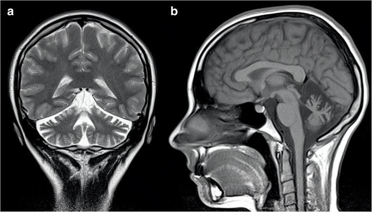
| Incidental | Cerebellar atrophy on neuroimaging |
Late Signs
- Falls, swallowing difficulty (dysphagia)
- Blurry vision
- Loss of hand dexterity (dressing, utensils)
Key Examination Domains (SARA Scale)
The Scale for Assessment and Rating of Ataxia (SARA) assesses: gait, stance, sitting, speech, finger-chase, nose-to-finger test, fast alternating movements, and heel-to-shin test (note: SARA does not include eye movements separately).
Topographic Signs
- Vermis/midline lesions: Wide-based lurching gait, truncal sway, titubation (3-Hz head and trunk tremor with anterior lobe degeneration); heel-shin test may appear normal supine
- Cerebellar hemisphere lesions: Ipsilateral limb ataxia - dysmetria, dysdiadochokinesis, decomposition of movement; little truncal instability if vermis spared
- Flocculonodular lobe lesions: Severe multidirectional body sway, sitting/standing may be impossible, but limb coordination can be relatively preserved
- Spastic ataxia: "Bouncing" gait seen in MS, Arnold-Chiari malformation, hydrocephalus
Classification by Onset (Temporal Pattern)
This is the most clinically useful initial classification:
Acute Onset (Minutes to Days)
- Ischemic or hemorrhagic cerebellar stroke
- Alcohol intoxication
- Toxins: mercury, thallium, toluene, solvents
- Medications: phenytoin, carbamazepine, phenobarbital, lithium
- Multiple sclerosis relapse
- Meningitis (especially basilar)
- Wernicke encephalopathy (thiamine deficiency)
- Biotinidase deficiency
- Acute postinfectious (e.g., varicella)
- Miller Fisher syndrome / Bickerstaff brainstem encephalitis (anti-GQ1b)
- Tick paralysis; labyrinthitis
Subacute Onset (Weeks to Months)
- Paraneoplastic cerebellar degeneration (anti-Yo, anti-Hu, anti-Ri) - common tumors: ovary, breast, lung
- Autoimmune: anti-GAD65, anti-CASPR2, gluten ataxia
- Prion disease (CJD)
- Infections (HIV, EBV, abscess)
- Nutritional: Wernicke, vitamin E deficiency
Chronic Progressive
- Genetic ataxias (see below)
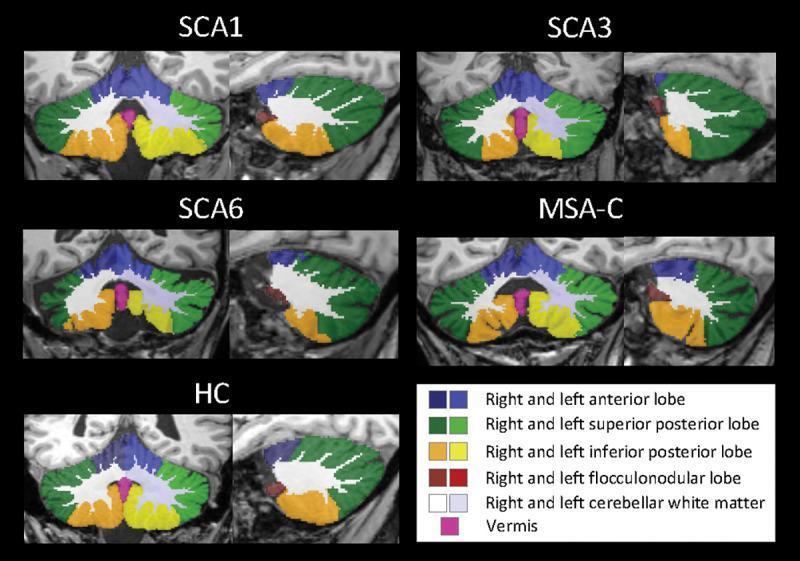
- Multiple system atrophy - cerebellar type (MSA-C)
- Normal pressure hydrocephalus
- Hypothyroidism
- Superficial CNS siderosis
- CANVAS (cerebellar ataxia, neuropathy, vestibular areflexia syndrome)
Episodic
- Episodic Ataxia type 1 (EA-1): KCNA1 mutation (potassium channel); myokymia interictal; episodes last seconds to minutes
- Episodic Ataxia type 2 (EA-2): CACNA1A mutation (calcium channel); gaze-evoked/downbeat nystagmus interictal; episodes last hours; responds to 4-aminopyridine
- Migraine with brainstem aura
- Metabolic: urea cycle defects, maple syrup urine disease, Hartnup disease
Genetic Causes
Autosomal Dominant - Spinocerebellar Ataxias (SCAs)
Over 40 SCA subtypes are recognized. Most common:
| SCA | Gene/Mutation | Clinical Clues |
|---|
| SCA1 | ATXN1 (CAG repeat) | Pyramidal signs, neuropathy |
| SCA2 | ATXN2 (CAG repeat) | Slow saccades, neuropathy |
| SCA3 (Machado-Joseph) | ATXN3 | Most common SCA worldwide; dystonia, bulging eyes |
| SCA6 | CACNA1A | Late onset, pure cerebellar |
| SCA7 | ATXN7 | Cerebellar ataxia + retinal degeneration |
| SCA17 | TBP | Resembles Huntington's disease |
Autosomal Recessive Cerebellar Ataxias (ARCAs)
| Condition | Gene | Key Features |
|---|
| Friedreich ataxia | FXN (GAA repeat) | Onset < 25 years; areflexia, cardiomyopathy, diabetes, Babinski |
| Ataxia-telangiectasia | ATM | Childhood onset; telangiectasias, immune deficiency, ↑AFP |
| Ataxia with oculomotor apraxia 1 (AOA1) | APTX | Oculomotor apraxia, neuropathy, ↓albumin |
| AOA2 | SETX | Oculomotor apraxia, ↑AFP, neuropathy |
| ARCA1 | SYNE1 (Nesprin-1) | ~5% of recessive ataxias |
| Cerebrotendinous xanthomatosis | CYP27A1 | Tendon xanthomas, cataracts, dementia; ↑plasma cholestanol |
| Ataxia with vitamin E deficiency | TTPA | Resembles Friedreich ataxia; ↓vitamin E; treatable |
| Abetalipoproteinemia | MTTP | Acanthocytosis, fat malabsorption; ↓vitamin E; treatable |
| POLG-ataxia | POLG | Mitochondrial; ↑SDH on muscle biopsy |
X-linked
- Fragile X-associated tremor/ataxia syndrome (FXTAS): FMR1 premutation (55-200 CGG repeats); late-onset in males; tremor + ataxia + parkinsonism + cognitive decline; MRI shows T2 hyperintensity in middle cerebellar peduncles
Acquired (Potentially Treatable) Causes
Always exclude these first, as they may be reversible:
| Category | Cause | Treatment |
|---|
| Nutritional | Wernicke (thiamine deficiency) | IV thiamine |
| Nutritional | Vitamin E deficiency | Vitamin E supplementation |
| Autoimmune | Anti-GAD65, CASPR2, AMPAR ataxia | Immunotherapy (steroids, IVIG, rituximab) |
| Paraneoplastic | Anti-Yo (PCA-1), anti-Hu | Treat underlying cancer |
| Gluten ataxia | Anti-gliadin/anti-TG6 antibodies | Gluten-free diet |
| Metabolic | Hypothyroidism | Thyroid hormone replacement |
| Infectious | Whipple disease | Antibiotics (TMP-SMX) |
| Toxic | Medications (phenytoin, etc.) | Discontinue offending agent |
| Vascular | Cerebellar stroke | Stroke management |
Neuroimaging
Brain MRI Findings
| Finding | Condition |
|---|
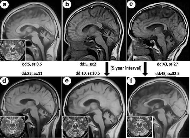
| Cerebellar cortical atrophy (vermis, paravermis, hemispheres) | Most genetic and degenerative ataxias |
| T2 hyperintensity - middle cerebellar peduncles | FXTAS |
| T2 hyperintensity - inferior olivary nuclei | POLG-ataxia, adult-onset Alexander disease, gluten ataxia |
| Hot-cross-bun sign (T2 cross in pons) | MSA-C |
| Linear T2 hyperintensity outer striatum | MSA-P |
| Cortical ribboning on DWI | CJD |
| T2 hyperintensity - mamillary bodies, periaqueductal gray, paraventricular thalamus | Wernicke encephalopathy |
| Surface hypointensity (GRE/SWI) | Superficial siderosis |
| Enlarged fourth ventricle | Associated with cerebellar atrophy |
Important note: In early Friedreich ataxia, vitamin E deficiency ataxia, and POLG-ataxia, cerebellar atrophy may be absent as these primarily affect sensory neurons first.
Additional Investigations
- Electromyography / Nerve conduction studies: Assess associated neuropathy (Friedreich, POLG)
- Autonomic testing + sleep study (REM behavior disorder): Suggests MSA
- Muscle biopsy (SDH staining): POLG-ataxia shows increased SDH (mitochondrial proliferation)
- EEG: Periodic sharp wave complexes in CJD
- Dopamine transporter (DAT) scan: Dopaminergic involvement in MSA
- Serum antibodies: Anti-Yo, anti-Hu, anti-Ri (paraneoplastic); anti-GAD65; anti-gliadin/TG6 (celiac/gluten ataxia); anti-GQ1b (Miller Fisher)
- Genetic testing: Next-generation sequencing panels for hereditary ataxias
- Metabolic labs: Cholestanol (cerebrotendinous xanthomatosis), vitamin E levels, AFP (ataxia-telangiectasia, AOA2), albumin (AOA1), lactate (mitochondrial)
Management
Treat Underlying Cause (where possible)
- Thiamine for Wernicke; vitamin E supplementation; gluten-free diet; immunotherapy for autoimmune/paraneoplastic; antibiotics for Whipple disease; treat neoplasm in paraneoplastic
Symptomatic / Disease-Modifying Pharmacotherapy
| Drug | Indication / Evidence |
|---|
| 4-Aminopyridine | EA-2 (episodic ataxia type 2) - blocks K channels, restores Purkinje cell pacemaking; reduces attack frequency significantly |
| Acetazolamide | EA-1 and EA-2 - reduces attack frequency |
| Riluzole | Multiple SCAs - normalizes Purkinje cell firing via SK channel modulation; showed improvement in SCA1/2/3/6 at 8 weeks and 12 months |
| Valproic acid | SCA3 - HDAC inhibitor; improved stance at 12 weeks in one RCT |
| Thyrotropin-releasing hormone | Some improvement in speech/gait in older uncharacterized ataxia cohorts |
| Coenzyme Q10 / Idebenone | Friedreich ataxia (cardiomyopathy benefit) |
Non-pharmacological (important for all)
- Physical therapy and balance training
- Speech therapy for dysarthria and dysphagia
- Occupational therapy for ADLs
- Assistive devices (walkers, wheelchairs)
- Fall prevention strategies
Key Differentials: Ataxia vs. Other Gait Disorders
| Feature | Cerebellar Ataxia | Sensory Ataxia | Vestibular Ataxia |
|---|
| Romberg test | Mildly positive | Markedly positive (falls with eyes closed) | Positive (falls to one side) |
| Effect of eye closure | Mild worsening | Marked worsening | Worsening |
| Nystagmus | Common (gaze-evoked, horizontal) | Absent | Common (unidirectional) |
| Proprioception | Normal (unless mixed) | Lost | Normal |
| Gait character | Wide-based, lurching | Stamping, high-stepping | Veering to one side |
Recent Evidence (2024-2026)
- A 2025 review in Nature Reviews Neurology (PMID 40983776) discusses progress and ongoing challenges in sporadic late-onset cerebellar ataxias - an important category where ~50% of adult-onset progressive ataxias remain genetically uncharacterized.
- A 2026 review in Neurodegenerative Disease Management (PMID 42057699) covers current management of spinocerebellar ataxia, including emerging disease-modifying strategies.
Sources: Bradley and Daroff's Neurology in Clinical Practice; Localization in Clinical Neurology, 8e; Adams and Victor's Principles of Neurology, 12e; Harrison's Principles of Internal Medicine 22E