
Oxygen dissociation curve 10 mark answer
I'll start by reading the medical library skill, then search for comprehensive content on the oxygen dissociation curve.
Reading File
Now I'll search the library and find images simultaneously:
Finding Sources
Searching Images
oxygen dissociation curve sigmoid haemoglobin
Excellent - I found dedicated sections. Let me read all three sources in parallel:
Reading File
Reading File
Reading File
Excellent content retrieved. Let me fetch the ODC diagram from Fishman's and also read more of Guyton's content for the Bohr effect details:
Loading Image
Loading Image
Finding Sources
I have the diagrams and core content. Let me now read the Bohr effect section from Fishman's and the 2,3-DPG / fetal haemoglobin content:
Reading File
Finding Sources
I have very rich content from the textbooks. Now let me fetch the third diagram (the shift curves from Fishman's) and read the Bohr effect section:
Loading Image
Reading File
I now have all the material needed for a comprehensive 10-mark answer. Let me compile everything.
Oxygen Dissociation Curve - 10 Mark Answer
Definition
The oxygen dissociation curve (ODC) represents the relationship between the partial pressure of oxygen (PO2) and the oxygen content (or % saturation) of haemoglobin in blood. It is plotted at standard conditions: pH 7.40, temperature 37°C, and atmospheric pressure 760 mmHg.
- Fishman's Pulmonary Diseases and Disorders, p. 256
Shape of the Curve and Why It Is Sigmoid
The curve is characteristically S-shaped (sigmoid) rather than hyperbolic. This is due to the cooperative binding of oxygen to the four haem groups of the haemoglobin tetramer. When one haem group binds O2, it induces a conformational change in the quaternary structure of haemoglobin that increases the O2 affinity of the remaining unbound haem groups. Successive binding steps therefore become progressively easier, producing the sigmoid shape.
Haemoglobin exists in two states:
- T (tense/deoxy) state - low O2 affinity
- R (relaxed/oxy) state - high O2 affinity
As O2 binds, the molecule shifts from T to R state, explaining cooperativity.
The Normal Curve and Key Values
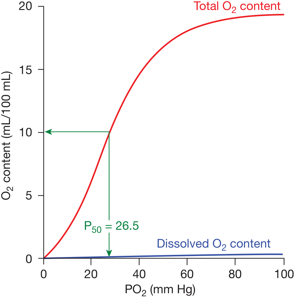
Figure: Oxygen dissociation curve - total O2 content (red) and dissolved O2 (blue). P50 = 26.5 mmHg. (Fishman's Pulmonary Diseases and Disorders)
| Point | PO2 (mmHg) | SaO2 (%) | Significance |
|---|---|---|---|
| Arterial blood | ~100 | ~98 | Lungs - loading |
| Mixed venous blood (rest) | ~40 | ~75 | Tissues - unloading |
| P50 (half-saturation) | 26.5 | 50 | Index of Hb-O2 affinity |
Two functional zones of the curve:
-
Flat upper portion (PO2 60-100 mmHg): Here the curve is nearly flat, so even moderate falls in PO2 (e.g., in lung disease) cause little drop in saturation. This is a protective "safety zone" for loading in the lungs.
-
Steep lower portion (PO2 20-60 mmHg): Here a small drop in PO2 releases large amounts of O2 to tissues - ideal for efficient unloading at capillary level.
At rest, only ~25% of delivered O2 is extracted; during exercise, extraction can rise substantially due to a rightward shift of the curve.
- Fishman's Pulmonary Diseases and Disorders, pp. 255-256
Dissolved Oxygen
While most O2 is bound to haemoglobin (~20 mL/100 mL at Hb 15 g/dL), a small amount is dissolved in plasma (obeying Henry's law). Though present in tiny concentrations, dissolved O2 is physiologically essential because it is the form that diffuses across the alveolar-capillary membrane, erythrocyte membrane, and into the cell; haemoglobin itself cannot cross these membranes.
Shifts of the Curve and the P50
The P50 (PO2 at 50% saturation) measures haemoglobin-oxygen affinity:
- Increased P50 = rightward shift = reduced affinity = more O2 released to tissues
- Decreased P50 = leftward shift = increased affinity = O2 held tighter by Hb
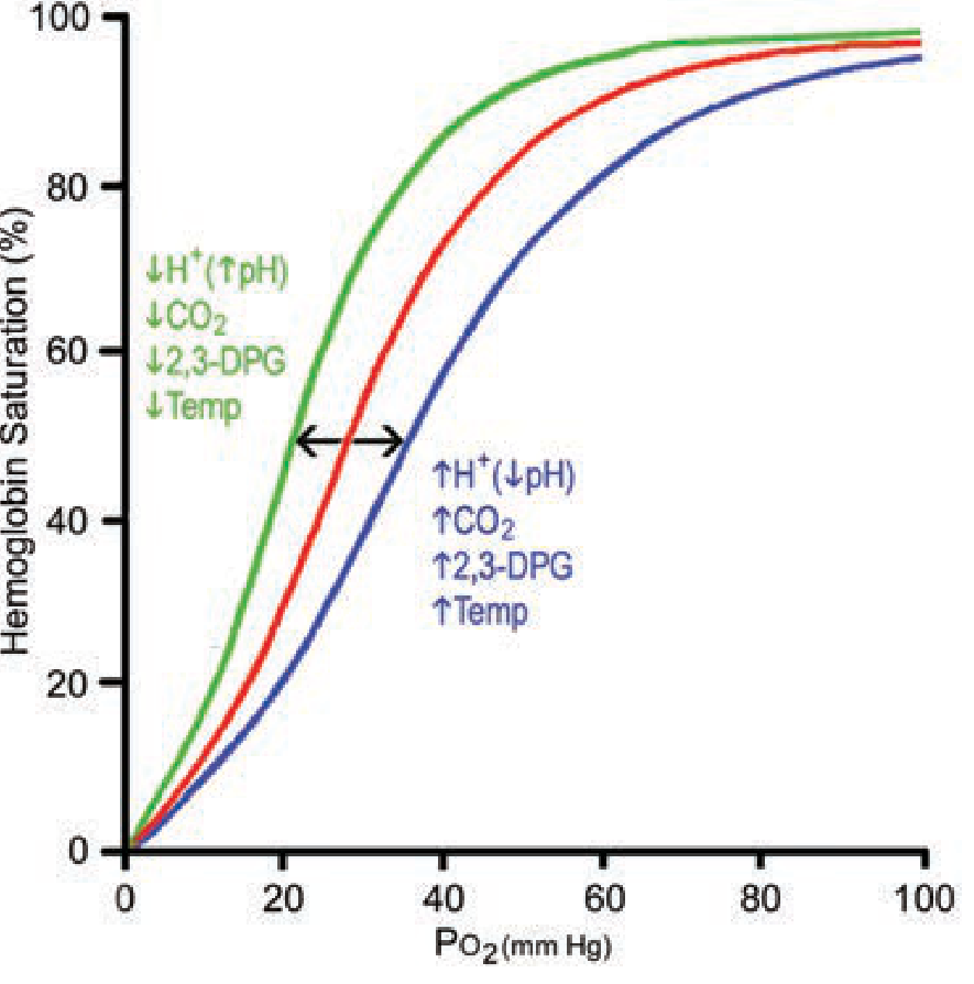
Figure: Factors causing left (green) and right (blue) shifts of the ODC. (Fishman's Pulmonary Diseases and Disorders)
Causes of Right Shift (CADET - mnemonic):
| Factor | Physiological Setting |
|---|---|
| ↑ CO2 | Actively metabolising tissues |
| ↑ H+ (↓ pH, acidosis) | Exercise, sepsis, lactic acidosis |
| ↑ Temperature | Exercising muscle |
| ↑ 2,3-DPG | Chronic hypoxia, anaemia |
| Exercise | All above factors combined |
Causes of Left Shift:
| Factor | Physiological Setting |
|---|---|
| ↓ CO2, ↑ pH (alkalosis) | Pulmonary capillary (Haldane effect) |
| ↓ Temperature | Hypothermia |
| ↓ 2,3-DPG | Stored bank blood |
| Carbon monoxide (CO) | CO poisoning |
| Fetal haemoglobin (HbF) | Placental O2 transfer |
| Methaemoglobin | Oxidation of Fe2+ to Fe3+ |
- Mulholland and Greenfield's Surgery, p. 619; Fishman's Pulmonary Diseases and Disorders, p. 256
The Bohr Effect
The rightward shift caused specifically by increased PCO2 and decreased pH (increased H+) is called the Bohr effect, named after Christian Bohr who first described it.
Mechanism: CO2 and H+ bind to sites on the haemoglobin molecule distinct from the O2-binding (haem) sites. As the tertiary and quaternary configuration changes during O2 release, 16 buffering groups (14 histidine residues + 2 terminal amino groups) change their pKa values, increasing haemoglobin's buffering power. This is the alkaline Bohr effect and it:
- Facilitates O2 release in metabolically active tissues (high CO2, low pH)
- Buffers ~50% of the H+ released in aerobic metabolism, minimising the fall in blood pH
In the lungs, the reverse occurs: CO2 diffuses out, pH rises, the curve shifts left, and more O2 is loaded onto haemoglobin. This is physiologically ideal.
- Fishman's Pulmonary Diseases and Disorders, pp. 257-258
Role of 2,3-Diphosphoglycerate (2,3-DPG)
2,3-DPG is a product of glycolysis inside red blood cells. It binds between the beta chains of deoxyhaemoglobin (which are more widely separated in the T state) via electrostatic interaction with four positively charged amino acids. This stabilises the T (deoxy) state, reducing O2 affinity and shifting the curve to the right.
Conditions increasing 2,3-DPG (rightward shift):
- Chronic hypoxia (high altitude, chronic lung disease)
- Anaemia
- Thyroid hormone excess
Conditions decreasing 2,3-DPG (leftward shift):
- Stored bank blood (2,3-DPG falls within 1-2 weeks of storage)
- Acidosis acutely inhibits glycolysis
During exercise: Increased CO2 + H+ + temperature + 2,3-DPG all combine to force a large rightward shift, maximising O2 delivery to active muscles.
- Fishman's Pulmonary Diseases and Disorders, p. 256; Guyton and Hall, p. 546
Fetal Haemoglobin (HbF)
HbF has gamma-chains instead of beta-chains. 2,3-DPG binds less effectively to gamma-chains, so HbF has a higher O2 affinity than adult HbA. This shifts the HbF ODC to the left relative to maternal blood, enabling the fetus to extract O2 across the placenta from maternal blood (which has a lower PO2 than alveolar blood).
Carbon Monoxide Poisoning
CO binds to haemoglobin with an affinity ~250 times that of O2, forming carboxyhaemoglobin (COHb). Two damaging effects occur:
- Functional anaemia - CO occupies O2-binding sites, reducing O2 capacity
- Leftward shift - CO bound to haem groups increases the affinity of remaining haem groups for O2, making it harder to release O2 in the tissues
This combination makes CO poisoning particularly dangerous.
Clinical Relevance
| Scenario | Effect on ODC |
|---|---|
| Exercise | Right shift - more O2 to muscles |
| Altitude acclimatisation | ↑ 2,3-DPG → right shift → more unloading |
| Stored blood transfusion | Left shift → impaired tissue O2 delivery |
| CO poisoning | Left shift + reduced O2 capacity |
| Respiratory alkalosis | Left shift → O2 held tightly (e.g., hyperventilation) |
| Sepsis / lactic acidosis | Right shift → more O2 available to tissues |
Summary
The sigmoid ODC reflects cooperative O2 binding by haemoglobin and is ideally suited for efficient O2 loading in the lungs (flat zone) and unloading in the tissues (steep zone). The P50 (normally 26.5 mmHg) quantifies haemoglobin-O2 affinity. The Bohr effect (CO2 and H+ shifting the curve right at tissues) and Haldane effect (reverse in the lungs) are physiologically complementary mechanisms. 2,3-DPG, temperature, abnormal haemoglobins, and CO all further modulate O2 delivery to match metabolic demand.
Sources: Fishman's Pulmonary Diseases and Disorders (pp. 255-258); Guyton and Hall Textbook of Medical Physiology (p. 546); Mulholland and Greenfield's Surgery (p. 619)
This is a shared conversation. Sign in to Orris to start your own chat.