

What is probable diagnosis and treatment in detail
impetigo secondary bacterial infection eczema hand crusting pustules
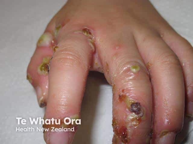
Clinical photograph of the dorsum and interdigital surfaces of the hand demonstrating extensive crusted skin lesions with yellow-brown crusts, eroded margins, and surrounding erythema. The appearance is characteristic of crusted (Norwegian) scabies with superimposed bacterial infection; in this case the crusts and purulent debris are more accentuated by a secondary pyococcal infection. Numerous discrete plaques are distributed across several digits, including the proximal and middle phalanges, with fissuring in interdigital spaces. In addition to crusts, there are vesicles or pustules and areas of tissue breakdown, suggesting active inflammation and possible secondary necrosis. The overall picture emphasizes a high infectious burden and contagiousness, requiring urgent dermatologic and infectious disease management. The clinical relevance includes recognizing crusted scabies as a risk factor for disseminated bacterial colonization and sepsis if untreated. Management implications entail combination therapy with specific scabicides (permethrin cream, ivermectin) and broad-spectrum antibiotics active against Pseudomonas aeruginosa or equivalent pyogenic organisms; strict contact precautions are advised in clinical settings. The image provides educational value for diagnosing crusted scabies, secondary impetigo, and differential diagnoses such as eczema with crusting or bullous impetigo, and supports teaching on infection control, wound care, and outpatient vs inpatient management.
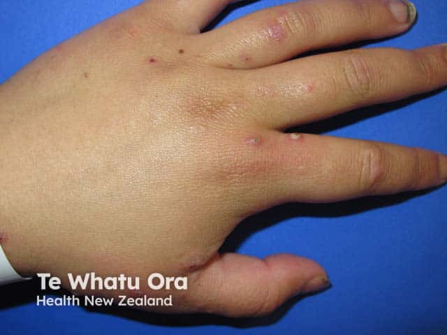
A high-resolution clinical photograph captures the dorsum of the hand demonstrating scattered pustular papules and small erosions on an erythematous base. The image was acquired using clinical photography in visible light with a neutral background to ensure color fidelity; no contrast agents or staining were used. The primary anatomical focus is the skin of the dorsal hand, including the metacarpal dorsum and interdigital spaces. Pustules are nonfollicular, approximately 2–4 mm in diameter, with surrounding mild erythema and shallow crusts, consistent with secondary bacterial infection superimposed on a scabies eruption. The appearance is compatible with infected scabies (scabies with impetiginization) and may show superficial crusting or pustulation at burrow sites. Clinically significant features include pruritus, linear burrows that may be subtle, and a crusted/pustular infestation pattern. Diagnostic significance lies in identifying secondary infection that necessitates antibiotic therapy (e.g., anti-staphylococcal coverage) in addition to scabicidal treatment. This image is valuable for dermatology education, clinical case repositories, and differential diagnosis training (impetigo, eczema herpeticum, papular urticaria). Potential use cases include teledermatology consultations, image-based teaching modules, and research on mite-related skin lesions and superinfection dynamics.
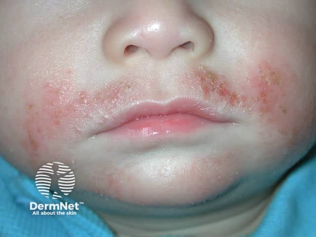
Clinical dermatology photograph of a pediatric patient demonstrates perioral and malar facial involvement consistent with atopic dermatitis (eczema) complicated by secondary Staphylococcus aureus infection. Modality and technique: noninvasive clinical photography, frontal close-up view of the lower face to document surface changes, crusting, and distribution; no staining or contrast. Anatomical context: integumentary system; skin of the cheeks and perioral region; bilaterally symmetric involvement with inflammatory erythema, scaling, and excoriations. Visual features: erythematous papules and plaques with golden-yellow crusts over the vermilion border and adjacent cheeks; superficial fissuring and oozing are evident; skin texture appears rough with edema and surrounding dry patches. Pathological findings (clinical): secondary bacterial colonization with Staphylococcus aureus on compromised atopic dermatitis skin, leading to impetiginous crusts; may show mild edema and superficial pustules; no overt necrosis or lymphangitis seen in image. Diagnostic significance: presence of secondary infection increases risk of spreading, prolongs inflammation, and necessitates antibiotic therapy and intensified skin care; supports diagnosis of infected atopic dermatitis rather than isolated dermatitis. Differential considerations: uncomplicated eczema, impetigo, contact dermatitis with bacterial colonization, or eczema herpeticum to be ruled out clinically. Clinical correlation: guide treatment decisions including topical antibiotics (mupirocin/fusidic acid) depending on severity, antiseptic cleansing, barrier emollients, and follow-up to assess resolution.
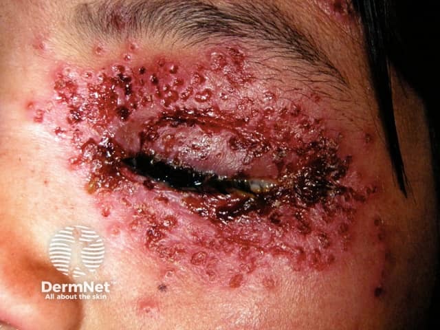
This clinical photograph shows a severe periorbital eruption in a patient with dermatitis, clinically consistent with eczema herpeticum (Kaposi varicelliform eruption). The image is a frontal close‑up of the left periorbital skin with numerous vesicles, pustules, and crusted erosions surrounding the eyelid margin and adjacent cheek. The involved skin is erythematous and edematous, with moist surfaces and dark hemorrhagic crusts. Vesicles have coalesced into crusted plaques and the ocular adnexa appear threatened by surface breakage, increasing risk of keratoconjunctivitis. The presentation reflects acute HSV infection superimposed on underlying atopic dermatitis or eczema, a pattern that can mimic impetigo or other bacterial dermatitis but is distinguished by vesiculopustular morphology and rapid crusting. Diagnostic considerations include HSV or other herpetic viruses; bacterial superinfection; varicella; and, less commonly, dermatitis herpetiformis is not typical. Clinical significance is high due to potential ocular involvement and systemic risk; urgent antiviral therapy and broad-spectrum antibiotics if secondary infection are considered. This image serves as a teaching example for dermatology, infectious disease, and ophthalmology contexts, illustrating periorbital HSV eruption on compromised skin, and highlighting the importance of early recognition, swab PCR confirmation, and prompt management to prevent complications. Further documentation should include sequential photographs documenting lesion evolution carefully.
Probable Diagnosis
Primary Diagnosis: Chronic Irritant/Allergic Contact Dermatitis of the Hand with Secondary Bacterial Infection (Impetiginization)
- Diffuse lichenification - markedly thickened skin with exaggerated skin creases across the entire dorsum, indicating a prolonged itch-scratch cycle
- Widespread scaling and desquamation - dry, rough, powdery texture
- Post-inflammatory hyperpigmentation - diffuse dark brown/grayish discoloration consistent with a dark phototype and chronic inflammation
- Mild edema of the hand and fingers
- Grouped vesicopustules (tense, yellowish-white, purulent fluid) - dorsal hand and inter-knuckle areas
- Honey-colored/dark brown crusts (golden crusts - pathognomonic for impetiginization with Staphylococcus aureus or Streptococcus pyogenes)
- Erosions and excoriations with active oozing and fresh blood at the lateral index finger
Clinical Pattern Explanation
- The chronic eczematous background (likely occupational irritant contact dermatitis or atopic hand eczema) has disrupted the skin barrier, allowing colonization and infection
- Secondary bacterial infection then leads to impetiginization - the formation of pustules, oozing, and honey-colored crusting on top of preexisting eczema
- As described in Fitzpatrick's Dermatology: "Secondary infection with pyogenic micrococci leads to impetiginization" following chronic eczematous disease
Differential Diagnoses to Consider
| Diagnosis | Supporting Features | Against |
|---|---|---|
| Crusted (Norwegian) Scabies | Dorsal hand/knuckle predilection, crusting, high mite burden | No visible burrows, no inter-digital web space burrows described |
| Dyshidrotic eczema | Vesicles on hands/fingers | Typically palmar/lateral finger vesicles, no lichenification this diffuse |
| Bullous impetigo | Pustules, crusts | No bullae; chronic lichenification suggests underlying eczema |
| Psoriasis | Scaling plaques on dorsal hands | No silvery plaques, no nail pitting visible |
| Tinea manuum | Scaling on dorsal hand | Typically unilateral, fine scaling without lichenification or pustules |
Treatment in Detail
STEP 1: Treat the Secondary Bacterial Infection
Topical Antibiotics (mild-to-moderate infection, no systemic signs)
- Mupirocin 2% ointment (Bactroban) - apply TID x 7-10 days to crusted/pustular areas
- Fusidic acid 2% cream - apply BD x 7-10 days; good Gram-positive coverage
- Gently remove crusts with saline-soaked gauze before application to improve penetration
Systemic Antibiotics (moderate-to-severe infection OR spreading cellulitis)
- Flucloxacillin 500 mg QID x 5-7 days (first-line, anti-staphylococcal)
- Cefalexin 500 mg QID x 5-7 days - if penicillin-allergic (first-generation cephalosporin)
- Co-amoxiclav (amoxicillin-clavulanate) 625 mg TID - if mixed flora suspected
- If MRSA is suspected (recurrent/hospital-acquired): Doxycycline 100 mg BD or Trimethoprim-sulfamethoxazole (co-trimoxazole) DS
Wound care
- Soak with dilute potassium permanganate (1:10,000) solution for 10-15 min BD - astringent, antimicrobial, dries oozing areas
- Alternatively, use 0.9% saline wet compresses to soften and remove crusts
- Keep wounds clean and loosely covered
STEP 2: Manage the Underlying Chronic Eczema/Contact Dermatitis
A. Identify and Eliminate the Trigger
- Take a detailed occupational and environmental history - chemicals, detergents, solvents, rubber gloves, metals (nickel, cobalt), wet work
- Patch testing is essential once the acute infection resolves to identify allergic contact sensitizers
- Avoid wet work exposure; if unavoidable, use cotton inner gloves under vinyl/nitrile outer gloves
- Avoid soap/detergents on affected skin; use soap substitutes (emulsifying ointment, aqueous cream)
B. Topical Corticosteroids (after infection clears)
- Moderate-to-potent corticosteroid needed for lichenified skin on hands:
- Betamethasone valerate 0.1% cream or ointment - apply OD/BD x 2-4 weeks
- Mometasone furoate 0.1% OD
- Clobetasol propionate 0.05% (ultrapotent) for severely lichenified areas - use short-course, BD x 1-2 weeks max
- Ointment base preferred for dry, lichenified, fissured skin (better penetration, occlusive)
- Reduce frequency to alternate days once improvement occurs
C. Emollients and Skin Barrier Repair (cornerstone of management)
- Apply bland emollients (white soft paraffin, Vaseline, or ceramide-containing moisturizers) multiple times daily
- Apply immediately after soaking hands ("soak and seal" method)
- Ceramide-based moisturizers (CeraVe, Cetaphil RestoraDerm) help restore barrier function
- Urea 10-20% cream helps reduce lichenification/hyperkeratosis
D. Tacrolimus/Pimecrolimus (Calcineurin Inhibitors)
- Use tacrolimus 0.1% ointment BD as a steroid-sparing agent for maintenance
- Particularly useful on dorsal hands for long-term management to avoid steroid-induced atrophy
STEP 3: Ancillary Measures
| Measure | Details |
|---|---|
| Antihistamines | Oral cetirizine 10 mg OD or hydroxyzine 25 mg nocte to reduce itch-scratch cycle |
| Wound protection | Fingertip bandaging/cotton gloves at night to prevent scratching |
| Occupational modification | Reduce wet work, chemical exposure; use protective gloves with caution (occlusion worsens ICD) |
| Glove selection | Nitrile gloves (not rubber/latex) to avoid rubber allergen sensitization |
| Patient education | Emollient use, trigger avoidance, avoiding harsh soaps |
STEP 4: Follow-Up
- Review at 2 weeks to assess infection resolution
- Introduce or escalate topical steroid therapy if lichenification persists
- Patch testing at 4-6 weeks (once acute inflammation settles) to identify contact allergens
- If chronic dermatitis is refractory: consider phototherapy (NB-UVB), oral methotrexate, or dupilumab (for atopic component)
- If MRSA or resistant infection suspected: send wound swab for culture and sensitivity
Summary
| Issue | Treatment |
|---|---|
| Active bacterial infection | Systemic flucloxacillin/cefalexin + topical mupirocin/fusidic acid |
| Oozing/crusting | KMnO₄ soaks, saline compresses |
| Lichenification | Potent topical corticosteroid (betamethasone/clobetasol) once infection clears |
| Barrier repair | Emollients multiple times daily |
| Pruritus | Oral antihistamines |
| Underlying cause | Patch testing, trigger identification, occupational modification |
Clinical note: This is an image-based assessment only. A formal skin examination, history (occupation, atopy, duration), and possibly skin swab/patch testing are needed to confirm the diagnosis and tailor therapy. If there are signs of spreading cellulitis (warmth, erythema tracking up the arm, fever), systemic antibiotics and possible hospital review are warranted.


More photos of that pt
scabies abdomen waistband papules burrows nodular trunk
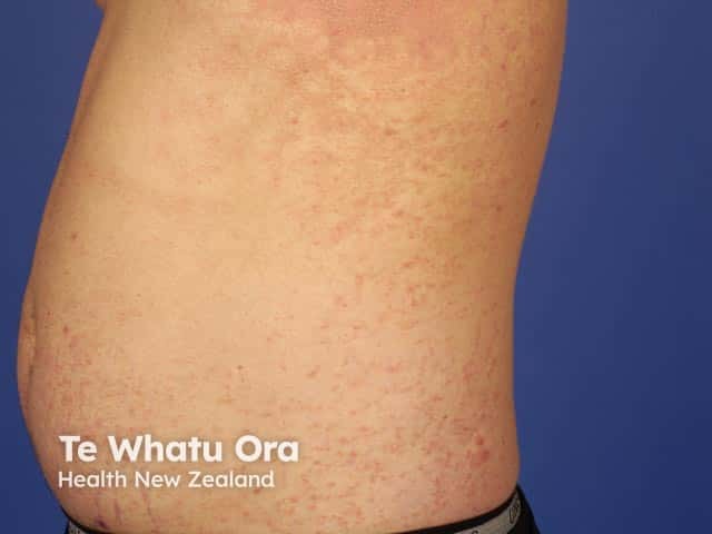
Clinical photography of an adult torso showing a widespread papular eruption consistent with scabies. Modality: Dermatologic clinical photograph using a high‑resolution digital camera under diffuse, color‑calibrated lighting. Viewpoint: lateral/anterior trunk exposure of the left hemithorax and adjacent abdomen. Lesion morphology: numerous small, erythematous papules (approximately 2–5 mm) with occasional faint vesicles and minimal crusting; surface appears slightly excoriated from scratching. Distribution: diffuse involvement of the trunk with higher density on the flanks and intertriginous areas; legs and feet not shown. Texture and color features: discrete, flat or slightly raised papules with a dull reddish hue; no surrounding edema; no pustules observed in the frame. Impression: cutaneous eruption compatible with scabies infestation, where the pruritic papules reflect mite‑related inflammatory response. Ancillary notes: Burrows may be present but are often inconspicuous in standard photographs; confirmation may require skin scraping or dermoscopy. Clinical relevance: aids diagnosis in dermatology teaching, primary care screening, and teledermatology triage; supports differentiation from other papular rashes such as eczema, papular urticaria, pityriasis rosea, or insect bites. Potential clinical applications: education, exam preparation, case review, and patient counseling. The image serves as a teaching reference for recognizing trunk‑dominant papular scabies and communicating suspected diagnosis to patients.
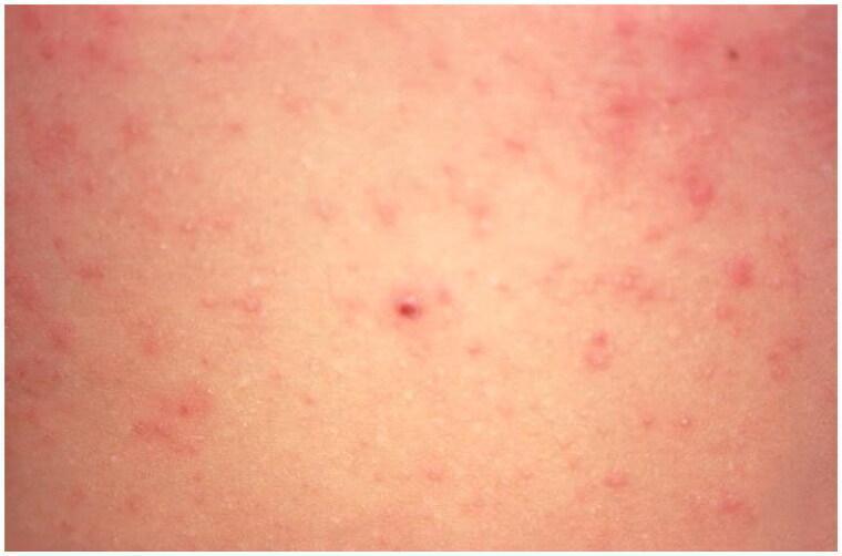
This clinical photograph displays a close-up view of a cutaneous eruption on the human abdomen, characteristic of a scabies infestation (Sarcoptes scabiei). The image demonstrates numerous small, erythematous papules (red, raised bumps) scattered across a light-toned skin background. The lesions exhibit a polymorphic distribution with varying density, accompanied by a diffuse, mild background erythema suggesting a generalized hypersensitivity reaction to the scabies mites, eggs, or scybala (feces). Some papules appear slightly excoriated, which is a common secondary finding due to intense pruritus. The absence of clearly visible linear burrows in this specific frame is typical, as they are often obscured by secondary inflammatory changes or scratching. This image serves as an educational tool for identifying the primary morphology of inflammatory scabietic lesions in common predilection sites like the trunk.
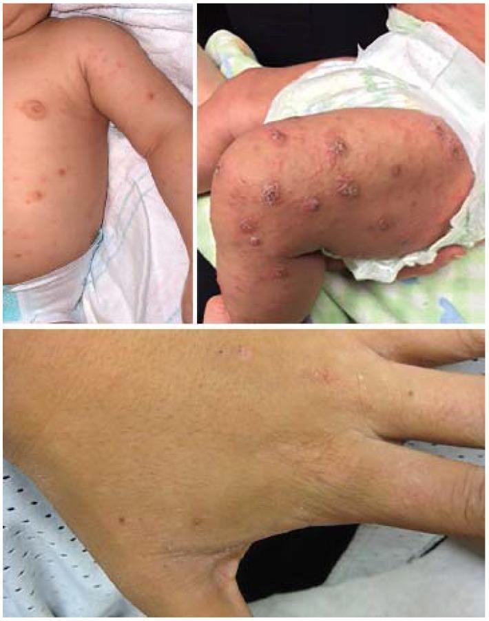
A composite of three clinical photographs demonstrating the dermatological manifestations of scabies in an infant and an adult. The top-left panel shows an infant's trunk and arm with scattered, erythematous papules. The top-right panel displays the infant's lower extremity, showing a higher density of larger, inflammatory papulovesicles and nodules on the thigh and leg, some of which appear crusted or excoriated, which is a common presentation in infantile scabies. The bottom panel provides a close-up of an adult hand, focusing on the dorsal surface and finger webs. This view reveals small, discrete erythematous papules and subtle, linear, thread-like structures consistent with Sarcoptes scabiei burrows. The skin also shows signs of generalized xerosis. Collectively, these images illustrate the age-dependent distribution and morphology of scabies, highlighting the more inflammatory, nodular response in infants versus the classic acral distribution and burrowing seen in adults.
REVISED DIAGNOSIS: SCABIES (with secondary impetiginization of hands)
Why the Diagnosis Has Changed
| Feature | What Was Seen |
|---|---|
| Waistband/beltline papules | Peri-umbilical and waistband-line excoriated papules - a pathognomonic site for scabies |
| Diffuse scattered pruritic papules on trunk/flanks | Hypersensitivity reaction to mite proteins |
| Linear/serpiginous track on the lateral thigh/hip | Highly suspicious for a scabies burrow (8-10 cm serpiginous elevated track) |
| Linear excoriations in diagonal arrangement on flanks | Itch-scratch cycle with secondary lichenification |
| Clustered linear papules/nodules near groin | Nodular scabies - characteristic of the groin/genitalia/axilla |
| Dorsal hand pustules + impetiginization (first photo) | Secondary bacterial infection of scabietic lesions |
| Widespread body distribution | Trunk, flanks, groin, thighs, hands - typical scabies spread pattern |
| Post-inflammatory hyperpigmentation throughout | Consistent with dark phototype + chronic infestation |
- Intense pruritus (implied by widespread excoriations)
- Peri-umbilical and waistband distribution (almost diagnostic)
- Linear/serpiginous burrow track on thigh
- Nodular lesions near groin
- Secondary bacterial infection (impetiginization) on dorsal hand
COMPREHENSIVE TREATMENT PLAN
STEP 1: Confirm Diagnosis (if possible)
- Dermoscopy of suspected burrow on thigh - look for the "jet with contrail" sign (mite body at the end of a white serpiginous track)
- Skin scraping from the burrow/inter-digital web spaces: mineral oil mount, microscopy for mites, eggs, or scybala
- If unavailable in the field: treat on clinical grounds - the distribution is highly characteristic
STEP 2: Scabicidal Treatment (first-line)
Option A - Permethrin 5% cream (first-line, safest)
- Apply from neck to toes (include all skin folds, genitalia, soles, between toes, under nails, navel)
- Leave on for 8-12 hours (overnight), then wash off
- Repeat at 1 week (day 8) to kill newly hatched mites
- Massage well into lichenified/thick areas (more product needed)
Option B - Oral Ivermectin (for extensive disease, compliance issues, or treatment failure)
- 200 mcg/kg single dose orally
- Repeat at 2 weeks (day 14)
- This patient has extensive/widespread disease - ivermectin is strongly preferred as compliance with full-body permethrin application may be difficult
- Avoid in children <15 kg, pregnancy (Category C)
- Best taken with food (fatty meal increases absorption by 2.5x)
STEP 3: Treat Secondary Bacterial Infection (the hand lesions)
Systemic antibiotics (for the impetiginized hand):
- Cefalexin 500 mg QID x 7 days (first-line for skin infection)
- Or Flucloxacillin 500 mg QID x 7 days (anti-staphylococcal)
- If MRSA suspected: Co-trimoxazole DS BD x 7 days
Local wound care (hands):
- Potassium permanganate (1:10,000) soaks BD to dry and debride oozing areas
- Mupirocin 2% ointment to crusted/pustular areas TID
STEP 4: Treat Residual Itch and Nodular Lesions
- Oral antihistamine: Cetirizine 10 mg OD (non-sedating) or hydroxyzine 25 mg nocte (sedating - better for night itch)
- Note: Itch can persist for 4-6 weeks after successful treatment due to residual mite antigens - this does NOT indicate treatment failure
- Nodular scabies (the groin nodules): intralesional triamcinolone 10 mg/mL, or potent topical corticosteroids (betamethasone 0.1% ointment OD) applied after scabicidal treatment is complete
- Post-scabies eczema: moderate topical steroid once infection and infestation are cleared
STEP 5: Contact Tracing and Household Treatment (MANDATORY)
- All household members and close contacts (especially sleeping partners, family members) must be treated on the same day, even if asymptomatic
- All clothing, bedding, towels used in the past 3 days: wash in hot water (>60°C) and machine dry on high heat, OR seal in a plastic bag for 72 hours (mites die without host contact)
- Furniture/carpets/mattresses: spray with permethrin or vacuum thoroughly; mites survive off the body only 48-72 hours
STEP 6: Follow-Up
| Timeframe | Action |
|---|---|
| Day 7-8 | Second permethrin application |
| Day 14 | Second ivermectin dose if used; check for new lesions |
| Week 4-6 | Review - persistent itch alone does NOT mean treatment failure |
| Week 6+ | Persistent new burrows/papules = retreatment or crusted scabies screen |
Summary Table
| Problem | Treatment |
|---|---|
| Scabies infestation | Ivermectin 200 mcg/kg day 1 + 14 AND/OR permethrin 5% day 1 + 8 |
| Secondary bacterial infection (hands) | Cefalexin 500 mg QID x 7 days + mupirocin ointment topically |
| Pruritus | Cetirizine 10 mg OD + hydroxyzine 25 mg nocte |
| Nodular scabies (groin) | Potent topical steroid after infestation cleared |
| Contacts | Treat all household contacts on same day |
| Clothing/bedding | Hot wash (>60°C) same day |
Important: Given the body distribution (trunk, waistband, thighs, groin, and both hands), this is likely a chronic/well-established infestation of several weeks duration. The previous diagnosis of "contact dermatitis" on the hand alone was incomplete - the hand impetiginization was the secondary manifestation of the underlying scabies infestation. Treating scabies will resolve the underlying substrate; treating the infection will resolve the acute hand pustules.
He has no itching since start
leprosy skin lesions papules nodules trunk hands non-pruritic
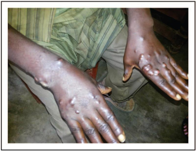
This clinical photograph displays the bilateral hands and distal forearms of a patient with lepromatous leprosy, showcasing characteristic dermatological manifestations. The image reveals numerous disseminated, firm, skin-colored to slightly hyperpigmented nodules and papules. These lesions are most prominent on the dorsal surfaces of the hands, particularly overlying the metacarpophalangeal joints and along the extensor aspects of the forearms. The nodules vary in size, with some coalescing into larger, dome-shaped masses. The surrounding skin appears thickened and exhibits a subtle waxy or shiny texture, consistent with diffuse infiltration or localized edema common in the lepromatous spectrum of the disease. The symmetric distribution and nodular morphology are hallmark features of a high bacillary load and a Th2-mediated immune response. This visual provides a classic example of multibacillary leprosy for clinical education in infectious diseases and dermatology, emphasizing the importance of recognizing nodular cutaneous presentations in endemic regions.
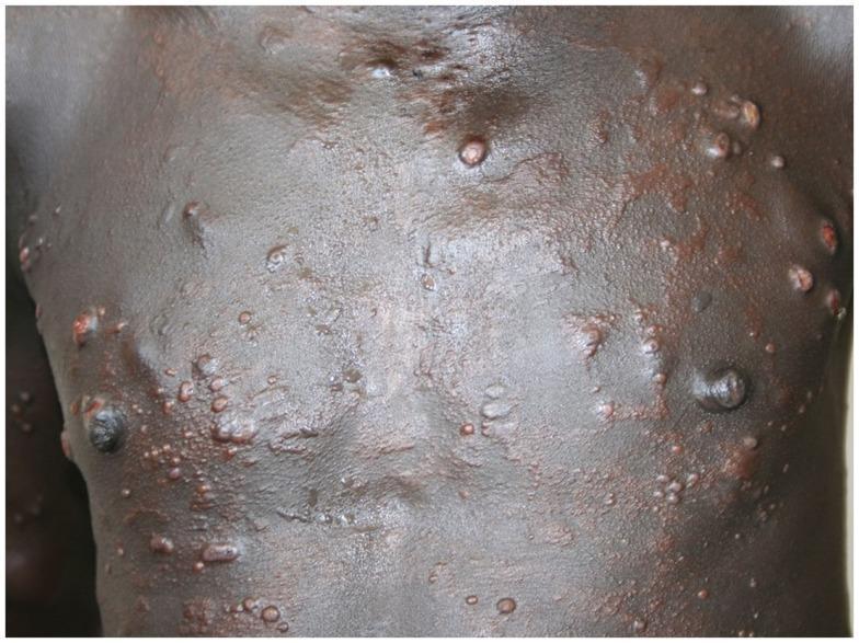
This clinical photograph displays a widespread cutaneous eruption on the anterior trunk of a patient with darkly pigmented skin. The lesions consist of numerous firm, well-demarcated, dome-shaped papules and nodules that vary in size from 2 mm to over 1 cm. Many lesions exhibit a smooth, glistening, succulent surface characteristic of histoid leprosy, a variant of multibacillary leprosy. The distribution is symmetric across the chest and abdomen, with a tendency toward clustering. Colors of the lesions range from flesh-colored to reddish-brown or copper tones. While most nodules are intact without scaling or ulceration, the background skin shows focal areas of thickening and subtle textural changes. This image serves as a classic diagnostic example of the cutaneous manifestations of histoid leprosy (Mycobacterium leprae), often seen in patients following inadequate treatment or developing de novo resistance.
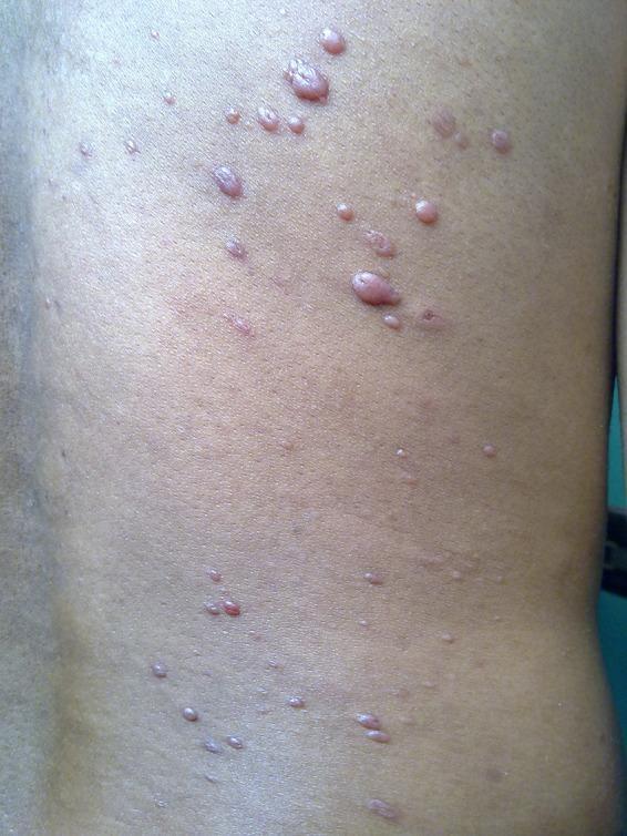
This clinical photograph shows the trunk of a patient featuring numerous discrete, skin-colored to slightly erythematous nodules. The lesions are characteristic of histoid leprosy, a distinct variant of lepromatous leprosy. The nodules are well-demarcated, varying in size from approximately 0.5 to 1.0 cm, with a notably shiny, smooth, and tense surface. Their distribution is scattered across the back and flank, with morphology ranging from firm, dome-shaped papules to larger, raised, succulent nodules. Unlike typical lepromatous leprosy lesions which may be ill-defined, these nodules appear sharply circumscribed and arise from otherwise normal-looking skin. The visual presentation is a classic example of cutaneous manifestations in multibacillary leprosy, used in medical education to differentiate histoid leprosy from other nodular skin conditions such as neurofibromatosis or dermatofibromas.
CRITICAL DIAGNOSTIC REVISION: Leprosy (Hansen Disease) - Lepromatous/Borderline Lepromatous Type
Why Leprosy Fits This Case
| Clinical Feature | This Patient | Lepromatous Leprosy (LL/BL) |
|---|---|---|
| Pruritus | ABSENT from start | Sensation in LL lesions is NOT affected (table above from Dermatology 5e) |
| Multiple papules and nodules | Trunk, flanks, abdomen, thighs | Macules, papules, nodules - numerous, symmetric |
| Distribution | Bilateral, symmetric trunk, thighs | Symmetric distribution characteristic of LL |
| Waistband/beltline lesions | Present | Lepromatous lesions favor cooler areas of skin |
| Dorsal hand involvement | Yes - severe, with superinfection | Dorsal hands - classic LL predilection site |
| Post-inflammatory hyperpigmentation | Present throughout | Common in dark skin phototypes with lepromatous disease |
| No defined hypopigmented patches | Lesions are skin-colored/dark | LL lesions - vague borders, difficult to distinguish normal vs affected skin |
| Lichenification/skin thickening | Present on hands | Diffuse skin infiltration in LL |
| Serpiginous track on thigh | Seen | Could represent infiltrated plaque or nerve involvement |
| Geography | India/tropical area (date on photo, language context) | Endemic region - India has highest global burden |
From Dermatology 2-Volume Set 5e (Table 75.2): In lepromatous leprosy (LL), sensation in lesions is "Not affected" early on, and lesions are numerous with symmetric distribution - macules, papules, and nodules. This perfectly matches the clinical picture.
Cardinal WHO diagnostic criterion: "Anesthesia in a hypopigmented or erythematous skin lesion" - but in LL, sensation may be normal early since the mite load is high and nerve damage comes later or is diffuse. The absence of itch specifically (not just reduced sensation) points toward a non-inflammatory etiology rather than the hypersensitivity-driven itch of scabies.
Revised Differential Diagnosis
| Rank | Diagnosis | Key Supporting Feature |
|---|---|---|
| 1. Lepromatous/BL Leprosy | No itch, symmetric papules/nodules, trunk + hands, endemic region | |
| 2. Crusted (Norwegian) Scabies | Minimal itch due to immunosuppression, hyperkeratosis on hands - but hand lesions here look superinfected rather than hyperkeratotic | |
| 3. Histoid Leprosy (variant of LL) | Smooth, firm, shiny nodules on normal-looking skin - matches the trunk nodules | |
| 4. Secondary syphilis | Papular rash, trunk + palms - but no itch, consider if sexually active | |
| 5. Prurigo nodularis | Nodules but these are ALWAYS intensely pruritic |
Immediate Investigations Required
Mandatory (at point of care):
-
Slit-skin smear (SSS) - from earlobes, chin, and the most active skin lesion edge
- Look for acid-fast bacilli (AFB) using Ziehl-Neelsen or Wade-Fite stain
- Positive smear = lepromatous disease
-
Sensory testing of lesions - use a wisp of cotton or pin-prick on the papules and nodules on the trunk
- If reduced/absent sensation in lesions: cardinal sign of leprosy
- In LL, sensation may be reduced diffusely rather than in individual lesions
-
Peripheral nerve examination:
- Palpate: ulnar nerve at elbow, common peroneal at fibular head, great auricular nerve, radial cutaneous nerve
- Look for nerve thickening and tenderness
-
Skin biopsy - from the active edge of a trunk papule/nodule
- Wade-Fite stain for AFB
- Look for: foamy macrophages (Virchow cells), globi (clumps of AFB), perineural inflammation
-
Lepromin test (Mitsuda) - if available: negative in LL (positive in tuberculoid)
-
Additional tests to consider: VDRL/RPR (to exclude secondary syphilis), CBC (anaemia common in leprosy), LFTs (if starting dapsone)
Treatment: WHO Multi-Drug Therapy (MDT) for Multibacillary Leprosy
WHO MDT - Multibacillary (MB) Regimen - 12 months
| Drug | Dose | Frequency |
|---|---|---|
| Rifampicin 600 mg | Supervised | Once monthly |
| Clofazimine 300 mg | Supervised | Once monthly |
| Clofazimine 50 mg | Self-administered | Daily |
| Dapsone 100 mg | Self-administered | Daily |
- Duration: 12 months (previously 24 months, now WHO-recommended 12 months for MB leprosy)
- Available free of charge through WHO and national leprosy programs in India
- Refer to a leprosy/dermatology specialist or national leprosy elimination program
Also treat the secondary hand infection concurrently:
- Cefalexin 500 mg QID x 7 days for the impetiginized dorsal hand
- Mupirocin 2% ointment TID to crusted areas
Important Clinical Points
- Notify the national disease surveillance - leprosy is a notifiable disease in India and most countries
- Screen household contacts - close contacts need to be examined and placed under surveillance
- BCG vaccination of contacts - some protection against leprosy
- Disability prevention - assess for foot/hand anaesthesia and refer for physiotherapy/protective footwear if needed
- Do NOT give corticosteroids until leprosy is confirmed - could worsen if diagnosis is wrong
- If a lepra reaction (Type 1 reversal or Type 2 ENL) develops during treatment: prednisolone under specialist supervision
Bottom Line
The absence of itch from the very start is the diagnostic pivot. Scabies causes intense pruritus from sensitization - this is its cardinal feature. No itch = think leprosy in a tropical/endemic setting with:
- Symmetric widespread papules/nodules on trunk and extremities
- Dorsal hand involvement with secondary infection
- No defined pruritus
Let me give you detailed history a 18 yrs old boy otherwise healthy present history of illness as smaal vesicles in inner thighs and dorsum of hand and he aaplued juice of papaya on lesions present on hand later he used steroid plus antifungal plus antibiotics cream and get secondary infection since four to five days
scabies vesicles inner thigh young male no itch initial presentation
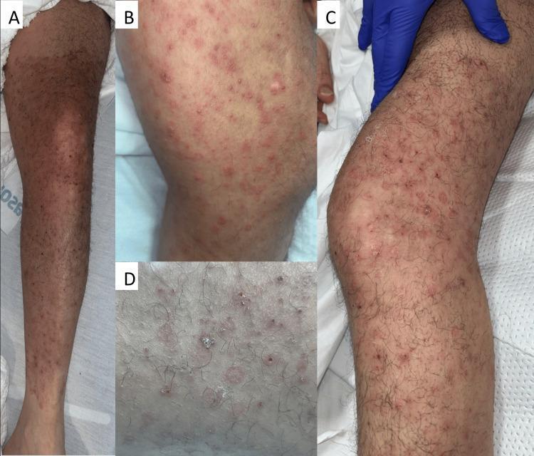
Clinical photograph of a 45-year-old male's left lower extremity demonstrating a complex dermatologic presentation involving co-infection of Herpes Zoster (HZ) and Majocchi's granuloma (MG). Panels A and B show the initial presentation, characterized by a well-demarcated, confluent erythematous plaque extending from the proximal thigh to the ankle. Within the plaque are visible erythematous papules, vesicles, and crusted erosions. Panels C and D illustrate the progression after 24 hours of IV acyclovir treatment, highlighting more prominent erythematous, scaly, annular papules and crusted red perifollicular lesions. The morphology exhibits both a dermatomal distribution suggestive of Varicella-Zoster Virus (VZV) and follicular involvement indicative of a deep fungal infection (Majocchi's granuloma). This comparison illustrates the diagnostic challenge when viral and fungal pathologies coexist in an immunocompromised patient, demonstrating how treatment for one condition (acyclovir) can make the features of the underlying fungal component more visually distinct.
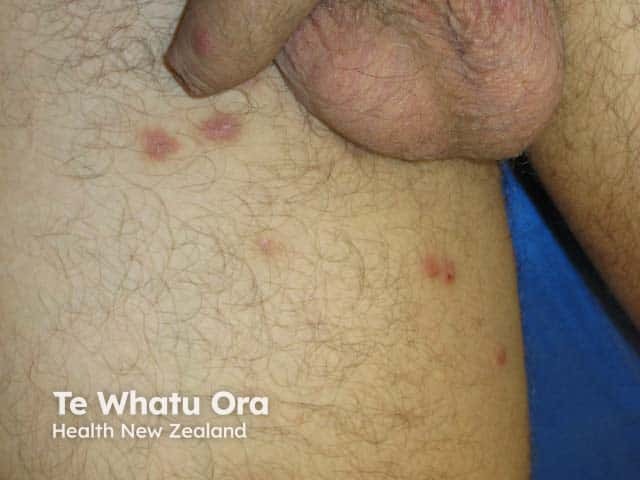
Imaging modality: Clinical photography. Technique: High-resolution external view with frontal close-up of the genital region and proximal thighs under standard ambient lighting; no dermoscopy or magnification. Anatomical location: penile foreskin and adjacent proximal thigh skin. Visual features: multiple erythematous papules and small nodules on the foreskin and inner thigh; some lesions show shallow central pits, mild scaling, and subtle surface edema. The lesions are non-vesicular in this view; color ranges from pink to red; surface is slightly excoriated with superficial capillary prominence. Distribution: localized to genito-femoral skin with bilateral involvement of proximal thighs. Inferred pathology: lesions compatible with nodular scabies and cutaneous hypersensitivity reaction to Sarcoptes scabiei mites; burrows are not clearly visible in a single clinical photograph. Diagnostic significance: supports diagnosis of cutaneous scabies or nodular scabies in a male patient and warrants antiparasitic treatment and contact tracing. Clinical correlation: presents with intense nocturnal pruritus and history of exposure to an infested individual; secondary bacterial infection risk if scratched. Management implications: topical permethrin 5% or oral ivermectin per guidelines, treat all household close contacts, wash clothing and bedding. Differential considerations: folliculitis, contact dermatitis, tinea cruris, eczema, sexually transmitted skin infections. Educational use: clinical recognition of scabies nodules on genital skin.
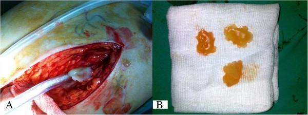
This composite clinical photograph depicts the intraoperative findings and macroscopic pathology of a hydatid cyst in a 56-year-old male. Image A shows an open surgical field in the proximal thigh, revealing a large incision with deep, erythematous soft tissue. A suction tube is visible within the wound, positioned near a whitish, gelatinous mass emerging from the muscular or bursa planes. Image B displays three macroscopic daughter vesicles or cysts placed on a sterile white gauze for examination. These vesicles are translucent, yellowish-amber in color, and possess a gelatinous texture. They exhibit a characteristic irregular, lobulated, or 'snow-shaped' morphology typical of Echinococcus granulosus infection. The educational focus is on the clinical presentation of musculoskeletal hydatid disease, which can mimic bursitis or soft tissue tumors around orthopedic implants. Key concepts illustrated include the visual identification of daughter cysts and the surgical management of cystic soft tissue masses in the extremities.