
Deny drash syndrome
Reading File
Denys-Drash Syndrome (DDS) is a rare congenital disorder caused by mutations in the WT1 (Wilms Tumor 1) gene on chromosome 11p13. It is characterized by a classic triad: Wilms tumor (nephroblastoma), diffuse mesangial sclerosis leading to nephrotic syndrome, and disorders of sexual differentiation (pseudohermaphroditism). Most cases present in infancy or early childhood with heavy proteinuria progressing rapidly to end-stage renal disease.
Searching sources...
Finding Sources
Finding Sources
Searching PubMed
"Denys-Drash syndrome" WT1
Good hits in the textbooks. Let me read the key sections in parallel.
Reading File
Reading File
Reading File
Good. Let me now read more from Robbins for the Wilms tumor/DDS section and from Campbell Walsh for the Frasier comparison, plus the nephrology textbook for the renal aspects.
Reading File
Reading File
Excellent - I now have comprehensive information from multiple authoritative textbooks. Let me compile the full answer.
Denys-Drash Syndrome (DDS)
Overview
Denys-Drash syndrome is a rare genetic disorder first described by Denys et al. (1967) and further characterized by Drash et al. (1970). It is caused by dominant-negative missense mutations in the WT1 (Wilms Tumor 1) gene on chromosome 11p13, encoding a zinc-finger DNA-binding transcription factor critical for renal and gonadal embryogenesis.
Classic Triad
| Component | Details |
|---|---|
| Nephropathy | Early-onset proteinuria, hypertension, diffuse mesangial sclerosis (DMS) - the common denominator of the syndrome |
| Wilms Tumor (Nephroblastoma) | ~90% lifetime risk; high incidence of bilateral tumors; usually favorable triphasic histology |
| Gonadal/Genital Abnormalities | Male pseudohermaphroditism (46,XY), gonadal dysgenesis, genital ambiguity, hypospadias, cryptorchidism |
Incomplete forms exist; the nephropathy is considered the minimum diagnostic criterion.
Genetics and Molecular Mechanism
- Gene: WT1, chromosome 11p13
- Mutation type: Dominant-negative missense mutation in the zinc-finger region of the WT1 protein
- This disrupts DNA-binding properties and interferes with the remaining wild-type allele
- The heterozygous mutation is sufficient to cause genitourinary abnormalities but not tumorigenesis alone
- Wilms tumors in DDS require biallelic inactivation of WT1 (i.e., a second hit is needed for tumor development)
- WT1 is expressed in the kidney and gonads during embryogenesis; it regulates glomerular podocyte-specific proteins and genes involved in renal differentiation
This contrasts with WAGR syndrome (Wilms tumor + Aniridia + Genitourinary anomalies + intellectual disability), where the mechanism involves a germline deletion of WT1 at 11p13.
Renal Pathology
- Diffuse mesangial sclerosis (DMS) - the hallmark glomerular lesion
- Presents as congenital or infantile nephrotic syndrome (steroid-resistant)
- Rapidly progressive to end-stage renal disease (ESKD), often by age 3 years
- DMS is distinct from focal segmental glomerulosclerosis (FSGS), which characterizes the related Frasier syndrome
An interesting radiological finding is caliceal blunting without obstruction (Jadresic et al., 1990).
Gonadal Features
- Patients with 46,XY karyotype have gonadal dysgenesis with streak gonads and male pseudohermaphroditism - female or ambiguous external genitalia
- Mixed gonadal dysgenesis is common
- Gonadoblastoma risk: ~40% in DDS - streak gonads must be removed prophylactically
- Patients who reach ESKD may present phenotypically female but be genotypically XY
Wilms Tumor Features
- Risk approximately 90% (much higher than WAGR at ~33%)
- May be diagnosed before, simultaneously with, or after the nephropathy
- High incidence of bilateral Wilms tumor
- Favorable triphasic histology (blastemal, stromal, epithelial elements) is most common
- Risk of germ cell tumors (gonadoblastomas) is also increased due to disrupted gonadal development
DDS vs. Frasier Syndrome (Related WT1 Disorder)
| Feature | Denys-Drash Syndrome | Frasier Syndrome |
|---|---|---|
| Mutation | Dominant-negative missense (exon 8/9 zinc-finger) | Splice-site mutation in intron 9 (KTS splice donor) |
| Nephropathy | Diffuse mesangial sclerosis (DMS); early, rapid | Focal segmental glomerulosclerosis (FSGS); later onset, slower |
| Wilms tumor | ~90% risk | No known predisposition |
| Gonadoblastoma risk | ~40% | ~60% |
| Karyotype (usual) | 46,XY, DSD | 46,XY, female phenotype |
| Onset of ESKD | Infancy/early childhood | Adolescence/adulthood |
Wilms Tumor Predisposition Syndromes Summary
| Syndrome | Chromosomal Locus | Risk of Wilms Tumor |
|---|---|---|
| WAGR | WT1 deletion, 11p13 | ~33% |
| Denys-Drash | WT1 missense, 11p13 | ~90% |
| Beckwith-Wiedemann | WT2/IGF2 region, 11p15.5 | ~5-10% |
Management
- Nephrectomy - prophylactic bilateral nephrectomy has been used given the high Wilms tumor risk and to delay or manage ESKD
- Wilms tumor treatment - surgery, chemotherapy (vincristine, actinomycin D), +/- radiotherapy per stage
- Renal replacement therapy - dialysis and renal transplantation for ESKD
- Gonadectomy - bilateral prophylactic removal of streak gonads due to ~40% gonadoblastoma risk
- Gender assignment - in XY DSD cases, involves multi-disciplinary team; historically most raised female; decisions guided by anatomy and degree of virilization
- Wilms tumor surveillance - abdominal ultrasound every 3 months until age 7 in WT1 mutation carriers
Sources:
- Robbins, Cotran & Kumar Pathologic Basis of Disease, p. 454
- Campbell Walsh Wein Urology (3-Volume Set), p. 1275
- Comprehensive Clinical Nephrology, 7th Edition
- Brenner and Rector's The Kidney, 2-Volume Set
Recent reviews (2024-2025) confirm ongoing expansion of the WT1 disorder spectrum: Lopez-Gonzalez & Ariceta, Pediatr Nephrol 2024 and Nagano & Nozu, Clin Exp Nephrol 2025 emphasize that DDS represents one end of a broad WT1-related continuum.
Wagers comples
WAGR complex (also written WAGR syndrome) is a contiguous gene deletion syndrome on chromosome 11p13 characterized by Wilms tumor, Aniridia, Genitourinary anomalies, and intellectual Retardation/disability. It results from deletion of both the WT1 and PAX6 genes on 11p13.
Searching sources...
Finding Sources
Finding Sources
Good hits. Let me read the key sections in parallel.
Reading File
Reading File
Reading File
Reading File
I also have a useful diagram image. Let me fetch it for the answer.
Loading Image
I now have all the information needed. Here is the comprehensive answer:
WAGR Complex (WAGR Syndrome)
What Does WAGR Stand For?
| Letter | Feature |
|---|---|
| W | Wilms tumor (nephroblastoma) |
| A | Aniridia (absence of the iris) |
| G | Genitourinary anomalies |
| R | intellectual Retardation / developmental disability |
Chromosomal Basis - Contiguous Gene Deletion Syndrome
WAGR is a contiguous gene deletion syndrome caused by a constitutional (germline) interstitial deletion of chromosome 11p13. Two adjacent genes are co-deleted:
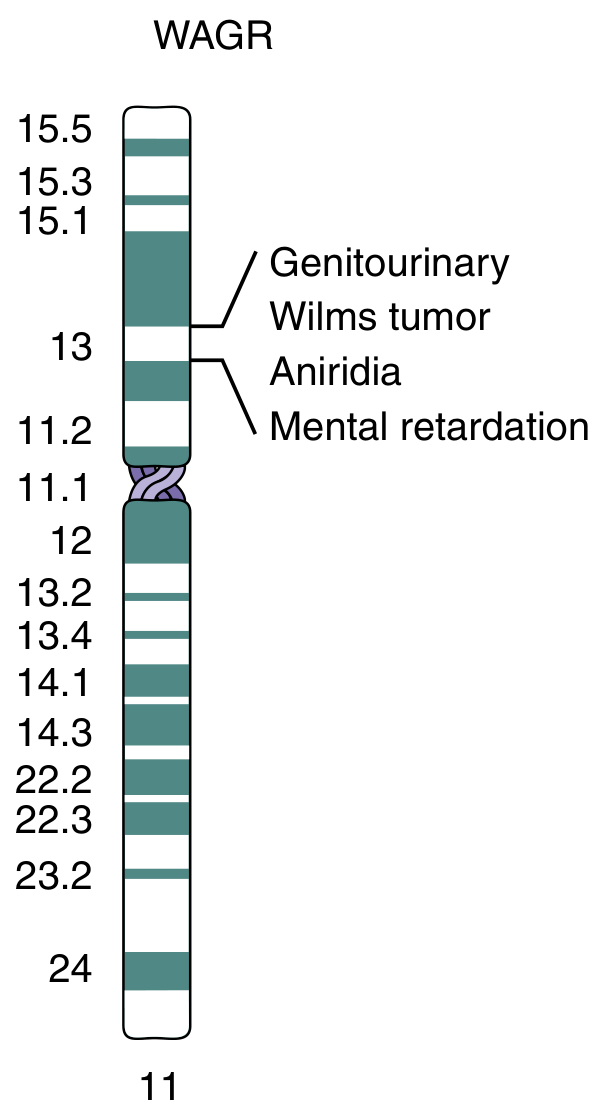
| Gene | Location | Consequence of deletion |
|---|---|---|
| WT1 | 11p13 | Wilms tumor predisposition + genitourinary anomalies |
| PAX6 | 11p13 (~0.6 Mb from WT1) | Aniridia |
The deletion is detected by chromosomal microarray (CMA) or FISH. In ~40-70% of aniridia patients found to have a deletion encompassing WT1, Wilms tumor will develop (usually by age 4 years in 90% of cases).
Key point: patients with deletions restricted to PAX6 (with intact WT1) develop aniridia but have no increased Wilms tumor risk.
Molecular Mechanism: Two-Hit Model
- First hit - germline heterozygous deletion of one WT1 allele (constitutional, inherited or de novo)
- Second hit - somatic nonsense or frameshift mutation in the remaining WT1 allele in a renal cell
- This biallelic inactivation of WT1 triggers Wilms tumor formation
This follows Knudson's two-hit model for tumor suppressor genes.
Clinical Features
1. Wilms Tumor (Nephroblastoma)
- Lifetime risk ~33% (lower than Denys-Drash syndrome's ~90%)
- Peak incidence age 2-5 years; 90% before age 4 in WAGR
- Can be bilateral (synchronous or metachronous)
- Belongs to the "type I Wilms tumor" subset: stromal-predominant favorable histology, intralobar nephrogenic rests, early onset, genitourinary anomalies in males
2. Aniridia
- Complete or partial absence of the iris
- Caused by PAX6 haploinsufficiency
- Associated with nystagmus, photophobia, reduced visual acuity, cataracts, glaucoma
- Aniridia alone (without WT1 deletion) does NOT predispose to Wilms tumor
3. Genitourinary Anomalies
- Cryptorchidism, hypospadias, streak gonads, renal anomalies
- Males are more severely affected
- Result from WT1 haploinsufficiency (WT1 is required for normal renal and gonadal embryogenesis)
4. Intellectual Disability / Developmental Delay
- Due to deletion of additional contiguous genes at 11p13 beyond WT1 and PAX6
- Variable in severity; generally mild to moderate
- The larger the deletion, the more severe the phenotype
WT1 Gene - Role in Development
- Encodes a zinc-finger transcription factor
- Critical for normal kidney and gonadal development
- Regulates mesenchymal-to-epithelial transition during nephrogenesis
- Required for ureteric bud outgrowth
- Knockout in mice results in complete failure of kidney and gonadal development
- Also regulates podocyte-specific proteins in glomeruli
Wilms Tumor Predisposition Syndromes - Comparison
| Syndrome | Locus | Mechanism | Wilms Tumor Risk | Extra Features |
|---|---|---|---|---|
| WAGR | 11p13 (deletion) | WT1 + PAX6 deletion; 2-hit for tumor | ~33% | Aniridia, GU anomalies, ID |
| Denys-Drash | 11p13 (point mutation) | Dominant-negative WT1 missense; 2-hit for tumor | ~90% | DMS nephropathy, pseudohermaphroditism, gonadoblastoma |
| Beckwith-Wiedemann | 11p15.5 | IGF2 overexpression / imprinting defect | ~5-10% | Macroglossia, organomegaly, omphalocele, hemihypertrophy |
Management
- Wilms tumor surveillance - renal ultrasound every 3 months until age 8 (given high risk)
- Ophthalmology - regular review for aniridia-related glaucoma, cataract
- FISH/CMA testing - all children with aniridia should be screened for 11p13 deletion to identify those with WT1 co-deletion (i.e., WAGR vs. isolated PAX6 aniridia)
- Developmental support - early intervention for intellectual disability
- Nephrology follow-up - monitoring renal function (especially post-nephrectomy)
- Wilms tumor treatment - surgery + chemotherapy per COG/SIOP protocols; prognosis is generally excellent with ~90% cure rates overall
Sources:
- Robbins, Cotran & Kumar Pathologic Basis of Disease, p. 453-454
- Campbell Walsh Wein Urology (3-Volume Set), p. 1600-1604
- Emery's Elements of Medical Genetics and Genomics, p. 192
- Henry's Clinical Diagnosis and Management by Laboratory Methods, p. 1644
VHL SYNDROME
Von Hippel-Lindau (VHL) syndrome is an autosomal dominant tumor suppressor syndrome caused by germline mutations in the VHL gene on chromosome 3p25. It predisposes to a characteristic set of tumors including clear cell renal cell carcinoma, hemangioblastomas of the CNS/retina, pheochromocytoma, and pancreatic neuroendocrine tumors. The VHL protein normally targets HIF-1α for degradation; loss of VHL leads to constitutive HIF activation and upregulation of angiogenic factors like VEGF.
Searching sources...
Finding Sources
Finding Sources
Excellent - found dedicated sections. Let me read all three key sources in parallel.
Reading File
Reading File
Reading File
Excellent. Now let me read the rest of the Campbell Walsh section on VHL (the HIF pathway and clinical management) and search for Robbins for the molecular mechanism and RCC pathology.
Reading File
Finding Sources
Reading File
Reading File
Reading File
There's a great VHL/HIF pathway diagram. Let me fetch it.
Loading Image
I now have comprehensive information from multiple authoritative textbooks plus an excellent pathway diagram. Here is the full answer:
Von Hippel-Lindau (VHL) Syndrome
Overview
VHL syndrome is an autosomal dominant tumor suppressor syndrome with a frequency of approximately 1 in 30,000-40,000 people. It is caused by germline loss-of-function mutations in the VHL tumor suppressor gene on chromosome 3p25-26. It predisposes to a characteristic spectrum of highly vascular tumors across multiple organ systems.
Genetics
| Feature | Detail |
|---|---|
| Gene | VHL, chromosome 3p25-26 |
| Inheritance | Autosomal dominant |
| Mechanism | Tumor suppressor - two-hit model |
| Gene structure | 3 exons; encodes a 213 amino acid protein |
| Somatic role | VHL mutations also occur in ~80% of sporadic clear cell RCC |
Mutation Types and Genotype-Phenotype Correlation
| Mutation Type | Protein Effect | Associated Phenotype |
|---|---|---|
| Type 1 - deletions/truncating | Truncated non-functional protein | VHL without pheochromocytoma |
| Type 2 - missense | Full-length but dysfunctional protein | VHL with pheochromocytoma |
Molecular Mechanism - The VHL/HIF Pathway
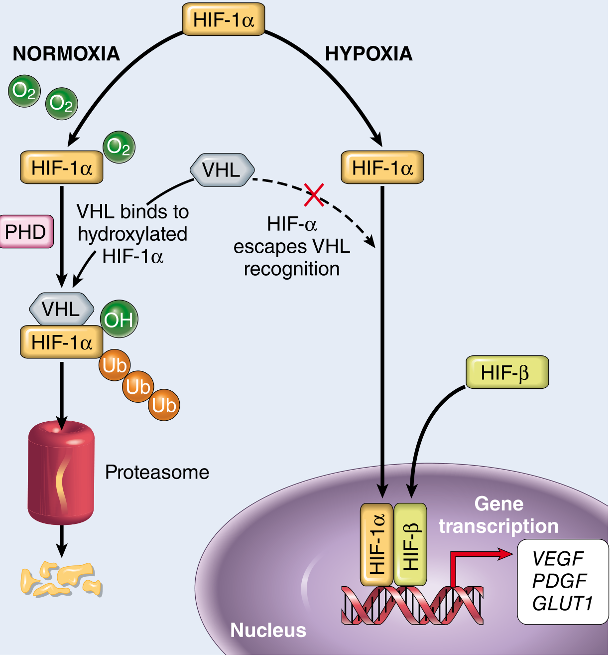
Under normal (normoxic) conditions:
- HIF-1α is hydroxylated by HIF prolyl hydroxylase (PHD) in the presence of oxygen
- Hydroxylated HIF-1α is recognized and bound by the VHL protein
- VHL is part of an E3 ubiquitin ligase complex (also containing elongins B and C)
- This complex ubiquitinates HIF-1α, targeting it for proteasomal degradation
- HIF levels remain low → no upregulation of angiogenic/growth genes
When VHL is mutated/lost:
- HIF-1α and HIF-2α cannot be ubiquitinated and degraded (even under normoxia)
- HIF accumulates and enters the nucleus, dimerizing with HIF-β
- The HIF-α/HIF-β complex drives transcription of target genes:
- VEGF - vascular endothelial growth factor (angiogenesis)
- PDGF - platelet-derived growth factor (mitogen)
- GLUT1 and glycolytic enzymes (Warburg metabolism)
- IGF-1 - insulin-like growth factor-1 (cell growth)
- Erythropoietin - can cause paraneoplastic polycythemia
- Result: constitutive "pseudo-hypoxic" state → uncontrolled angiogenesis and tumor growth
This explains why all VHL-associated tumors are highly vascular.
Clinical Manifestations
Major Tumor Types
| Organ/System | Lesion | Key Features |
|---|---|---|
| Kidney | Clear cell RCC | ~50% of VHL patients; bilateral, multifocal; early onset (3rd-5th decade); leading cause of mortality in VHL |
| Kidney/Pancreas | Renal & pancreatic cysts | Renal cysts in 50-75%; complex cysts raise suspicion for RCC |
| Cerebellum/CNS | Hemangioblastoma | Most common neurologic manifestation; cystic lesion with mural nodule; can also occur in brainstem, spinal cord, nerve roots |
| Retina | Retinal angioma (hemangioblastoma) | Can cause blindness if untreated; treated with laser ablation |
| Adrenal | Pheochromocytoma | Clustered in Type 2 families; secretes catecholamines |
| Pancreas | Neuroendocrine tumors + cysts | Pancreatic islet cell tumors |
| Epididymis/Broad ligament | Papillary cystadenoma | Benign; bilateral in males; can cause infertility |
| Inner ear | Endolymphatic sac tumor | Papillary tumor; can cause hearing loss, vertigo |
Hemangioblastoma - Histology
- Highly vascular neoplasm
- Presents as a mural nodule within a large fluid-filled cyst
- Microscopically: numerous thin-walled capillary vessels separated by stromal cells with vacuolated, lipid-rich cytoplasm
- Stromal cells express inhibin (TGF-β family member) - useful diagnostic marker
Clear Cell RCC - Molecular Note
- In virtually all clear cell RCCs (sporadic, familial, or VHL-associated): deletion of one copy of chromosome 3p25 (VHL locus)
- In up to 80% of sporadic clear cell RCC: the second VHL copy is inactivated by somatic mutation or epigenetic silencing (hypermethylation)
- This confirms VHL as a classic two-hit tumor suppressor for this histologic subtype
VHL Syndrome Classification (Types)
| Type | Pheochromocytoma | RCC/Hemangioblastoma | Mutation |
|---|---|---|---|
| Type 1 | Absent | Present | Large deletions / truncating |
| Type 2A | Present | Low RCC risk | Missense |
| Type 2B | Present | High RCC risk | Missense |
| Type 2C | Present only | No RCC/HB | Specific missense |
Screening and Surveillance
Given the high morbidity of CNS and renal lesions if undetected:
| Organ | Modality | Interval |
|---|---|---|
| Kidney | MRI/CT abdomen | Annual |
| CNS | MRI brain + spine | Annual |
| Retina | Ophthalmoscopy | Annual |
| Adrenal | Plasma/urine catecholamines + MRI | Annual |
| Pancreas | MRI abdomen | Annual |
Management
- Renal tumors - active surveillance until tumor reaches 3 cm; then nephron-sparing surgery is preferred to preserve renal function (given bilateral/multifocal nature)
- CNS hemangioblastomas - surgical resection for symptomatic lesions
- Retinal angiomas - laser photocoagulation or cryotherapy
- Pheochromocytoma - alpha-blockade first, then surgical excision
- Targeted therapy - VHL loss → HIF overactivation → VEGF pathway is a key therapeutic target; agents such as belzutifan (HIF-2α inhibitor) are now approved specifically for VHL-related tumors
Comparison with Other Hereditary RCC Syndromes
| Syndrome | Gene/Locus | RCC Histology | Other Features |
|---|---|---|---|
| VHL | VHL, 3p25 | Clear cell | Hemangioblastomas, pheochromocytoma, cysts |
| Hereditary Papillary RCC | MET, 7q31 | Papillary type 1 | Bilateral papillary tumors |
| Hereditary Leiomyomatosis RCC | FH (fumarate hydratase) | Aggressive papillary | Cutaneous/uterine leiomyomas |
| Birt-Hogg-Dubé | FLCN, 17p11.2 | Chromophobe/oncocytoma | Fibrofolliculomas, lung cysts, pneumothorax |
Sources:
- Robbins, Cotran & Kumar Pathologic Basis of Disease, pp. 453, 880
- Robbins & Kumar Basic Pathology, p. 866
- Campbell Walsh Wein Urology (3-Volume Set), pp. 2849-2850
- Quick Compendium of Clinical Pathology, 5th ed., p. 408
Dietyl crisis
Dietl's crisis is an episodic condition characterized by sudden, severe colicky flank pain, nausea, vomiting, and hematuria caused by intermittent obstruction of the ureteropelvic junction (UPJ), typically due to a mobile ("floating") kidney or intermittent UPJ obstruction. Episodes are often triggered by high fluid intake or physical activity and resolve spontaneously.
Searching sources...
Finding Sources
Finding Sources
Excellent hits. Let me read the key sections in parallel.
Reading File
Reading File
Reading File
Reading File
Also read the Sabiston section and Bailey & Love for surgical management details.
Reading File
Reading File
I have comprehensive information from multiple authoritative sources. Here is the complete answer:
Dietl's Crisis
Definition
Dietl's crisis is a clinical syndrome of sudden, severe episodic flank pain associated with nausea, vomiting, and sometimes hematuria, caused by intermittent obstruction at the ureteropelvic junction (UPJ). It is a classic presentation of pelviureteric junction (PUJ) obstruction, particularly in older children and adults.
The hallmark is that episodes are precipitated by high fluid intake (caffeine, alcohol, large volumes of water) or diuretics and relieved after voiding a large volume of urine as the obstructed renal pelvis drains.
Pathophysiology
The underlying mechanism involves intermittent or partial UPJ/PUJ obstruction:
- High fluid intake or diuretics → brisk diuresis → rapid increase in urine flow rate
- Urine reaches the partially obstructed UPJ faster than it can drain into the ureter
- Renal pelvis and calyces acutely distend → rise in intrapelvic pressure
- This distension causes severe colicky flank pain
- After a few hours, urine eventually overcomes the partial obstruction or backs up until voiding drives a large urinary output
- The pelvis decompresses → pain resolves + large volume of urine is passed + the renal swelling visibly reduces
Physiologically, at high flow rates the UPJ block ceases and a 1:1 pacemaker-to-ureteral contraction correspondence develops - but if the UPJ segment is intrinsically or extrinsically narrowed, this compensation fails and pelvic dilatation occurs.
Causes of the Underlying UPJ Obstruction
Intrinsic (more common)
- Idiopathic UPJ stenosis - most common; aperistaltic/atretic segment with abnormal musculature
- Deficiency of c-KIT-positive interstitial pacemaker cells
- Increased collagen deposition replacing normal smooth muscle
- Mucosal folds or valves at the UPJ
Extrinsic
- Aberrant/accessory lower pole renal artery - crosses and compresses the UPJ
- Adhesions
- Mobile/ptotic kidney (nephropexy) - historically considered a cause
Key Anatomic Points
- More common in males and on the left side
- Bilateral in 10-40% of cases
- Approximately 10% of UPJ obstruction patients also have vesicoureteral reflux (VUR)
Clinical Features
| Feature | Detail |
|---|---|
| Pain | Severe, episodic, colicky flank/loin pain |
| Triggers | Large fluid intake, caffeine, alcohol, diuretics |
| Associated symptoms | Nausea, vomiting |
| Relief | Passing a large volume of urine - with reduction in visible/palpable flank swelling |
| Hematuria | May occur, especially after mild trauma |
| Hypertension | Can occur (rarely) |
| UTI/stones | Recurrent pyelonephritis or nephrolithiasis may develop |
| Age | Older children and adults (neonates present differently with abdominal mass) |
Investigations
During an Acute Episode (Most Informative)
- Renal ultrasound - best performed during the painful episode to capture acute dilatation of the renal pelvis and calyces without a dilated ureter (distinguishes UPJ from ureteral obstruction)
Between Episodes
| Investigation | Findings |
|---|---|
| Renal ultrasound | Hydronephrosis, dilated renal pelvis; may look normal between episodes |
| CT urogram / MRI | Demonstrates level and degree of obstruction; identifies crossing vessels |
| Diuretic renogram (MAG3 / DTPA + Furosemide) | Gold standard for functional assessment - measures excretion half-life (T½max) |
MAG3 Lasix Renogram Interpretation
- Normal: T½max < 10-15 minutes
- Obstruction: T½max > 20 minutes after furosemide administration + rising curve
- Also provides split renal function (affected vs. contralateral kidney)
- Differential function < 40% on the affected side indicates significant obstruction
- Helps differentiate true obstruction from congenital ectasia (non-obstructed dilatation - does not need surgery)
Management
Conservative (Observation)
- Mild symptoms + good renal function + low-grade obstruction
- Monitor with serial ultrasound and renography
Decompression (Temporary/Emergency)
- Percutaneous nephrostomy tube - for acute obstructive uropathy or infected hydronephrosis
- Ureteral (DJ) stent insertion
Definitive Surgery - Pyeloplasty
Indicated when:
- Symptomatic (Dietl's crises)
- Obstructive excretory curve on diuretic renography
- Renal function deterioration (>10% decrease in split function)
- Progressive hydronephrosis
- Recurrent UTI, stones
Techniques
| Technique | Approach | Notes |
|---|---|---|
| Anderson-Hynes dismembered pyeloplasty | Open / laparoscopic / robotic | Gold standard; atretic UPJ segment is excised and renal pelvis-ureter widely spatulated and re-anastomosed over a stent; preferred when a crossing vessel is present |
| Culp-DeWeerd (spiral flap) | Open | Non-dismembered; flap of redundant pelvis used as onlay |
| Foley Y-V plasty | Open | For high ureteral insertion |
| Endopyelotomy / ureteroscopic incision | Endoscopic | Less invasive; lower success rates |
- Overall long-term patency rate for pyeloplasty: > 90%
- A stent is left across the anastomosis; Foley catheter placed to prevent reflux during the renogram
Summary Table
| Feature | Detail |
|---|---|
| Cause | Intermittent UPJ obstruction |
| Trigger | Brisk diuresis (caffeine, alcohol, large fluids) |
| Symptom | Severe colicky flank pain + N/V |
| Relief | Large-volume void + reduction of flank swelling |
| Investigation of choice | MAG3 diuretic renogram |
| Definitive treatment | Anderson-Hynes dismembered pyeloplasty |
Sources:
- Campbell Walsh Wein Urology (3-Volume Set), pp. 521, 3199-3206
- Sabiston Textbook of Surgery, p. 134
- Comprehensive Clinical Nephrology, 7th Edition, p. 840
- Bailey & Love's Short Practice of Surgery, 28th Edition, p. 3484
- S. Das: A Manual on Clinical Surgery, 13th Edition, p. 4299
Potters syndrome
Potter sequence (Potter syndrome) is a fetal condition caused by severe oligohydramnios, most commonly due to bilateral renal agenesis. The absence of amniotic fluid leads to a characteristic cluster of findings: compressed facial features (Potter facies), limb deformities, and fatal pulmonary hypoplasia. It is lethal at birth due to respiratory failure.
Searching sources...
Finding Sources
Finding Sources
Good hits. Let me read the key sections in parallel - Robbins pathology for pathogenesis, Comprehensive Nephrology for renal agenesis, Emery's genetics, and Creasy & Resnik for the clinical image and antenatal management.
Reading File
Reading File
Reading File
Reading File
Let me now fetch the pathogenesis diagram and the Potter facies image.
Loading Image
Loading Image
Finding Sources
Reading File
Reading File
I now have comprehensive information and excellent images. Here is the complete answer:
Potter Sequence (Potter Syndrome)
Terminology - Important Distinction
The term "Potter syndrome" is technically a misnomer but is widely used. The correct terminology is:
| Term | Meaning |
|---|---|
| Potter sequence | The full chain of events: any cause of severe oligohydramnios → fetal compression → characteristic features |
| Bilateral renal agenesis | The most classic cause, but responsible for only ~20% of Potter sequence cases |
| "Potter syndrome" | Loosely used to refer to bilateral renal agenesis specifically, but this is semantically imprecise |
Pathogenesis
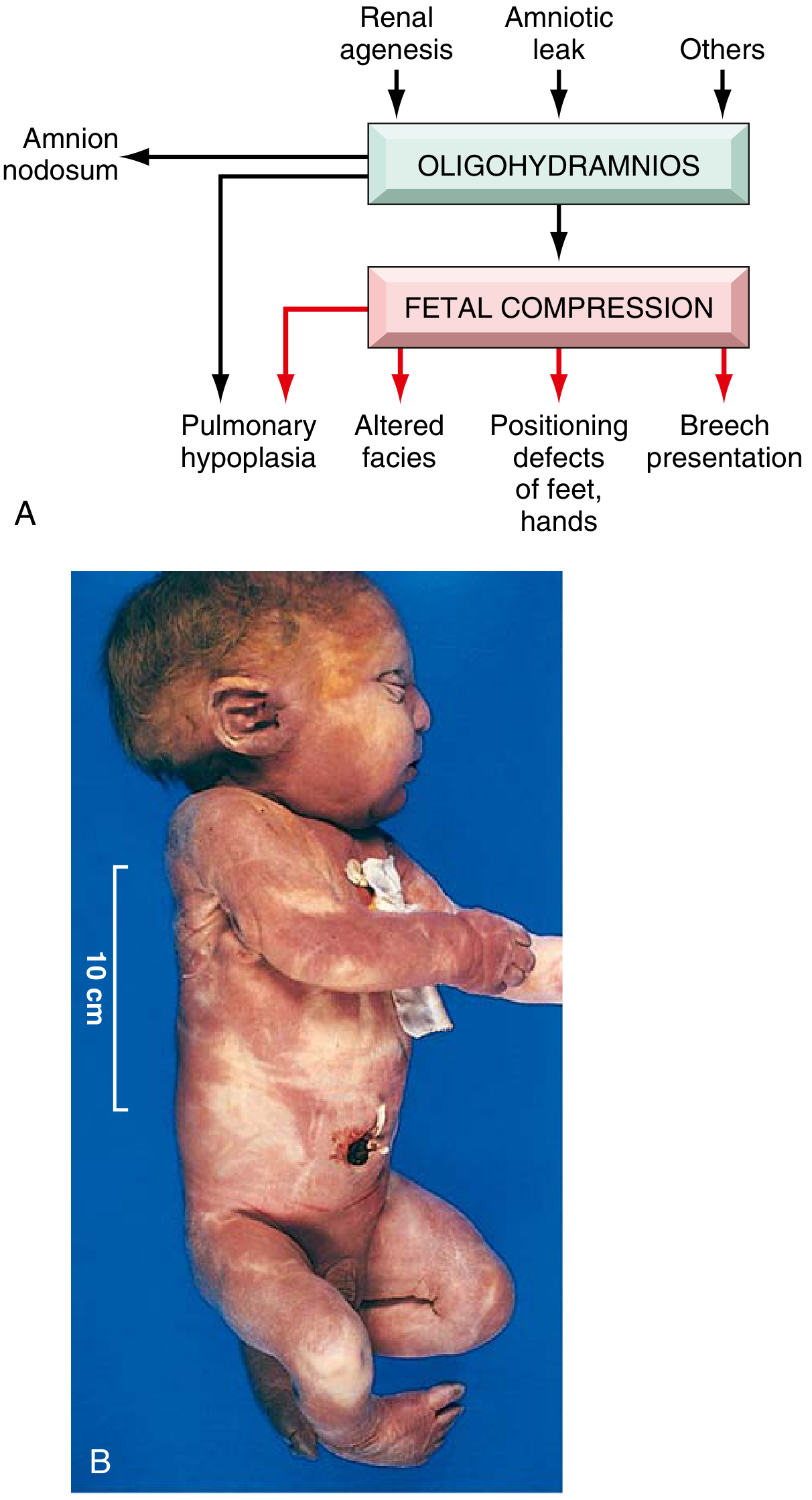
The central mechanism is oligohydramnios (severe reduction in amniotic fluid), which leads to chronic fetal compression in utero.
Why is amniotic fluid important?
- From the second trimester onward, fetal urine is the primary source of amniotic fluid
- Amniotic fluid provides:
- Mechanical cushion - protects against compression/deformity
- Lung development - fetal breathing movements of amniotic fluid are essential for bronchial branching and alveolar development
Consequences of oligohydramnios
| Effect of Fetal Compression | Clinical Manifestation |
|---|---|
| Absent lung fluid movements | Pulmonary hypoplasia - the lethal component |
| Facial compression | Potter facies (flattened nose, retrognathia, low-set ears, epicanthic folds) |
| Limb compression | Talipes equinovarus (clubfoot), hip dislocation, positional limb deformities |
| Abnormal lie | Breech presentation |
| Amnion abrasion from fetal skin | Amnion nodosum - nodules on the amnion surface made of squamous cell aggregates from vernix caseosa |
Potter Facies (Classic Facial Appearance)
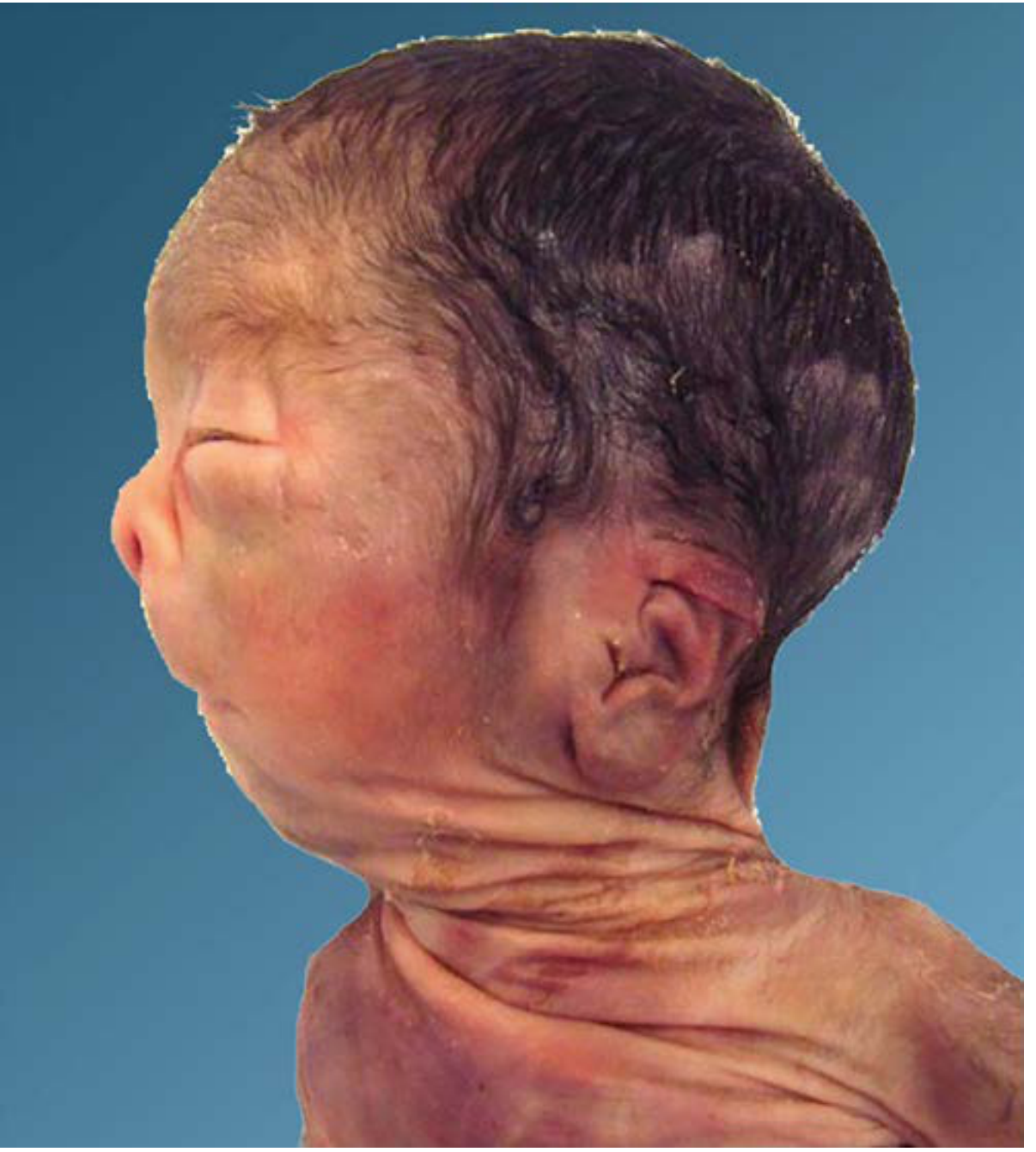
Characteristic features:
- Flattened/broad nose
- Retrognathia (receding chin)
- Low-set ears - severely malformed, bilateral
- Epicanthal folds (palpebronasal folds)
- Widely separated eyes
- Skin appears redundant and compressed
Causes of Potter Sequence
Any condition causing severe oligohydramnios can produce the Potter sequence:
| Cause | Notes |
|---|---|
| Bilateral renal agenesis | Classic cause; ~1 in 3000 births; responsible for ~1 in 5 (20%) of Potter sequence cases |
| Bilateral multicystic dysplastic kidney | Dysplastic nonfunctional kidneys; can cause the full sequence if bilateral |
| Obstructive uropathy | Posterior urethral valves, urethral atresia → bladder outlet obstruction → back pressure → renal dysplasia + anuria |
| Chronic amniotic fluid leak | Loss of fluid through amniotic membrane |
| Infantile (autosomal recessive) polycystic kidney disease | Massively enlarged non-functional kidneys |
Bilateral Renal Agenesis - In Detail
Embryology
- Renal agenesis results when ureteric buds fail to develop from the mesonephric duct or degenerate early
- Without the ureteric bud, the metanephrogenic blastema receives no inductive signal → no nephron formation
- Failure of bud penetration → no kidney development
- Likely multifactorial causation; rarely biallelic variants in ITGA8 or GFRA1 reported
- Also theorized: complete in utero involution of polycystic kidneys → apparent agenesis with a blind-ending ureter
Epidemiology
- Incidence: ~1 in 3,000-10,000 births (varies by source)
- More common in males
- Usually sporadic; sibling recurrence risk ~3%, rising to 15-20% with a family history of agenesis
- 15% risk of congenital renal abnormalities in first-degree relatives of the index patient
Prenatal Diagnosis
- Antenatal ultrasound: oligohydramnios/anhydramnios + absent/non-visualized kidneys + contracted/non-visualized bladder (no urine produced)
- Fetal MRI can confirm the diagnosis
Clinical Features at Birth
| System | Feature |
|---|---|
| Respiratory | Pulmonary hypoplasia - most infants die immediately from respiratory failure/insufficiency |
| Face | Potter facies (see above) |
| Ears | Low-set, severely malformed |
| Limbs | Talipes equinovarus (clubfoot), hip dislocation |
| Abdomen | No palpable kidneys; absent ureteric orifices |
| Incidence of absent ureter | ~50% with bilateral renal agenesis also have absent ureters and lower urinary tract |
Prognosis: nearly universally fatal due to pulmonary hypoplasia; most die during delivery or shortly after.
Prenatal Management
| Situation | Management |
|---|---|
| Suspected bilateral renal agenesis | Fetal MRI to confirm; discuss lethal nature with parents |
| Termination | Should be offered and considered |
| Serial amnioinfusion | Proposed to prevent pulmonary hypoplasia (canalicular phase 16-24 weeks) and reduce cord compression risk; very rarely achieves long-term survival; only within approved experimental protocols |
| Delivery | Unmonitored labor and vaginal delivery appropriate; cesarean section reserved for obstetric indications only |
| Unilateral renal agenesis | Serial ultrasound to monitor fetal growth, contralateral kidney, amniotic fluid; maternal renal assessment |
Unilateral Renal Agenesis (Contrast)
- Occurs ~1 in 1000 neonates; males more affected; left kidney usually absent
- Usually asymptomatic - contralateral kidney undergoes compensatory hypertrophy
- Associated with single umbilical artery - should prompt investigation
- Long-term complications: hypertension, recurrent infection, renal insufficiency
Summary
| Feature | Detail |
|---|---|
| Core mechanism | Oligohydramnios → fetal compression |
| Most classic cause | Bilateral renal agenesis |
| Lethal feature | Pulmonary hypoplasia (respiratory failure) |
| Facial features | Flattened nose, retrognathia, low-set ears, epicanthal folds |
| Limb features | Clubfoot, hip dislocation |
| Amnion finding | Amnion nodosum |
| Incidence | ~1 in 3000-4000 live births |
| Prognosis | Lethal - nearly 100% mortality |
Sources:
- Robbins & Kumar Basic Pathology, p. 135
- The Developing Human: Clinically Oriented Embryology, p. 696
- Quick Compendium of Clinical Pathology, 5th ed.
- Comprehensive Clinical Nephrology, 7th Edition, p. 775
- Creasy & Resnik's Maternal-Fetal Medicine, p. 6130
- Emery's Elements of Medical Genetics and Genomics, p. 242
- Grainger & Allison's Diagnostic Radiology
Horse shoe kidney
Horseshoe kidney is the most common renal fusion anomaly, occurring in ~1 in 400-600 births. The two kidneys are fused at their lower poles by a parenchymatous or fibrous isthmus that crosses the midline. Ascent is arrested by the inferior mesenteric artery. It is associated with PUJ obstruction, VUR, nephrolithiasis, and an increased risk of Wilms tumor and renal carcinoid.
Searching sources...
Finding Sources
Finding Sources
Excellent - dedicated sections found across multiple textbooks. Let me read all in parallel.
Reading File
Reading File
Reading File
Reading File
Good - let me now get the dedicated Campbell Walsh section on horseshoe kidney (block3) for complete clinical details, and fetch the horseshoe kidney illustration.
Reading File
Reading File
Loading Image
Now let me read the remaining Campbell Walsh section for complications, associated conditions, and management.
Reading File
I now have comprehensive information from multiple authoritative textbooks. Here is the complete answer:
Horseshoe Kidney
Definition
Horseshoe kidney (HSK) is the most common renal fusion anomaly, consisting of two distinct renal masses lying vertically on either side of the midline, connected at their respective lower poles (95% of cases) by a parenchymatous or fibrous isthmus that crosses the midline anterior to the great vessels.
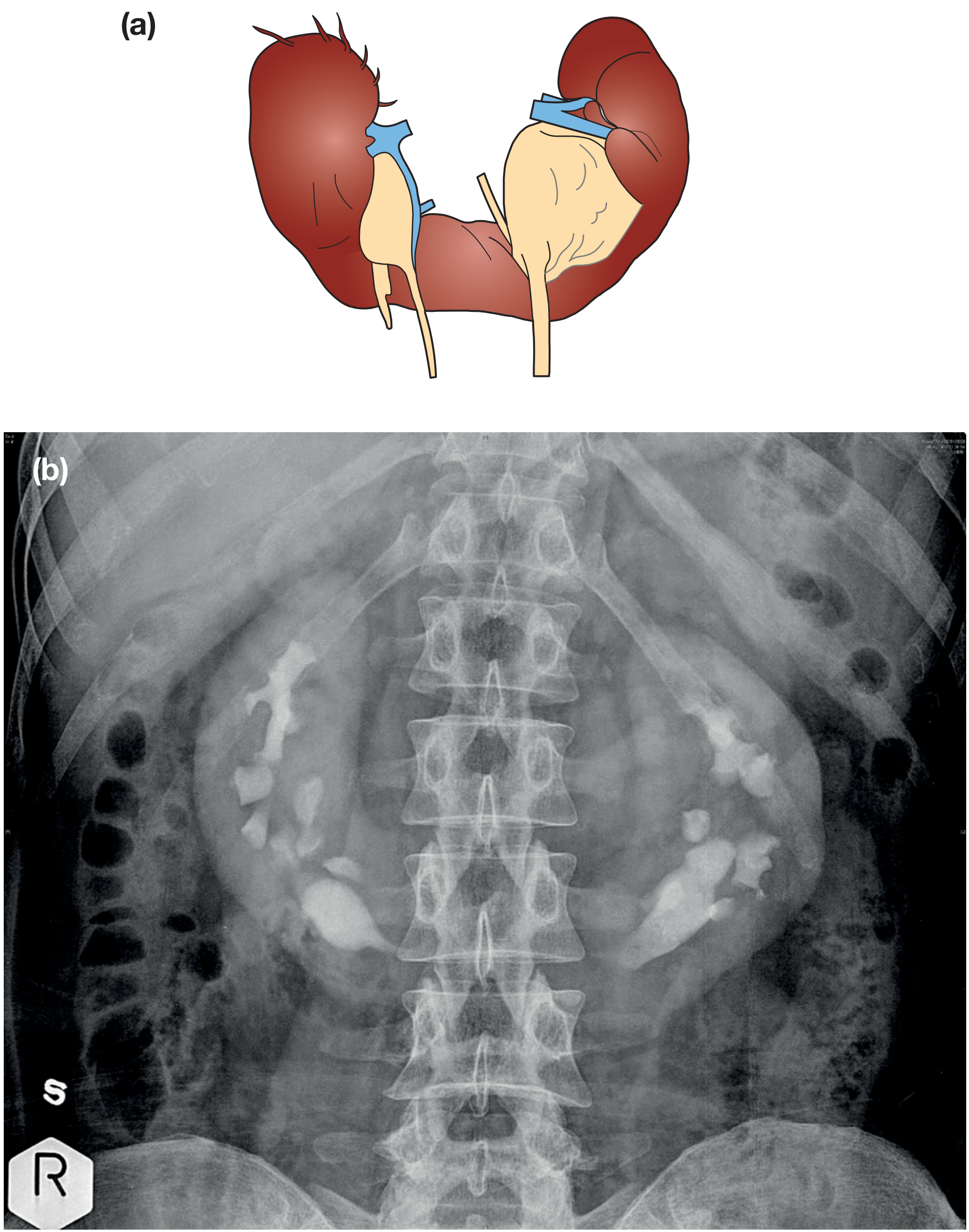
Epidemiology
| Feature | Detail |
|---|---|
| Incidence | ~1 in 400 individuals (~0.25%); range 1:400-1:800 |
| Sex | Males > females (2:1) |
| Familial occurrence | Reported in twins and siblings; likely multifactorial with low penetrance |
| Most common fusion | Lower pole (95%); upper pole fusion is rare |
Embryology and Pathogenesis
- During ascent from the pelvis, the developing metanephric kidney masses normally rotate medially and migrate cranially
- In HSK, the lower poles of the two metanephric masses fuse before the kidneys have completed rotation and ascent (at the 4-6 week stage)
- Further ascent is arrested when the isthmus becomes entrapped beneath the inferior mesenteric artery (IMA) as it arises from the aorta
- The isthmus typically lies at the level of L3-L4 vertebrae, just below the IMA origin
Why Does Rotation Fail?
- Because fusion occurs before rotation, the renal pelvis faces anteriorly (instead of medially) and the calyces point posteriorly and medially - the opposite of the normal orientation
Anatomy
Isthmus
- Located anterior to the aorta and inferior vena cava (rarely passes between or behind them)
- Usually bulky with functioning parenchyma and its own blood supply
- Sometimes just fibrous tissue
- Position: anterior to L3/L4, below the IMA, occasionally as low as the sacral promontory or true pelvis
Collecting System and Ureters
- Calyces are normal in number but atypically oriented - pointing posteriorly
- The lowermost calyces extend caudally/medially, may overlie the vertebral column
- Ureter inserts high on the renal pelvis and lies laterally (due to incomplete rotation)
- The ureter drapes ventrally over the renal isthmus (where it may be compressed by lower pole vessels) - this creates the risk of obstruction
- Lower ureter enters the bladder normally
- Occasionally associated with retrocaval ureter, ureteral ectopia, or duplication
Blood Supply - Highly Variable
- Only ~5-20% have a single artery to each moiety
- Usually multiple aberrant arteries from:
- Aorta (above or below isthmus)
- Inferior mesenteric artery
- Common or external iliac arteries
- Sacral arteries
- The isthmus itself may have separate arterial supply from the aorta
- This aberrant vasculature is a key consideration for surgery and PCNL
Clinical Features
Most horseshoe kidneys are asymptomatic - discovered incidentally on imaging. When symptoms occur, they are due to complications:
| Symptom/Sign | Cause |
|---|---|
| Vague lower abdominal/flank pain | Intermittent obstruction |
| Recurrent UTI | Urinary stasis |
| Hematuria | Stones, trauma, or tumor |
| Palpable abdominal mass | Large horseshoe isthmus |
Rovsing's Sign
- Abdominal pain, nausea, and vomiting upon hyperextension of the spine - occasionally described due to pressure on the isthmus against the aortic plexus
Complications and Associated Conditions
Urological Complications
| Complication | Incidence/Notes |
|---|---|
| PUJ obstruction (PUJO) | Common; due to high ureteral insertion + ureter crossing over isthmus; may cause hydronephrosis |
| Nephrolithiasis (stones) | Up to 20%; due to urinary stasis from incomplete drainage |
| Vesicoureteral reflux (VUR) | 8-32% of patients |
| Recurrent UTI/pyelonephritis | Secondary to obstruction and stasis |
| Hydronephrosis | Due to PUJO; investigate with MAG3 renogram |
Neoplastic Risk
| Tumor | Notes |
|---|---|
| Wilms tumor | Increased incidence in HSK vs. general population |
| Renal cell carcinoma | Can occur; clear cell most common |
| Transitional cell carcinoma | Slightly increased risk |
| Renal carcinoid tumor | Particularly associated with HSK |
Associated Syndromes and Congenital Anomalies
- Turner syndrome - HSK present in ~60% of females with Turner syndrome (45,X) - the most common association
- VACTERL association - anorectal malformations, vertebral/tracheoesophageal anomalies
- Neural tube defects - HSK in ~3% of these patients
- Caudal regression syndrome
- Genitourinary anomalies: hypospadias, undescended testes (4% of males), bicornuate uterus, septate vagina (7% of females)
- 50% of patients with HSK have at least one associated extrarenal anomaly
Imaging
| Modality | Findings |
|---|---|
| Plain X-ray (KUB) | Renal axes diverge inferiorly (inverted V) instead of converging inferiorly |
| IVU / CT urogram | Lower pole fusion; medially directed lower calyces; anteriorly placed pelves; ureters crossing over isthmus |
| Ultrasound | Dilated renal pelvis; lower poles joined; bilateral low-lying kidneys |
| CT/MRI | Gold standard - defines anatomy, vascular supply, isthmus, and any pathology; essential preoperatively |
| MAG3 diuretic renogram | To differentiate true PUJO from urinary stasis; provides split renal function |
Characteristic Imaging Signs
- "Drooping lily" / inverted V sign - renal axes
- Lower pole calyces pointing medially toward the spine
- Flower vase sign on IVU - overall HSK appearance
Management
Asymptomatic HSK
- No treatment required; most patients remain asymptomatic lifelong
- Follow-up if associated anomalies or risk factors
Complications
| Complication | Management |
|---|---|
| PUJO | Pyeloplasty (Anderson-Hynes); division of the isthmus (isthmusectomy) may be added if tethering the UPJ |
| Nephrolithiasis | ESWL (less effective due to bone attenuation), ureteroscopy, or PCNL via upper pole access (most favored in HSK) |
| VUR | Depending on grade; medical or surgical |
| Wilms tumor | Surgical excision ± chemotherapy |
PCNL in Horseshoe Kidney
- Upper pole access is preferred - most posterior, lateral, often subcostal, and provides excellent access to the whole collecting system
- Lower pole calyces are usually not safely accessible by direct puncture
- Puncture is more medial than in normal kidneys; may pass through paraspinal musculature
- Pre-op CT/MRI mandatory to assess: aberrant vasculature, retro-renal colon (present in ~5/12 cases in one series), calyceal orientation
Summary Table
| Feature | Detail |
|---|---|
| Type | Most common renal fusion anomaly |
| Incidence | 1 in 400 |
| Sex | Male > female (2:1) |
| Fusion | Lower poles (95%), connected by isthmus |
| Arrested by | Inferior mesenteric artery |
| Isthmus level | L3-L4 |
| Pelvis orientation | Anterior (malrotated) |
| Calyces orientation | Posterior and medial |
| Blood supply | Highly variable; multiple aberrant vessels |
| Key associations | Turner syndrome (60%), VACTERL, Wilms tumor |
| Key complications | PUJO, stones (20%), VUR (8-32%), UTI |
| Prognosis | Excellent if asymptomatic; complications treated on their merits |
Sources:
- Campbell Walsh Wein Urology (3-Volume Set), pp. 2934-2960
- Comprehensive Clinical Nephrology, 7th Edition, p. 784-795
- Bailey & Love's Short Practice of Surgery, 28th Edition, p. 3316-3327
- The Developing Human: Clinically Oriented Embryology, p. 697-698
This is a shared conversation. Sign in to Orris to start your own chat.