
Intercellular connections
Reading File
Intercellular connections (also called cell junctions) are specialized structures that physically link adjacent cells, allowing tissues to maintain structural integrity and coordinate function. The main types are tight junctions, adherens junctions, desmosomes, gap junctions, and hemidesmosomes.
Searching sources...
Finding Sources
Finding Sources
Reading File
Reading File
Reading File
Reading File
Loading Image
Loading Image
Loading Image
Reading File
Here is a complete overview of intercellular connections, drawn from Ganong's Review of Medical Physiology and Junqueira's Basic Histology.
Intercellular Connections (Cell Junctions)
Intercellular junctions fall into two broad functional categories:
- Fastening/structural junctions - hold cells together and anchor them to their basal laminas
- Communicating junctions - permit transfer of ions and molecules between cells
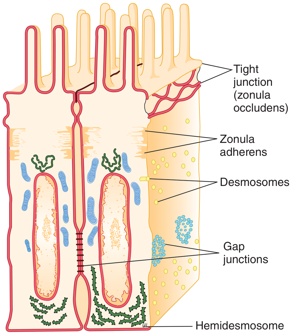
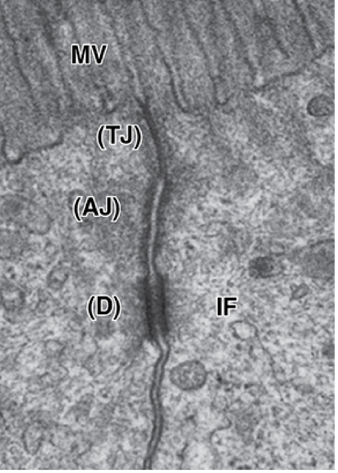
1. Tight Junction (Zonula Occludens)
- Location: Surrounds the apical margin of epithelial cells (intestinal mucosa, renal tubules, choroid plexus, blood-brain barrier endothelium)
- Structure: Ridges formed half from each cell; adjacent membranes appear fused in TEM. The seal results from interactions among three main transmembrane protein families: occludin, claudins, and junctional adhesion molecules (JAMs). Cytosolic scaffold proteins (e.g., ZO-1) anchor these to the actin cytoskeleton.
- Functions:
- Seals the paracellular space, forcing molecules to cross via the transcellular route
- Acts as a "fence" preventing lateral diffusion of membrane proteins between the apical and basolateral domains, maintaining cell polarity
- Degree of "leakiness" varies with claudin composition (e.g., proximal renal tubule = leaky; urinary bladder = very tight)
- Clinical relevance: Clostridium perfringens enterotoxin binds claudins, disrupting tight junctions and causing secretory diarrhea. Helicobacter pylori targets ZO-1 and disrupts gastric epithelial tight junctions, contributing to ulcer formation.
2. Adherens Junction (Zonula Adherens)
- Location: Immediately below the tight junction; encircles the cell as a continuous band
- Structure: Contains cadherins (Ca²⁺-dependent transmembrane glycoproteins). Cadherins bind homophilically across the intercellular space; their cytoplasmic tails bind catenins, which link to actin filaments via actin-binding proteins. These actin filaments contribute to the "terminal web" at the apical pole.
- Function: Major site of attachment for intracellular microfilaments; stabilizes and reinforces the occluding tight junction above it; firmly anchors cells to neighbors
3. Desmosome (Macula Adherens)
- Location: Scattered as spot-welds on lateral cell surfaces (not a continuous belt); abundant in skin, cardiac muscle, cervical epithelium
- Structure: Disc-shaped thickenings of apposed plasma membranes. Transmembrane proteins are desmogleins and desmocollins (larger cadherin family members). Their cytoplasmic ends bind plakoglobins → desmoplakins in an electron-dense plaque → intermediate filaments (cytokeratins/tonofilaments in epithelia, desmin in cardiac muscle)
- Function: Provides very strong mechanical adhesion; distributes tensile forces across a tissue via the intermediate filament network
- Clinical relevance: Pemphigus vulgaris - autoantibodies against desmogleins (especially desmoglein-3) disrupt desmosomes, causing intraepidermal blistering of skin and oral mucosa
4. Hemidesmosome
- Location: Basal surface of epithelial cells, anchoring them to the underlying basal lamina
- Structure: Resembles half a desmosome but uses integrins (not cadherins) as the transmembrane link proteins. Connected intracellularly to intermediate filaments; extracellularly binds laminin in the basal lamina.
- Function: Attaches epithelium to the extracellular matrix
- Clinical relevance: Mutations in hemidesmosomal proteins (e.g., BP180/collagen XVII, BP230) cause inherited or autoimmune blistering diseases such as bullous pemphigoid and junctional epidermolysis bullosa
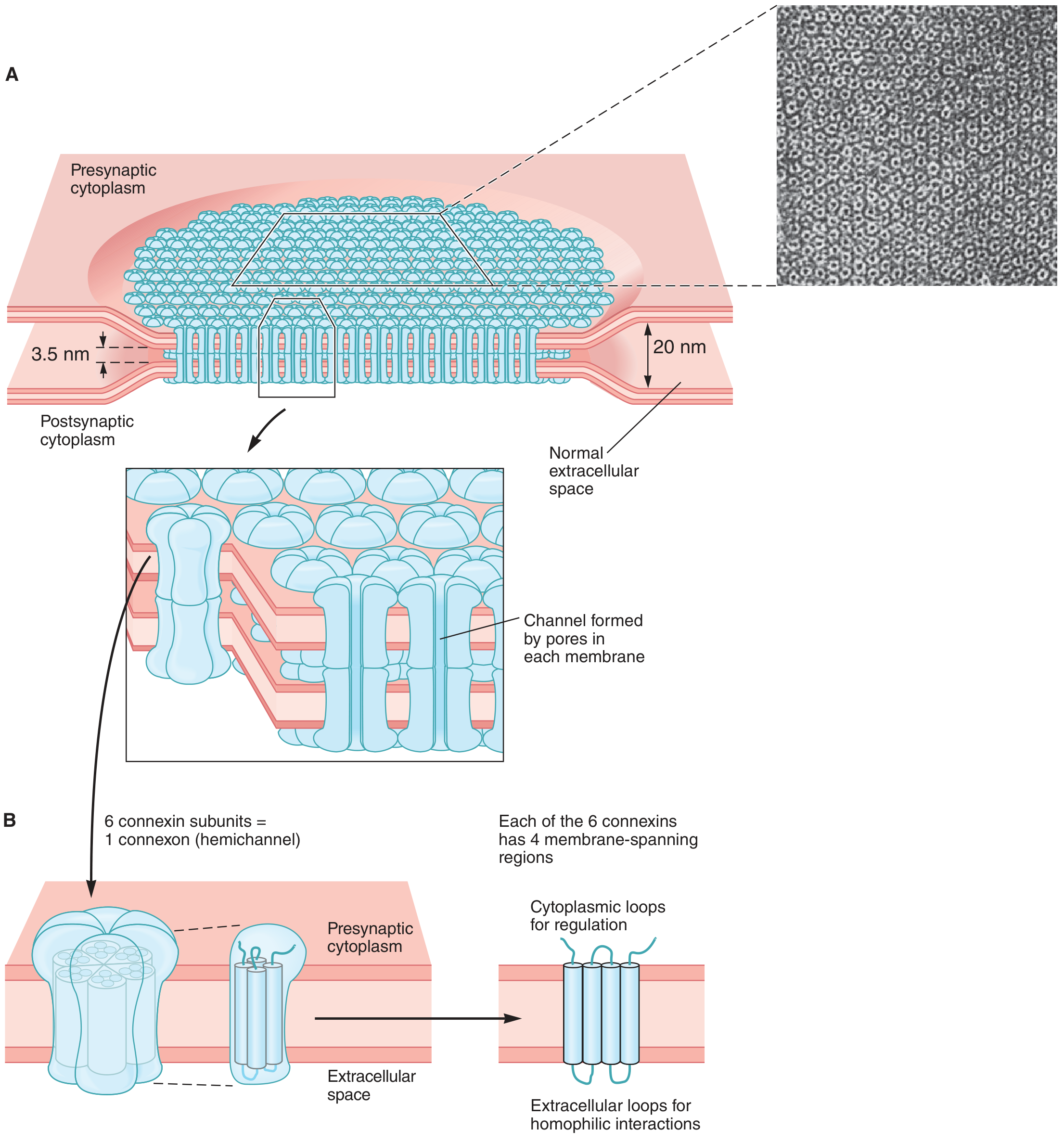
5. Gap Junction (Nexus)
- Location: Found in virtually all mammalian tissues; particularly important in cardiac muscle, smooth muscle, liver, lens of the eye
- Structure: The intercellular space is narrowed to ~4 nm. Each cell contributes a connexon (hemichannel) made of 6 connexin protein subunits (each with 4 membrane-spanning domains). Two connexons from adjacent cells align to form a complete dodecameric gap junction channel. Pore diameter ~0.8-1.4 nm; allows passage of molecules < 1000 Da. Humans have at least 20 different connexin-encoding genes.
- Function: Allows direct cell-to-cell passage of ions, sugars, amino acids, cAMP, IP₃, and other small signaling molecules. Enables electrical coupling (rapid propagation of action potentials in cardiac and smooth muscle) and chemical/metabolic coordination between cells.
- Regulation: Gap junction channels are gated; they close in response to increased intracellular Ca²⁺ or decreased pH (e.g., during cell injury).
- Clinical relevance: Connexin mutations cause:
- Connexin 26 (GJB2) mutations → hereditary sensorineural hearing loss (most common form)
- Connexin 32 mutations → Charcot-Marie-Tooth disease X (peripheral neuropathy)
- Cardiac connexin defects → electrophysiological arrhythmias and sudden cardiac death
Summary Table
| Junction | Alternative Name | Key Proteins | Intracellular Link | Function |
|---|---|---|---|---|
| Tight junction | Zonula occludens | Occludin, claudins, JAMs | ZO-1 → actin | Seal paracellular space; maintain cell polarity |
| Adherens junction | Zonula adherens | E-cadherin | Catenins → actin | Cell-cell adhesion; structural reinforcement |
| Desmosome | Macula adherens | Desmoglein, desmocollin | Desmoplakins → intermediate filaments | Strong spot-welds; tensile strength |
| Hemidesmosome | - | Integrins (α6β4) | Plectins → intermediate filaments | Attach cells to basal lamina |
| Gap junction | Nexus | Connexins | - | Intercellular communication; electrical coupling |
Sources: Ganong's Review of Medical Physiology, 26th ed., pp. 52-54 | Junqueira's Basic Histology, 17th ed., pp. 200-208
This is a shared conversation. Sign in to Orris to start your own chat.