
Lumbar plexues
Reading File
The lumbar plexus is a network of nerves formed from the anterior rami of L1-L4 (sometimes with contributions from T12), located within the psoas major muscle. It gives rise to key nerves supplying the lower abdominal wall, thigh, and groin.
Searching sources...
Finding Sources
Searching the Web
lumbar plexus anatomy branches clinical
Reading File
Reading File
Reading File
Reading File
Loading Image
Reading File
Loading Image
Reading File
Loading Image
Here is a complete overview of the Lumbar Plexus drawn from Gray's Anatomy for Students, Imaging Anatomy: Bones, Joints, Vessels and Nerves, and the THIEME Atlas of Anatomy.
Lumbar Plexus
Formation
The lumbar plexus is formed by the anterior rami of L1-L3 and most of L4, with a contributing branch from T12 (subcostal nerve). It is located within the substance of the psoas major muscle, anterior to the transverse processes of the lumbar vertebrae.
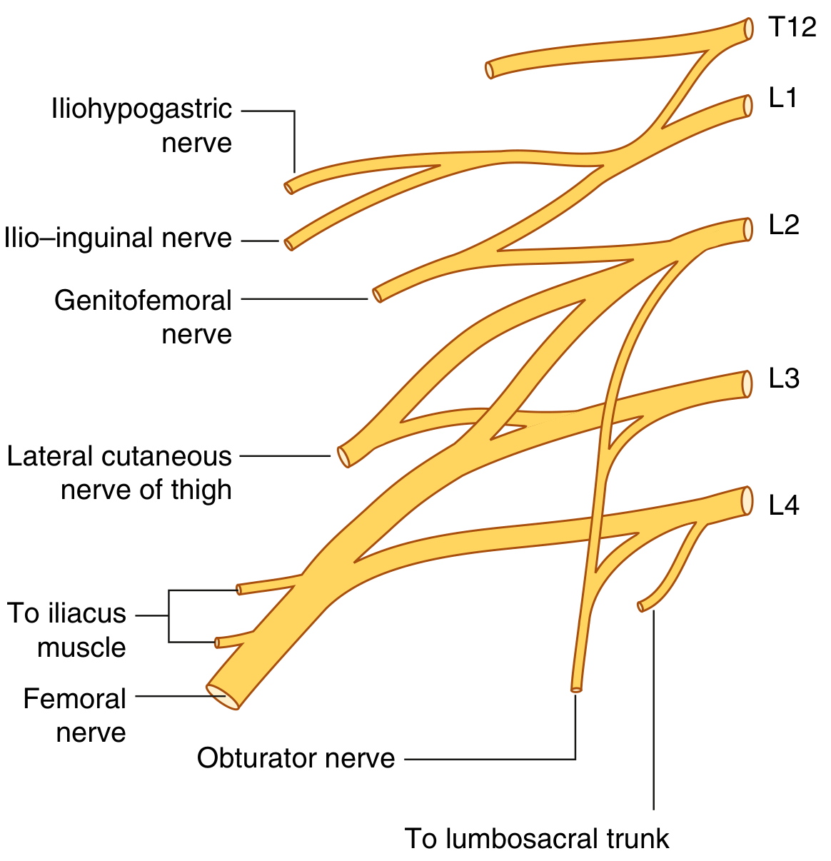
Fig. 4.177 - Lumbar Plexus (Gray's Anatomy for Students)
Branches - Emergence Relative to Psoas Major
Depending on where they exit the psoas muscle, branches emerge:
- Laterally - Iliohypogastric, ilio-inguinal, femoral nerve, lateral cutaneous nerve of thigh
- Anteriorly - Genitofemoral nerve
- Medially - Obturator nerve, lumbosacral trunk
Branches in Detail
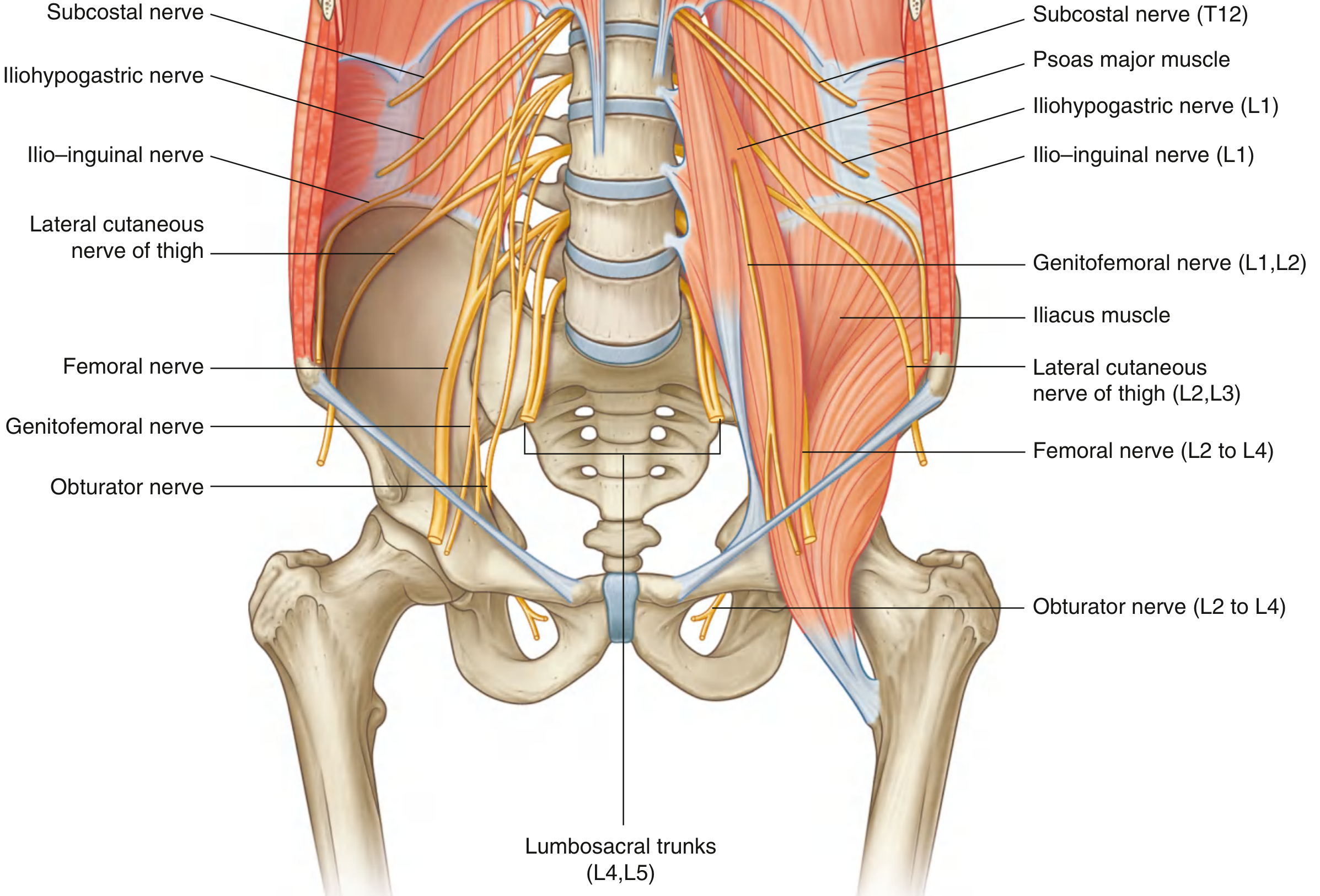
Fig. 4.178 - Lumbar Plexus in the Posterior Abdominal Region (Gray's Anatomy for Students)
1. Iliohypogastric Nerve (L1, ± T12)
- Emerges from the lateral border of psoas major, crosses quadratus lumborum, and pierces transversus abdominis.
- Lateral cutaneous branch - pierces internal and external oblique to supply posterolateral gluteal skin.
- Anterior cutaneous branch - passes above the superficial inguinal ring to supply skin of the pubic region.
- Motor: internal oblique and transversus abdominis.
2. Ilio-inguinal Nerve (L1)
- Smaller and inferior to the iliohypogastric nerve; enters the inguinal canal.
- Emerges through the superficial inguinal ring alongside the spermatic cord.
- Sensory: upper medial thigh, root of penis + anterior scrotum (men), or mons pubis + labium majus (women).
- Motor: internal oblique and transversus abdominis.
3. Genitofemoral Nerve (L1, L2)
- Descends through the substance of psoas major and emerges on its anterior surface, then divides into two branches:
- Genital branch - enters the inguinal canal through the deep inguinal ring; innervates the cremasteric muscle (men) or accompanies the round ligament (women); sensory to anterior scrotum / labium majus.
- Femoral branch - passes posterior to the inguinal ligament, enters the femoral sheath; sensory to the upper anterior thigh.
4. Lateral Cutaneous Nerve of Thigh (L2, L3)
- Purely sensory nerve.
- Emerges from the lateral border of psoas, crosses the iliacus muscle toward the anterior superior iliac spine (ASIS).
- Passes posterior to (or through) the inguinal ligament near ASIS.
- Supplies skin on the anterior and lateral thigh down to the knee.
- Clinical note: compression near the ASIS causes meralgia paresthetica (burning/tingling of the lateral thigh).
5. Obturator Nerve (L2-L4) - Anterior division
- Descends through psoas major, emerges from its medial border near the pelvic brim.
- Passes through the obturator canal to reach the medial compartment of the thigh.
- Divides into anterior and posterior branches (separated by obturator externus and adductor brevis):
- Motor: obturator externus, adductor longus, adductor brevis, adductor magnus, gracilis, pectineus.
- Sensory: medial aspect of the thigh; articular branches to the hip and knee joints.
6. Femoral Nerve (L2-L4) - Posterior division
- Largest branch; descends through psoas major, exits its lower lateral border.
- Lies between psoas and iliacus (iliacus fascia above), passes posterior to the inguinal ligament, lateral to the femoral artery, into the anterior compartment of the thigh.
- Immediately divides into multiple branches upon entering the thigh:
- Cutaneous: medial and intermediate cutaneous nerves (anterior thigh skin) + saphenous nerve (medial leg and foot - longest branch of lumbar plexus).
- Motor: iliacus, pectineus, sartorius, rectus femoris, vastus medialis, vastus intermedius, vastus lateralis.
- Articular: hip and knee joints.
Summary Table (Gray's Anatomy for Students, Table 4.6)
| Branch | Spinal Segments | Motor | Sensory |
|---|---|---|---|
| Iliohypogastric | L1 | Internal oblique, transversus abdominis | Posterolateral gluteal skin, pubic region |
| Ilio-inguinal | L1 | Internal oblique, transversus abdominis | Upper medial thigh, anterior scrotum/labium majus |
| Genitofemoral | L1, L2 | Cremaster (genital branch) | Anterior scrotum/labium majus (genital); upper anterior thigh (femoral) |
| Lateral cutaneous nerve of thigh | L2, L3 | None (purely sensory) | Anterior and lateral thigh to knee |
| Obturator | L2-L4 | Adductors, obturator externus | Medial thigh |
| Femoral | L2-L4 | Anterior thigh muscles, iliacus | Anterior thigh, medial leg (via saphenous) |
Cutaneous Distribution
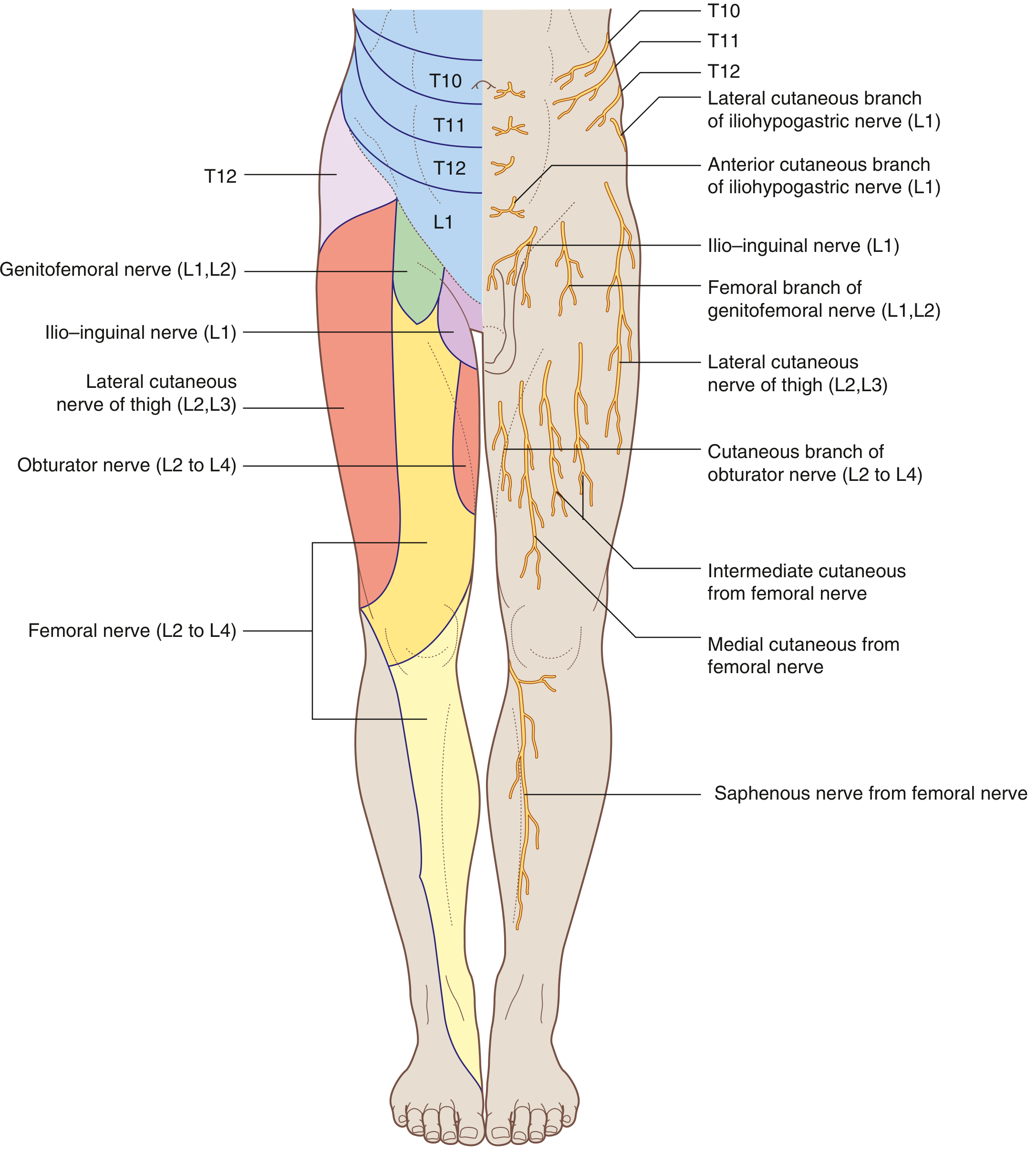
Fig. 4.179 - Cutaneous Distribution of Lumbar Plexus Nerves (Gray's Anatomy for Students)
Lumbosacral Trunk
The remaining part of L4 and all of L5 join to form the lumbosacral trunk, which descends into the pelvis to join the sacral plexus and eventually contributes to the sciatic nerve.
Key Clinical Points
| Nerve | Injury/Condition | Signs |
|---|---|---|
| Lateral cutaneous nerve of thigh | Meralgia paresthetica (entrapment near ASIS) | Burning, numbness, tingling on lateral thigh |
| Femoral nerve | Pelvic fracture, retroperitoneal hematoma, diabetic neuropathy | Weak knee extension, reduced patellar reflex, sensory loss anterior thigh |
| Obturator nerve | Hip surgery, pelvic mass | Weak thigh adduction, sensory loss medial thigh |
| Ilio-inguinal / Iliohypogastric | Inguinal hernia repair | Chronic groin/scrotal pain, numbness |
| Genitofemoral nerve | Appendectomy, retroperitoneal surgery | Loss of cremasteric reflex, groin numbness |
Sources: Gray's Anatomy for Students (9780323934237), Imaging Anatomy Vol. 3 (9781626239845), THIEME Atlas of Anatomy (9781626237186)
This is a shared conversation. Sign in to Orris to start your own chat.