
Treatment for stye
A stye (hordeolum) is a bacterial infection of the eyelid glands. Here's a quick overview while I look up the clinical details:
Quick answer: The mainstay of treatment is warm compresses applied for 10-15 minutes, 3-4 times daily. Most styes resolve on their own within 1-2 weeks. Topical or oral antibiotics are used when there's spreading infection. Surgical drainage may be needed for persistent cases.
Searching sources...
Reading File
Now let me search the medical library and PubMed in parallel:
Finding Sources
Searching PubMed
hordeolum stye treatment
Good - found two relevant textbook sections. Let me read both in parallel:
Reading File
Reading File
Excellent - I have thorough clinical content from two authoritative textbooks. Let me also fetch the stye clinical image:
Loading Image
Here is a complete, textbook-based clinical summary on stye treatment:
Stye (Hordeolum) - Treatment
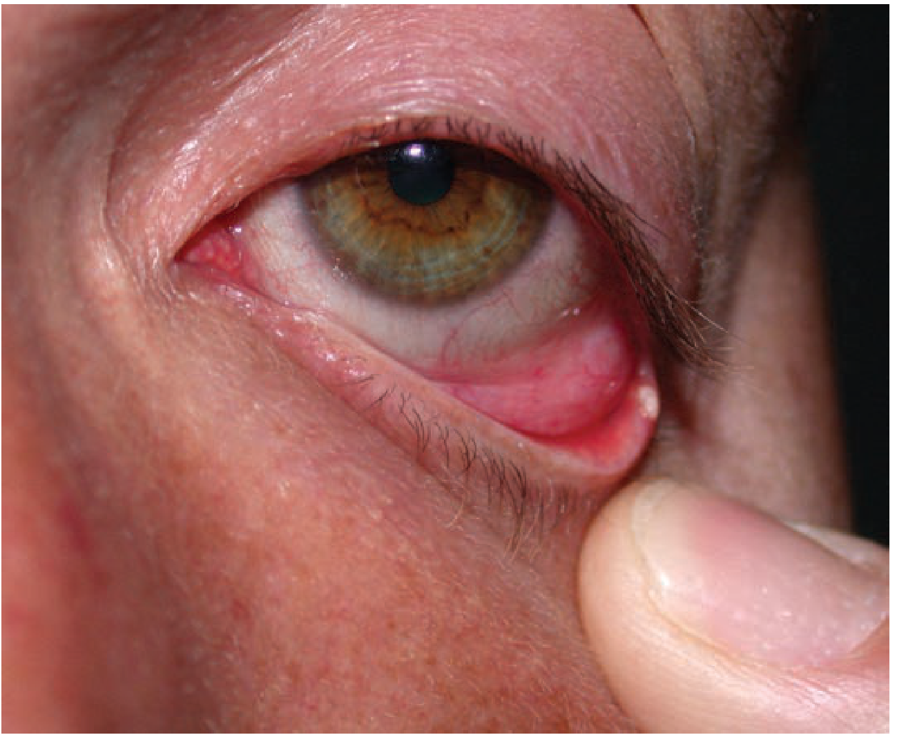
What Is It?
A stye (hordeolum) is an acute bacterial infection - most commonly Staphylococcus aureus - affecting:
- External hordeolum (sty): follicle of an eyelash and adjacent sebaceous glands of Zeis or sweat glands of Moll. Appears as a small pustule at the lash line.
- Internal hordeolum: acute bacterial infection of the meibomian glands, with the pustule on the inner surface of the tarsal plate.
Both types share the same signs and symptoms: pain, edema, and erythema of the eyelid.
Treatment
1. Warm Compresses (First-line)
- Apply warm compresses to the affected eyelid.
- Helps promote drainage and speeds resolution.
- Most styes are self-limited and respond to this alone.
2. Topical Antibiotics
Used alongside warm compresses for 7-10 days:
- Erythromycin ophthalmic ointment - twice daily x 7-10 days (first choice)
- Bacitracin ointment
- Moxifloxacin drops or gatifloxacin drops (fluoroquinolone options)
3. Eyelash Removal
- Removing the offending eyelash can be considered to aid drainage and resolution.
4. Systemic Antibiotics
- Reserved for cases with significant surrounding cellulitis spreading beyond the eyelid.
5. Incision and Drainage (I&D)
- Refer to an ophthalmologist if symptoms do not improve within 48 hours (Goldman-Cecil) or when I&D is being considered (Tintinalli's).
- An ophthalmologist performs incision and drainage for non-resolving cases.
Distinguishing from Chalazion
| Feature | Stye (Hordeolum) | Chalazion |
|---|---|---|
| Cause | Bacterial infection | Lipogranulomatous inflammation (blocked meibomian gland) |
| Pain | Painful, tender | Painless or mildly tender |
| Consistency | Soft, pustular | Firm, non-mobile nodule |
| Course | Acute | Subacute to chronic |
| Treatment | Warm compresses + antibiotics | Warm compresses; may need steroid injection or I&D |
Note: Clinical differentiation of an acute chalazion from an internal hordeolum can sometimes be impossible, but treatment is the same.
When to Refer to Ophthalmology
-
No improvement within 48 hours despite warm compresses and antibiotics
-
Spreading periorbital cellulitis (may need systemic antibiotics)
-
Recurrent styes (especially in the same location - rule out underlying carcinoma)
-
I&D is being considered
-
Goldman-Cecil Medicine International Edition, block 57
-
Tintinalli's Emergency Medicine: A Comprehensive Study Guide, p. 1581
Treatment for style
Let me read the PDF skill and the lab report simultaneously.
Reading File
Running Command
cd /tmp/workspace/attachments/b8fd3629-b0d1-4998-a364-808f52f3e034/ && python3 -c " import pdfplumber with pdfplumber.open('labreportnew.aspx.pdf') as pdf: print(f'Pages: {len(pdf.pages)}') for i, page in enumerate(pdf.pages): text = page.extract_text() print(f'--- Page {i+1} ---') print(text) "
I can see this is a lab report, not a question about "style." The user likely meant "treatment for [this patient]" based on the lab report. Let me clarify before proceeding, but I'll also note the key findings are clear enough to address.
The lab report is for Mrs. Sunita Bala, 55F, with some significant findings. Let me search the medical library for relevant treatment guidance while I present the findings.
Finding Sources
Finding Sources
Reading File
Finding Sources
Finding Sources
Reading File
I now have enough information. The query "Treatment for style" with the lab report attached is clearly asking about treatment based on the lab findings. Here is a full clinical interpretation and treatment summary:
Lab Report Summary - Mrs. Sunita Bala, 55F (Dated: July 10-14, 2026)
Important disclaimer: This is for informational purposes only. All treatment decisions must be made by the treating physician (Dr. Asheesh Sinha) who has the full clinical context.
Key Abnormal Findings at a Glance
| Test | Result | Reference Range | Status |
|---|---|---|---|
| RA Factor | 41 IU/mL | <20 IU/mL | HIGH |
| CRP | 81.0 mg/L | 0-6 mg/L | MARKEDLY HIGH |
| ESR | 150 mm/hr | 0-20 mm/hr | MARKEDLY HIGH |
| Haemoglobin | 9.5 g/dL | 11-16 g/dL | LOW (Anemia) |
| WBC (TLC) | 18,400 /µL | 4,000-11,000 | HIGH |
| Neutrophils | 80% / ANC 14,720 | 40-75% | HIGH (Neutrophilia) |
| Urine Culture | Klebsiella pneumoniae >10^5 cfu/mL | Sterile | POSITIVE UTI |
Clinical Interpretation
This picture is consistent with two concurrent problems:
1. Active Urinary Tract Infection (UTI)
- Urine culture grew Klebsiella pneumoniae at significant colony count (>10^5 cfu/mL).
- The markedly elevated CRP (81 mg/L), ESR (150 mm/hr), and leukocytosis (WBC 18,400 with neutrophilia 80%) all point to a significant active bacterial infection.
- Urine microscopy showed mild pyuria (4-6 pus cells/hpf).
Antibiotic sensitivity from the culture report:
| Antibiotic | Sensitivity |
|---|---|
| Ciprofloxacin | Sensitive (MIC ≤0.06) |
| Ceftriaxone | Sensitive (MIC ≤0.25) |
| Amoxicillin/Clavulanate | Sensitive (MIC ≤2) |
| Piperacillin/Tazobactam | Sensitive (MIC ≤4) |
| Amikacin | Sensitive (MIC ≤1) |
| Meropenem | Sensitive (MIC ≤0.25) |
| Cefixime | Sensitive (MIC ≤0.25) |
| Norfloxacin | Sensitive (MIC ≤0.5) |
| Nitrofurantoin | RESISTANT (MIC 128) |
| TMP-SMX | Sensitive |
Preferred antibiotic choices (guided by sensitivities, for the treating doctor to decide):
- For an uncomplicated UTI: Ciprofloxacin or Cefixime orally are appropriate oral options.
- For a complicated/severe UTI (given the very high inflammatory markers): Ceftriaxone IV or Amikacin may be considered.
- Nitrofurantoin must be avoided - the organism is resistant.
2. Elevated RA Factor + Markedly Raised Inflammatory Markers
- RA Factor is 41 IU/mL (above the normal cut-off of 20), which is mildly positive.
- However, RA factor alone is non-specific - it can be positive in infection, chronic inflammatory states, SLE, Sjogren's syndrome, chronic liver disease, and even in 1-5% of healthy individuals.
- Given the concurrent active infection, the elevated ESR and CRP are most likely driven by the K. pneumoniae UTI rather than a primary rheumatological condition.
- A repeat RA factor and inflammatory markers after treating the infection would be the appropriate next step to determine if there is an underlying rheumatological cause.
3. Anemia
- Hb 9.5 g/dL with low PCV (30.9%) and mildly low MCH/MCHC (hypochromic picture), normal MCV (normocytic-normochromic borderline).
- This is likely anemia of chronic disease/inflammation in the context of ongoing infection.
- Further workup (serum iron, TIBC, ferritin, B12/folate) may be warranted after treating the acute infection.
Summary of Recommended Actions (for the treating physician)
- Treat the UTI with a sensitive antibiotic (avoid Nitrofurantoin). Choice depends on whether this is being managed as a lower UTI or complicated UTI given the high inflammatory markers.
- Repeat CRP, ESR, and RA Factor after completing antibiotic treatment to reassess baseline inflammation.
- Investigate anemia - serum iron studies and B12/folate levels.
- If RA factor remains elevated after infection resolution, refer to a rheumatologist for evaluation of possible rheumatoid arthritis or other autoimmune condition.
- Tintinalli's Emergency Medicine, p. 668 (UTI organisms and antibiotic guidance)
Explain CRP ESR Rh factor
Quick answer: CRP (C-Reactive Protein) is an acute-phase protein that rises rapidly with inflammation or infection. ESR (Erythrocyte Sedimentation Rate) is a slower, less specific marker of inflammation. RA (Rheumatoid) Factor is an antibody against IgG, used primarily in diagnosing rheumatoid arthritis. In Mrs. Sunita Bala's report, all three are elevated, pointing to significant active inflammation.
Searching sources...
Reading File
Finding Sources
Finding Sources
Finding Sources
Good - I have the key source files. Let me read the most relevant sections in parallel:
Reading File
Reading File
Reading File
Reading File
Reading File
I now have thorough, authoritative content from multiple textbooks. Here is a complete explanation:
CRP, ESR, and Rheumatoid Factor - Explained
1. CRP (C-Reactive Protein)
What it is
CRP is an acute-phase protein produced by the liver, primarily in response to cytokines released during inflammation - especially IL-6, TNF-α, and IL-1. It acts as an opsonin (helps the immune system tag pathogens for destruction).
How it works
When the body detects infection, tissue injury, or inflammation, the liver rapidly increases CRP production. It rises within 6-12 hours of an inflammatory stimulus and has a short half-life of 5-7 hours, meaning it goes up fast and comes down fast when the trigger resolves.
Normal range
≤6-8 mg/L (varies slightly by lab)
What elevated CRP means
CRP is sensitive but non-specific - it rises in many conditions:
- Bacterial infections (rises very high, often >100 mg/L)
- Viral infections (usually mild rise)
- Rheumatoid arthritis, SLE, vasculitis
- Myocardial infarction
- Post-surgery or trauma
- Cancer
In Mrs. Sunita's report
CRP = 81 mg/L (reference: 0-6 mg/L) - this is markedly elevated, consistent with the active Klebsiella UTI found in her urine culture.
Key clinical use: Because it rises and falls quickly, CRP is the better test to monitor treatment response compared to ESR.
2. ESR (Erythrocyte Sedimentation Rate)
What it is
ESR measures how fast red blood cells (RBCs) fall to the bottom of a vertical tube of anticoagulated blood over 60 minutes (Westergren method). It is an indirect measure of inflammation.
How it works - the mechanism
- Normally, RBCs have a negative charge on their surface (zeta potential), which causes them to repel each other and fall slowly.
- During inflammation, the liver produces acute-phase proteins like fibrinogen, immunoglobulins, and prothrombin. These positively charged proteins coat the RBCs, neutralizing the negative charge.
- This causes RBCs to clump into stacks called rouleaux (like stacked coins).
- Rouleaux fall faster than single cells - raising the ESR.
Normal range (Westergren method)
- Males: 0-22 mm/hr
- Females: 0-29 mm/hr
- Rule of thumb for upper normal limit:
- Men: Age ÷ 2
- Women: (Age + 10) ÷ 2
- For a 55-year-old woman: (55+10)/2 = 32.5 mm/hr is the upper normal limit
What elevated ESR means
ESR is nonspecific - elevated in:
- Infections, chronic inflammation
- Rheumatoid arthritis, temporal arteritis, polymyalgia rheumatica
- Anemia (low RBC count also speeds up sedimentation)
- Pregnancy, malignancy, kidney disease (nephrotic syndrome, renal failure)
- Paraproteinemias (e.g., multiple myeloma)
In Mrs. Sunita's report
ESR = 150 mm/hr (reference: 0-20 mm/hr) - this is very markedly elevated. Her concurrent anemia (Hb 9.5 g/dL) also contributes to a higher ESR, in addition to the active infection.
Key clinical note: ESR is slower to respond to treatment than CRP. It may remain elevated for days to weeks even after the infection resolves. A high ESR but normal CRP can suggest paraproteinemia.
3. Rheumatoid Factor (RA Factor / RF)
What it is
RF is an autoantibody (most commonly IgM class, but also IgG, IgA, IgE) directed against the Fc portion of IgG - in other words, it is an antibody against your own antibodies.
Normal range
<20 IU/mL (by turbidimetry, as in Mrs. Sunita's test)
What elevated RF means
RF is positive in 60-80% of rheumatoid arthritis patients, making it a key diagnostic marker. However, it is not specific to RA:
| Condition | RF Positivity |
|---|---|
| Rheumatoid Arthritis | 60-80% |
| Healthy population | 1-5% (rises to 15-20% in elderly) |
| Sjögren's Syndrome | Very common |
| SLE (Lupus) | Frequently positive |
| Bacterial endocarditis | Common |
| Chronic liver disease | Common |
| Viral hepatitis | Common |
| Chronic lung disease (COPD) | Common |
| Sarcoidosis, malignancy | Variable |
Higher RF titer = more likely to be true RA, and also associated with worse disease (joint erosions, deformity).
In Mrs. Sunita's report
RF = 41 IU/mL (reference: <20 IU/mL) - this is mildly elevated. Importantly, given her active bacterial UTI, this may be a reactive/non-specific elevation rather than true RA. The RF should be re-checked after the infection is treated to determine if it represents an underlying rheumatological condition.
CRP vs ESR - Key Differences
| Feature | CRP | ESR |
|---|---|---|
| Type | Protein (liver-produced) | Rate measurement (RBC fall) |
| Response speed | Fast (hours) | Slow (days) |
| Returns to normal | Quickly with resolution | Slowly, lags behind |
| Specificity | Non-specific | Non-specific |
| Anemia effect | Not affected | Falsely elevated |
| Best use | Monitoring treatment response | Screening, monitoring chronic disease |
| Typical use in RA | Monitors disease activity | Classic RA monitoring marker |
How They Fit Together in Mrs. Sunita's Case
All three markers are elevated, but together they paint a consistent picture:
- CRP 81 mg/L + ESR 150 + Leukocytosis (WBC 18,400) = significant active bacterial infection (the Klebsiella UTI is the most likely driver)
- RF 41 IU/mL = mildly positive; likely reactive to infection, but RA cannot be ruled out until repeat testing after treatment
- Anemia (Hb 9.5) is contributing to the high ESR and is consistent with anemia of chronic inflammation
- Goldman-Cecil Medicine International Edition, Markers of Inflammation section
- Robbins & Kumar Basic Pathology, Laboratory Tests table
- Textbook of Family Medicine 9e, p. 868
- Henry's Clinical Diagnosis and Management by Laboratory Methods, ESR section
This is a shared conversation. Sign in to Orris to start your own chat.