
What do you mean by nodal? System of heart. Nodal system of heart. Uh, e- explain this, uh, please. If I got a long question answer. On this topic.
Reading File
Now let me search the medical library for the nodal system of the heart.
Finding Sources
Finding Sources
Good results. Now let me read the most comprehensive sources in parallel.
Reading File
Reading File
Reading File
Excellent content. Let me now read more of the physiology text and fetch the key diagram image.
Reading File
Reading File
Loading Image
I now have everything needed for a thorough long-answer response. Let me also fetch the conduction pathways diagram from Medical Physiology.
Loading Image
Loading Image
I now have comprehensive textbook content and three excellent diagrams. Here is the full long-answer response:
The Nodal (Conducting) System of the Heart
The term "nodal system" refers to the cardiac conducting system - a network of specialized muscle cells that generates and conducts electrical impulses, triggering the heart to beat in a coordinated, rhythmic way. These are not nerve cells - they are modified cardiac muscle cells that have given up most of their contractile function in favor of electrical signaling.
Overview Diagram
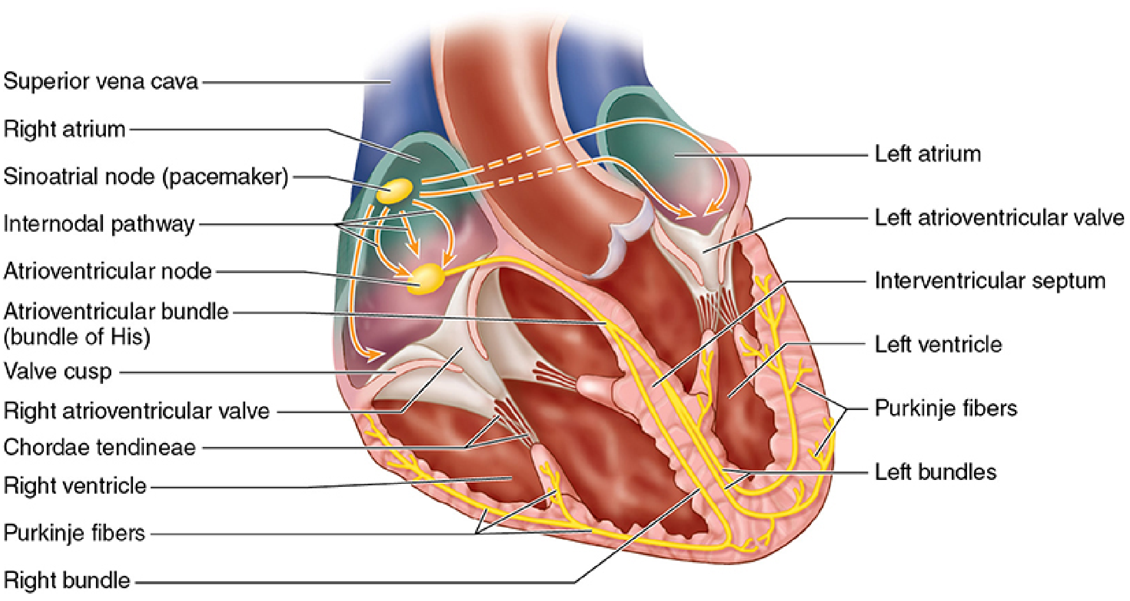
Components of the Nodal/Conducting System
The system has five main components, arranged in the order the impulse travels:
1. Sinoatrial (SA) Node - The Pacemaker
- Location: Beneath the epicardium on the posterior surface of the right atrium, near the opening of the superior vena cava, in the sulcus terminalis.
- Also called: Keith-Flack node, or simply the pacemaker.
- Size: Spindle-shaped, about 10 mm long.
- Function: Spontaneously generates electrical impulses at a rate of 60-80 (up to 100) per minute at rest. This automaticity (self-firing ability) makes it the primary pacemaker of the heart.
- All other parts of the conducting system can generate impulses too, but at slower rates - so the SA node normally dominates.
"The sinoatrial node...lies beneath the epicardium on the posterior surface of the right atrium, near the opening of the superior vena cava...referred to as the cardiac pacemaker, as it generates 60-80 impulses per minute." - Color Atlas of Human Anatomy Vol. 2
2. Internodal Pathways (Atrial Conduction)
- After the SA node fires, the impulse spreads through the right atrial myocardium cell to cell via gap junctions.
- There are three proposed internodal pathways that carry the signal to the AV node (anterior, middle, and posterior). The existence of discrete internodal tracts is still debated among physiologists.
- Bachmann's bundle (interatrial tract) carries the signal across to the left atrium, so both atria contract nearly simultaneously.
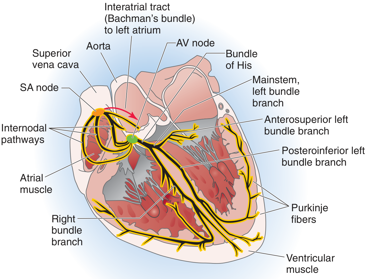
3. Atrioventricular (AV) Node
- Location: At the atrioventricular septum in the interatrial septum, between the opening of the coronary sinus and the septal cusp of the tricuspid valve.
- Also called: Aschoff-Tawara node.
- Size: About 5 mm long.
- Key function - the AV delay: The AV node slows the impulse (by about 0.1 seconds) before passing it to the ventricles. This delay is absolutely critical - it allows the atria to finish contracting and push blood into the ventricles before the ventricles contract.
- Electrical insulation: The fibrous atrioventricular ring electrically insulates the atria from the ventricles. The AV node is the only normal electrical connection between atria and ventricles.
- The AV node acts as a backup pacemaker at 40-60 beats/min if the SA node fails.
"Normally, the atrioventricular (AV) node, where conduction delay is physiologic, serves as the only electrical connection linking the atria and ventricles; the AV valve rings are insulated." - Goldman-Cecil Medicine
4. Bundle of His (Atrioventricular Bundle)
- Location: Arises from the AV node, penetrates the cardiac skeleton (the fibrous ring), and reaches the superior margin of the muscular interventricular septum.
- Structure: A bundle of specialized conducting cells (Purkinje-type fibers).
- Function: Carries the impulse rapidly from the AV node into the ventricular septum.
- At the top of the interventricular septum, the Bundle of His divides into two branches.
5. Right and Left Bundle Branches
- The Bundle of His splits into the right bundle branch and left bundle branch at the interventricular septum.
- Both branches run beneath the endocardium (subendocardially) down the septum toward the apex of the heart.
Right bundle branch:
- Runs down the right side of the septum.
- Enters the septomarginal trabecula (moderator band) to reach the anterior papillary muscle.
- Peripheral branches form a subendocardial plexus.
Left bundle branch:
- Fans out in flat bundles along the left side of the interventricular septum.
- Divides into two major fascicles: the anterosuperior and posteroinferior left bundle branches.
- Branches spread to the bases of both papillary muscles and then fan out across the ventricular wall.
6. Purkinje Fibers (Subendocardial Conducting Network)
- The terminal branches of the bundle system are the Purkinje fibers.
- They fan out throughout the subendocardial layer of both ventricles, reaching from the apex toward the base.
- Speed: Purkinje fibers conduct impulses at the fastest speed in the heart (up to 4 m/s) - much faster than ordinary myocardium (0.5 m/s). This ensures the entire ventricular wall is activated almost simultaneously.
- Histology: Purkinje fibers are modified cardiac myocytes. They are larger than regular myocardial cells, stain paler (because they are rich in glycogen and have relatively few myofibrils and organelles), and are joined by intercalated disks.
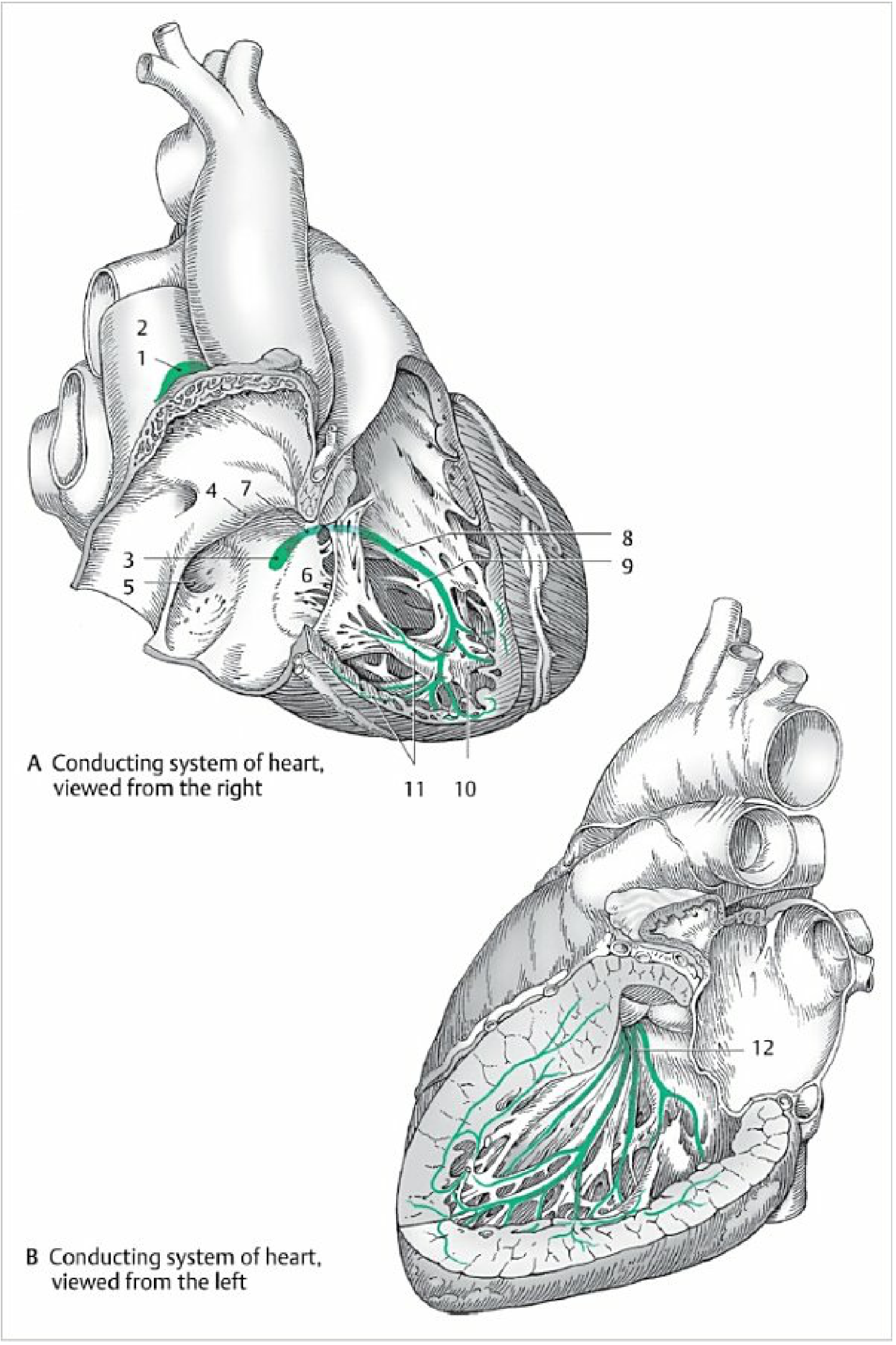
The Full Sequence of Impulse Conduction
Here is the complete pathway, in order:
SA Node (fires spontaneously)
    ↓
Internodal pathways → Right atrium depolarizes
    ↓ (also via Bachmann's bundle → Left atrium)
BOTH ATRIA CONTRACT
    ↓
AV Node (0.1 second delay - atria finish pumping)
    ↓
Bundle of His
    ↓ (splits into two)
Right Bundle Branch          Left Bundle Branch
    ↓                              ↓ (anterosuperior + posteroinferior fascicles)
    ↓                              ↓
Purkinje Fibers ←←←←←←←←←←←←←←←←←←
    ↓
BOTH VENTRICLES CONTRACT (from apex upward - efficient ejection)
The whole process from SA node firing to ventricular contraction takes about 0.2 seconds.
Why Does the Impulse Spread from Cell to Cell?
Cardiac cells are connected by gap junctions (intercalated disks). These are low-resistance channels that allow ions (electrical current) to pass directly from one cell to the next. When one cell depolarizes, it injects depolarizing current into the neighboring cell, which then reaches threshold and fires its own action potential. This is how a single impulse from the SA node can trigger the entire heart to contract in a coordinated wave. - Medical Physiology
Hierarchy of Pacemakers (Safety Net)
The conducting system has a backup hierarchy. If a higher pacemaker fails:
| Pacemaker | Intrinsic Rate |
|---|---|
| SA node | 60-100 beats/min (dominant) |
| AV node / junctional | 40-60 beats/min |
| Bundle of His / ventricle (Purkinje) | 20-40 beats/min |
If the SA node fails, the AV node takes over, but at a slower rate. If the AV node also fails, the ventricles can still beat on their own (idioventricular rhythm) at 20-40/min - slow, but life-sustaining.
Autonomic Modulation
The conducting system is not entirely autonomous - the autonomic nervous system can modify its rate and conduction speed:
- Sympathetic stimulation (e.g., exercise, stress): increases heart rate, increases force of contraction, speeds up conduction through the AV node.
- Parasympathetic stimulation (via vagus nerve, e.g., rest, sleep): slows heart rate, decreases force, slows AV node conduction.
Clinical Significance
Pathological disruption of the conducting system causes arrhythmias:
| Condition | Mechanism |
|---|---|
| SA node dysfunction (sick sinus syndrome) | SA node fails to fire or fires erratically |
| Heart block (1st, 2nd, 3rd degree) | Impaired conduction at AV node |
| Bundle branch block (LBBB/RBBB) | Blocked conduction in left or right bundle branch |
| Ventricular fibrillation | Chaotic, uncoordinated firing of ventricular myocardium |
| Wolff-Parkinson-White (WPW) syndrome | Abnormal accessory pathway bypasses AV node |
All of these are diagnosed using an ECG (electrocardiogram), which records the electrical activity of the conducting system from electrodes on the body surface.
Summary Table
| Component | Location | Rate if Pacemaker | Main Function |
|---|---|---|---|
| SA Node | Right atrium, near SVC | 60-100/min | Primary pacemaker |
| Internodal pathways | Right atrial wall | - | Conduct impulse to AV node + left atrium |
| AV Node | Interatrial septum | 40-60/min | Delay impulse; gate between atria & ventricles |
| Bundle of His | Interventricular septum | 20-40/min | Carry impulse to ventricular septum |
| Right & Left Bundle Branches | Interventricular septum | 20-40/min | Distribute impulse down each ventricle |
| Purkinje Fibers | Subendocardium of ventricles | 20-40/min | Rapid spread to all ventricular muscle |
Sources:
- Junqueira's Basic Histology, 17e, p. 542-543
- Color Atlas of Human Anatomy, Vol. 2, p. 60-62
- Medical Physiology (Boron & Boulpaep), p. 710-711
- Goldman-Cecil Medicine
So, the Nodal system is just the... Components that cause the cardiac cycle.
Yes, that's a good way to think about it — but with one small clarification worth making.
The nodal system is what initiates and coordinates the cardiac cycle, not what causes the actual pumping.
Here's the distinction:
- The nodal/conducting system = the electrical trigger. It generates and routes the electrical impulse through the heart in the right order and timing.
- The cardiac muscle (myocardium) = the mechanical response. It physically contracts when it receives that electrical signal, which is what actually pumps the blood.
So the relationship is:
Nodal System (electrical) → triggers → Myocardium (mechanical) → produces → Cardiac Cycle
Think of it like a car:
- The nodal system is the ignition and timing system
- The myocardium is the engine
- The cardiac cycle (blood moving in and out) is the car moving
Without the nodal system firing in the correct sequence, the heart muscle would either not contract at all, or contract chaotically (like in ventricular fibrillation) - and no effective pumping would happen even though the muscle itself is intact.
So yes - the nodal system is the organized electrical foundation that makes the cardiac cycle happen in a coordinated, rhythmic way.
What is the actual difference between nodal system and cardiac cycle and states both of their differences
Great question. These two are closely related but are completely different things. Let me search the textbooks to give you the most accurate comparison.
Finding Sources
Reading File
Reading File
Loading Image
Loading Image
Perfect. Now I have everything needed for a thorough, clear comparison.
Nodal System vs. Cardiac Cycle - The Actual Difference
These two are from completely different categories. One is a structure, the other is an event. Here is the full breakdown.
The Core Difference in One Line
| Nodal System | Cardiac Cycle | |
|---|---|---|
| What it is | A structural system inside the heart | A sequence of events the heart performs |
| Category | Anatomy + Electrophysiology | Physiology + Mechanics |
| Type of thing | A "thing" that exists permanently | A "process" that repeats every heartbeat |
1. The Nodal (Conducting) System
Definition: A network of specialized muscle cells that generates and conducts electrical impulses to make the heart beat.
It is a structure - it exists all the time, even between beats.
Components (already covered):
- SA Node - fires 60-100/min (pacemaker)
- Internodal pathways - carry impulse through atria
- AV Node - delays impulse (0.1 sec)
- Bundle of His - carries impulse to ventricles
- Right & Left Bundle Branches - distribute to each ventricle
- Purkinje Fibers - spread impulse through ventricular walls
What it does:
- Generates the electrical signal
- Routes it in the correct order (atria first, ventricles second)
- Controls the timing and rate of the heartbeat
- Ensures both ventricles contract simultaneously
Nature:
- Electrical in nature
- Always present - doesn't start and stop
- Works at the cellular / microscopic level
- Measured on an ECG (electrocardiogram)
2. The Cardiac Cycle
Definition: The complete sequence of mechanical events - filling and emptying of the heart - that occurs with each single heartbeat.
It is a process - it starts and ends with every beat (~0.8 seconds at rest).
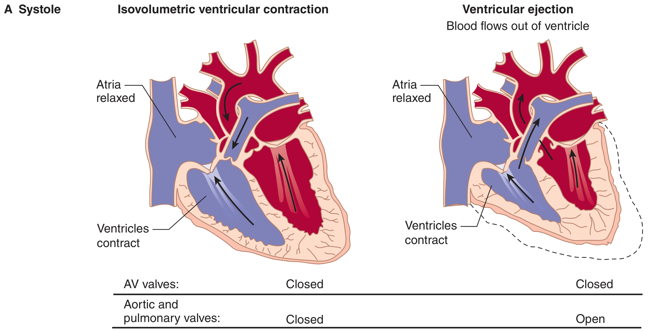
Phases of the Cardiac Cycle:
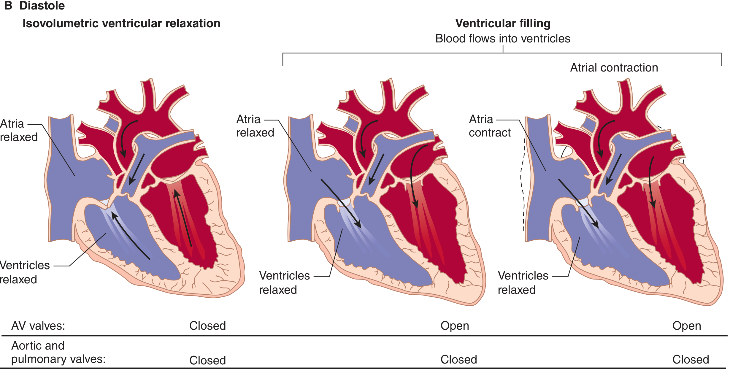
Phase 1 - Ventricular Filling (part of Diastole)
- AV valves (mitral + tricuspid) are open
- Aortic + pulmonary valves are closed
- Blood flows passively from atria into ventricles (~70% of filling is passive)
- At the end, atria contract (atrial systole) to push in the remaining ~30%
Phase 2 - Isovolumetric Contraction (start of Systole)
- All four valves are closed
- Ventricles start contracting - pressure builds rapidly
- No blood moves yet (volume stays the same - hence "isovolumetric")
- Lasts ~0.05 seconds
Phase 3 - Ventricular Ejection (Systole)
- Ventricular pressure exceeds aortic/pulmonary artery pressure
- Aortic + pulmonary valves open
- Blood is ejected into the aorta and pulmonary artery
- Ventricles shrink in size
Phase 4 - Isovolumetric Relaxation (start of Diastole)
- Ventricles stop contracting and begin relaxing
- All four valves are closed again
- Pressure in ventricles falls rapidly
- No blood moves yet
Then the cycle repeats from Phase 1.
Timing (at 75 beats/min, one cycle = 800 ms):
- Systole (contraction) = ~300 ms
- Diastole (relaxation) = ~500 ms
Nature:
- Mechanical in nature
- Repeating event - starts and ends with each beat
- Works at the organ / pressure-volume level
- Measured by blood pressure, heart sounds, echocardiography
Side-by-Side Differences Table
| Feature | Nodal System | Cardiac Cycle |
|---|---|---|
| Nature | Electrical | Mechanical |
| Type | A permanent structure | A repeating event/process |
| Function | Generate + conduct impulse | Fill and pump blood |
| Components | SA node, AV node, Bundle of His, Purkinje fibers | Systole, Diastole (4 phases) |
| Duration | Always present | ~0.8 seconds per cycle |
| Measured by | ECG | Blood pressure, heart sounds, Echo |
| Location of action | Specialized conducting cells | Entire heart muscle + valves |
| Failure result | Arrhythmia (heart beats wrongly or not at all) | Heart failure (heart beats but pumps inadequately) |
| Driven by | Spontaneous ion channel activity | Electrical signal from nodal system |
How They Relate to Each Other
NODAL SYSTEM                    CARDIAC CYCLE
(Electrical)         →          (Mechanical)

SA node fires        →   Atria contract  (atrial systole)
       ↓
AV node delays       →   AV valves close
       ↓
Bundle of His        →   Ventricles build pressure
       ↓
Purkinje fibers      →   Ventricles eject blood (ventricular systole)
       ↓
Next SA node fire    →   Ventricles relax, fill again (diastole)
The nodal system is the cause. The cardiac cycle is the effect.
One fires an electrical signal in a precise order. The other is the mechanical response - the filling, squeezing, and emptying of blood - that results from that signal reaching the heart muscle.
Without the nodal system, the cardiac cycle either doesn't happen or becomes chaotic. Without healthy cardiac muscle, the nodal system can still fire perfectly but the heart won't pump properly (as happens in heart failure).
Sources: Medical Physiology (Boron & Boulpaep), p. 744-745; Ganong's Review of Medical Physiology, 26e, p. 538-540
Then we can see that the nervous system is a structural system in the heart that, perform the sequence of events that causes the contraction of heart and causes the pump.
You're close, but there's one important word to correct there.
It's not the nervous system - it's the nodal (conducting) system.
These are two different things. Here's why the distinction matters:
The Nodal System ≠ The Nervous System
| Nodal/Conducting System | Nervous System | |
|---|---|---|
| Made of | Modified cardiac muscle cells | Nerve cells (neurons) |
| Location | Inside the heart itself | Outside the heart (brain, spinal cord, vagus nerve) |
| Job | Generates the heartbeat on its own | Only modifies the heart rate (faster or slower) |
| Can heart beat without it? | No - heart stops | Yes - heart still beats, just unregulated |
The Key Point
The heart is self-exciting - it does not need the brain or nervous system to beat. This is called automaticity.
Proof: A heart removed from the body and placed in a nutrient solution will keep beating on its own - because the SA node keeps firing independently. The nervous system has no role in that.
What the nervous system does do:
- Sympathetic (fight-or-flight) - speeds the heart up
- Parasympathetic (vagus nerve, rest) - slows the heart down
So Your Statement, Corrected:
"The nodal/conducting system is a structural system within the heart muscle that performs the electrical sequence of events that causes contraction of the heart and drives the pumping of blood."
That is exactly right. Just replace "nervous system" with "nodal/conducting system" and the statement is accurate.
This is a shared conversation. Sign in to Orris to start your own chat.