
Explain malignant melanoma surgery mbbs
malignant melanoma skin surgery wide local excision
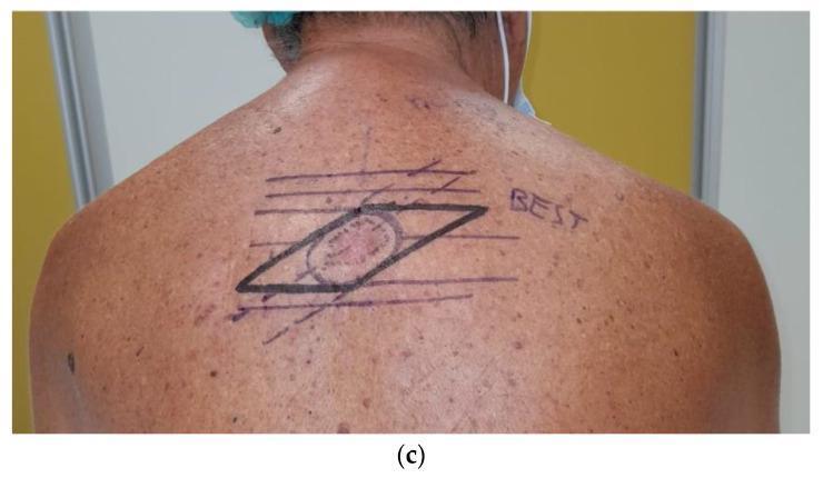
A clinical photograph of the upper back of a 67-year-old male demonstrating advanced preoperative surgical markings for the wide local excision of a malignant melanoma. The central focus is a pigmented skin lesion circumscribed by an elliptical marking. Surrounding this ellipse is a larger, prominent parallelogram drawn in black ink. This 'parallelogram excision' technique is a reconstructive surgical approach used when the lesion's major axis is not parallel to Relaxed Skin Tension Lines (RSTLs). The markings include multiple horizontal lines labeled as 'BEST' lines (Biodynamic Excising Skin Tension), which guide the orientation of the excision to optimize wound closure and minimize 'dog-ear' deformities. Diagonal tangent lines intersect the horizontal BEST lines to form the vertices of the parallelogram. The surrounding skin shows solar lentigines and diffuse actinic damage, typical for the patient demographic and clinical context of cutaneous melanoma.
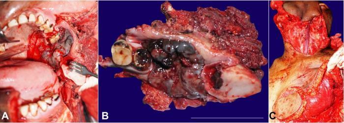
This composite of three clinical photographs documents the surgical management of oral malignant melanoma. Panel A provides an intraoperative view of a wide local excision involving a left segmental mandibulectomy and infrastructural maxillectomy. A large, irregular, darkly pigmented (melanotic) mass is visible in the retromolar trigone region, surrounded by surgically exposed anatomical landmarks including the tongue and remaining maxillary teeth. Panel B shows the gross surgical specimen, highlighting a 5.6 cm pigmented, lobulated tumor mass involving the alveolar process with two molar teeth attached to the resected bone. The specimen exhibits characteristic black-to-dark-blue pigmentation indicative of melanoma. Panel C demonstrates the reconstructive phase, specifically the harvesting of a left-sided pectoralis major myocutaneous (PMMC) flap. This panel shows the donor site on the chest with the vascularized muscle and skin paddle prepared for transposition to the oral cavity defect. These images illustrate the oncological resection and subsequent reconstructive strategy required for aggressive head and neck mucosal malignancies.
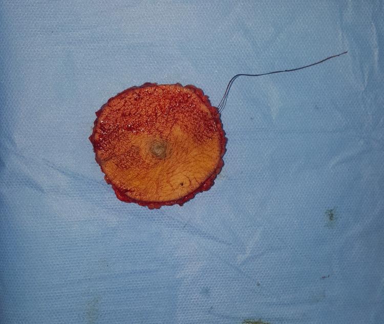
This surgical specimen photograph depicts a wide local excision of a skin lesion, contextually identified as a nodular melanoma. The specimen is roughly circular (discoid), consisting of a central skin island surrounded by deep soft tissue margins. The epidermal surface shows a distinct, centrally located, slightly raised, and pigmented nodule. The surrounding skin surface is tan with fine hair visible, while the peripheral surgical margins are erythematous and irregular, composed of subcutaneous adipose and connective tissue. A long, dark surgical suture is attached to the superior-right margin, serving as an orientation marker for pathological processing. This image illustrates the gross morphology of a radical resection aimed at achieving clear oncological margins for a high-thickness (Breslow 8mm, Clark Level V) malignant melanoma. The specimen is placed on a blue sterile surgical drape, highlighting the immediate postoperative context in a surgical oncology setting.
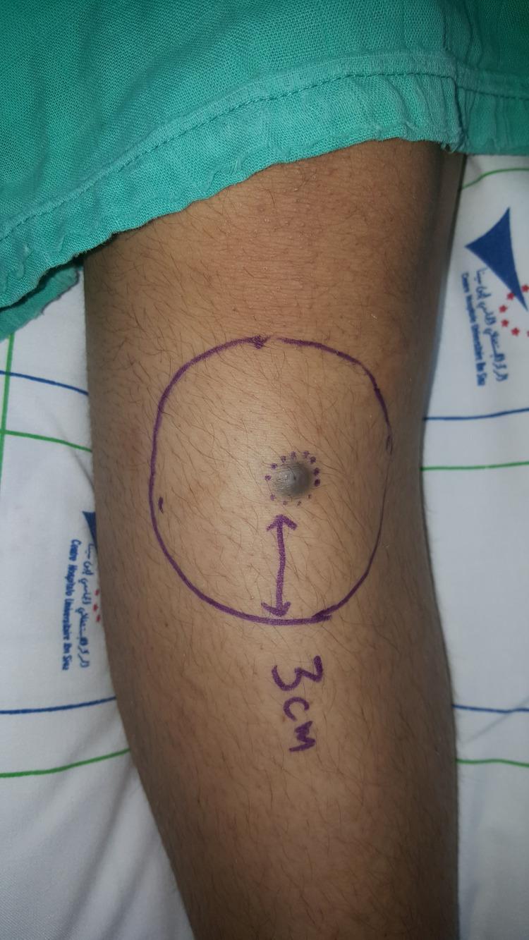
A clinical photograph of a patient's leg showing a preoperative view of a suspected malignant skin lesion, later identified as nodular melanoma. The primary lesion is a centrally located, dark-pigmented, nodular mass with irregular surface morphology. Surrounding the central nodule are several satellite dot-like pigmented spots. Surgical planning markings in purple ink delineate a planned 3 cm wide local excision margin, represented by a large circular boundary with an internal radial arrow and the text '3 cm' indicating the distance from the tumor edge. The surrounding skin appears normal with typical hair follicle distribution. This image illustrates the standard preoperative marking process for managing high-risk cutaneous malignancies to ensure clear histopathological margins.
Malignant Melanoma - Surgery (MBBS Level)
Overview
Diagnosis and Clinical Recognition
ABCDE Criteria (Clinical Screening)
| Feature | Description |
|---|---|
| A - Asymmetry | One half does not match the other |
| B - Border | Irregular, ragged, or notched edges |
| C - Color | Variegation (brown, black, red, white) |
| D - Diameter | >6 mm (approximately pencil eraser size) |
| E - Evolving | Any change in size, shape, or color |
Types (Histological Subtypes)
- Superficial spreading melanoma (SSM) - most common; radial growth phase predominates
- Nodular melanoma - vertical growth phase from the start; worst prognosis
- Lentigo maligna melanoma (LMM) - arises from lentigo maligna (in situ lesion) on sun-damaged skin; common on head/neck in elderly
- Acral lentiginous melanoma (ALM) - palms, soles, subungual; most common type in dark-skinned individuals
- Desmoplastic melanoma - head/neck; perineural infiltration; high local recurrence tendency
Biopsy - The First Surgical Step
- Incisional biopsy is occasionally used for large facial lesions where full excision would be disfiguring.
- Punch biopsy and partial-thickness biopsies should not be performed routinely as they may miss the deepest part of the tumour.
- For subungual melanoma, biopsy must be of the nail matrix, not just the nail plate.
Key Pathological Parameters Reported
| Parameter | Significance |
|---|---|
| Breslow thickness (mm) | Most important prognostic factor; measured from granular layer to base of tumour |
| Clark level | Depth of invasion (I-V); less used now due to interobserver variability |
| Ulceration | Upstages the tumour; worsens prognosis |
| Mitotic rate | Higher rate = worse prognosis |
| Microsatellites | Classified as Stage III even without nodal metastases |
| Lymphovascular invasion | Increases metastatic risk |
Staging
| Stage | Description |
|---|---|
| Stage 0 | In situ (confined to epidermis) |
| Stage I | Localised, thin (<2 mm), no ulceration or LN involvement |
| Stage II | Localised, thicker or ulcerated; no LN involvement |
| Stage III | Regional LN metastasis / in-transit / satellite lesions |
| Stage IV | Distant metastasis |
Definitive Surgery: Wide Local Excision (WLE)
Recommended Surgical Margins (Evidence-Based)
| Breslow Thickness | Recommended Margin |
|---|---|
| In situ (Stage 0) | 5 mm |
| <1 mm | 1 cm |
| 1-2 mm | 1-2 cm |
| >2 mm | 2 cm |
Multiple large RCTs have confirmed that margins wider than 2 cm offer no survival benefit. The WHO Melanoma Program (1988, n=612) showed no difference in local recurrence, DFS, or OS between 1 cm and 3 cm margins for tumours <2 mm. Historically, 5 cm margins were used - this is now abandoned.
- Fischer's Mastery of Surgery, p. 1825
Special Sites
- Digits/subungual: Partial amputation at the joint space proximal to the melanoma to obtain adequate margins
- Head and neck: Increased attention to cosmesis and reconstruction; lymphatic drainage is variable (lymphoscintigraphy is vital before SNB)
- Foot/acral: Skin is immobile, so skin grafting is often required after WLE
Sentinel Lymph Node Biopsy (SLNB / SNB)
Indications for SLNB
| Melanoma Thickness | Recommendation |
|---|---|
| <1 mm (T1) | Generally not indicated; consider if high mitotic rate, LVI, or young patient |
| 1-4 mm | Standard of care |
| >4 mm | Recommended (though yield of LN-positive is high) |
Technique
- Lymphoscintigraphy: Technetium-99m labelled colloid is injected intradermally around the primary site the day before (or on the day of) surgery; SPECT/CT used especially for head/neck lesions.
- Blue dye injection (isosulfan blue or patent blue V) at time of surgery around the primary site.
- In the operating room, a gamma probe detects the radioactive node; blue-stained nodes are also identified.
- The sentinel node(s) is excised and sent for permanent section (frozen section is less accurate for melanoma).
- If SLNB is positive, staging is confirmed as Stage III.
Important: WLE of the primary is performed before SLNB (if done simultaneously) to reduce background radioactive signal that can obscure nearby sentinel nodes.
- Bailey and Love's Short Practice of Surgery, 28th Ed., p. 679
Completion Lymph Node Dissection (CLND)
- MSLT-II trial: Showed that immediate CLND after positive SLNB did not improve melanoma-specific survival compared to close ultrasound surveillance of the nodal basin. CLND did provide better regional disease control.
- Current practice: CLND is not routinely performed after positive SLNB with micrometastasis (<0.2 mm in a single node). It is offered when macroscopic nodal disease is present or when regional control is clinically needed.
- Therapeutic lymphadenectomy: Performed when palpable nodes are confirmed by biopsy to contain melanoma.
Management of In-Transit Metastases
- Surgical excision if few and resectable
- Isolated limb perfusion (ILP) or isolated limb infusion (ILI) with melphalan - for unresectable in-transit disease in a limb
- Intralesional injections (e.g., talimogene laherparepvec - T-VEC, an oncolytic virus)
Management of Stage IV (Distant Metastasis)
- Resection of isolated distant metastases (e.g., single lung, brain, or adrenal metastasis) can achieve prolonged survival in selected patients.
- Palliative resection for bleeding or obstructing metastases.
- BRAF/MEK inhibitors (vemurafenib, dabrafenib + trametinib) for BRAF V600E mutant melanoma
- Immune checkpoint inhibitors: anti-PD-1 (pembrolizumab, nivolumab), anti-CTLA-4 (ipilimumab)
- Adjuvant immunotherapy after resection of Stage III/IV disease
Adjuvant Radiotherapy
- Not routinely used for primary melanoma.
- Indicated after WLE of desmoplastic melanoma where adequate margins are difficult to obtain (especially head/neck location with perineural invasion).
- Used for palliation of brain or bone metastases.
Mohs Micrographic Surgery
- Minimises normal tissue excised
- Is particularly useful for head/neck and acral melanomas where tissue conservation is important
- Has theoretical advantages for lentigo maligna melanoma with subclinical spread
Follow-Up
- Stage I: Clinical review every 3-6 months for 3 years, then annually
- Stage II-III: Imaging (CT/PET-CT) at intervals + clinical review
- Stage IV: Individualized
Summary for Exam Mnemonics
- In situ: 5 mm
- <1 mm: 1 cm
- 1-2 mm: 1-2 cm
-
2 mm: 2 cm