
Physiotherapy management of leprosy patient with bilateral claw hands for 10 marks to pt cvts subject
I'll look up the medical library skill first, then search for leprosy physiotherapy content in parallel.
Reading File
Now let me search the library and web in parallel for leprosy physiotherapy content.
Finding Sources
Finding Sources
Searching the Web
physiotherapy management leprosy bilateral claw hand rehabilitation exercises
Good results. Let me now read the relevant textbook sections and extract the web content in parallel.
Reading File
Finding Sources
Reading Web Page
https://www.physiotattva.com/blog/guide-to-leprosy-claw-hand…
Let me read the Bailey & Love claw hand section and search for more specific physiotherapy content.
Reading File
Finding Sources
I now have enough material to compose a comprehensive 10-mark answer. Let me fetch the bilateral claw hand image for the answer.
Loading Image
I now have all the material needed. Here is the full 10-mark structured answer for your PT CVTs subject:
Physiotherapy Management of Leprosy Patient with Bilateral Claw Hands
(10 Marks - PT CVTs)
1. Understanding the Deformity
Bilateral claw hand in leprosy results from damage to the ulnar nerve (ring and little finger clawing) and often the median nerve (index and middle finger clawing when both are affected). The nerve damage by Mycobacterium leprae causes:
- Paralysis of the intrinsic muscles (lumbricals, interossei, hypothenar and thenar muscles)
- Hyperextension at MCP joints (extensor digitorum dominates unopposed)
- Flexion at PIP and DIP joints (flexor digitorum superficialis and profundus act unopposed)
- Associated sensory loss over the ulnar border of the hand and palm
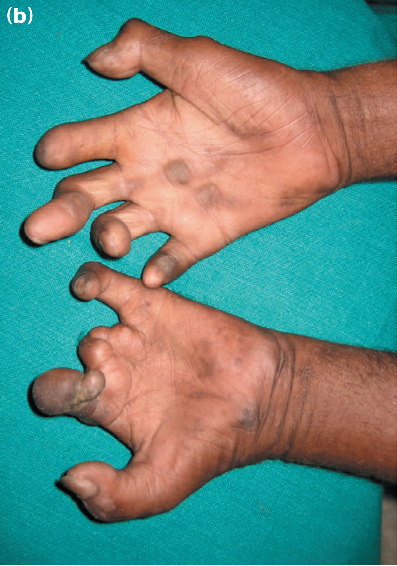
Typical bilateral claw hand in leprosy - Bailey & Love's Surgery, 28th Ed.
2. Goals of Physiotherapy
- Prevent fixed contractures and joint stiffness
- Maintain and improve range of motion (ROM)
- Strengthen weakened intrinsic muscles
- Prevent pressure injuries and ulcers (due to sensory loss)
- Restore functional hand use and grip
- Patient education for self-care and injury prevention
3. Assessment Before Treatment
The physiotherapist must assess:
- Manual Muscle Testing (MMT) - grade intrinsic muscle power
- ROM measurement at MCP, PIP, DIP joints bilaterally
- Sensory testing - light touch, pain, temperature (2-point discrimination)
- Voluntary Muscle Testing (VMT) - to detect early nerve function impairment
- Functional assessment - grip strength, pinch, ADL performance
- Check for soft tissue contracture vs. mobile clawing (mobile = better prognosis)
4. Physiotherapy Techniques
A. Splinting (Orthotic Management)
Splinting is a cornerstone of non-surgical management:
| Type | Purpose |
|---|---|
| Anti-claw splint (Lively splint / Knuckle-bender splint) | Blocks MCP hyperextension; passively corrects claw posture |
| Static cock-up splint | Maintains wrist in neutral; rest position for inflamed nerves |
| Dynamic outrigger splint | For mobile clawing; assists MCP flexion and IP extension |
| Gutter/finger splint | For individual finger deformities or post-surgical immobilization |
- Splints are worn during daily activities and removed for exercises
- Must be monitored closely due to sensory loss (risk of pressure sores)
B. Exercise Therapy
(i) Passive Stretching Exercises
- Gentle passive flexion and extension of MCP, PIP, DIP joints
- Stretch the intrinsic-plus position: flex MCP + extend IPs
- Prevents joint capsule tightening and soft tissue contracture
- Performed twice daily, 10 repetitions each joint
(ii) Active Range of Motion (AROM) Exercises
- Full fist making, hook fist, straight fist
- Finger abduction and adduction
- Opposition of thumb to each finger tip
- Tendon gliding exercises
(iii) Intrinsic Muscle Strengthening ("Anti-Claw" Exercises)
- Target the lumbricals and interossei (muscles that flex MCP + extend IPs)
- Lumbrical-position exercises: actively flex at MCP while keeping IPs straight
- Resistance training using therapy putty, rubber bands, resistance pegs
- Pinch and grip strengthening with graduated resistive tools
(iv) Tendon Transfer Re-Education (post-surgical)
- After sublimis transfer (Stiles-Bunnell operation) or FDS transfer to A2 pulley
- Patient is taught to re-educate the transferred tendon to perform new function
- Isolated contraction practice, followed by functional task training
C. Sensory Re-Education
Due to sensory loss, structured re-education is essential:
- Early phase: perception of moving touch and constant touch using erasers, cotton wool
- Late phase: object identification (stereognosis) - patient identifies objects by texture, shape, and size with eyes closed
- Graded difficulty: coarse to fine textures, familiar to unfamiliar objects
- Visual substitution training: teach patient to use vision to compensate for absent sensation
- Mirror therapy: watching reflection of normal hand performing exercises to stimulate motor cortex
D. Prevention of Secondary Complications (Wound Care & Self-Care)
Since both hands have sensory loss, the physiotherapist must educate on:
- Daily skin inspection of both hands (use a mirror for palm)
- Soaking and oiling dry skin to prevent cracking and entry of infection
- Avoid handling hot objects, sharp tools without protection
- Use of protective gloves during occupational tasks
- Pressure offloading - padded gloves, adapted tool handles
- Wound management - dressing of trophic ulcers; offloading pressure points
E. Electrotherapy (Adjunct)
| Modality | Indication |
|---|---|
| TENS (Transcutaneous Electrical Nerve Stimulation) | Nerve pain / neuritis |
| Faradic stimulation | Maintain muscle bulk; stimulate partially innervated muscles |
| Galvanic stimulation | Denervated muscles to retard atrophy |
| Wax bath / Hydrotherapy | Reduce joint stiffness; warm-up before exercises |
| Ultrasound therapy | Reduce fibrosis and promote nerve healing |
F. Occupational Therapy / Functional Training
- ADL training (dressing, eating, writing) with adapted equipment
- Vocational rehabilitation - modified tools for occupation
- Upper limb functional tasks: carrying, lifting, grasping objects
- Integration of bilateral hand function in coordinated activities
5. Surgical Physiotherapy (Pre- and Post-operative)
When mobile clawing does not respond to conservative treatment, tendon transfer surgery (e.g., Zancolli capsulorrhaphy, FDS transfer, Brand's EIP transfer) is performed.
Pre-operative physiotherapy:
- Maximize passive ROM - serial splinting to correct contractures
- Teach isolated contraction of donor tendon
- Strengthen donor muscle to Grade 4+ strength
Post-operative physiotherapy:
- POP cast immobilization (MCP at 90 degrees, IPs straight - lumbrical position) for 3-4 weeks
- After cast removal: gentle passive ROM, progressing to active ROM
- Tendon transfer re-education: retrain transferred tendon for new function
- Strengthening and functional integration over 3-6 months
6. Patient Education and Prevention
- Teach the warning signs of acute neuritis (pain, tenderness over nerve) - prompt referral for nerve decompression or corticosteroids
- Importance of continuing MDT (multi-drug therapy) under NLEP
- Home exercise program (HEP) with illustrated handouts
- Psychological support and counselling for stigma-related distress
- Ergonomic advice and workplace modifications
Summary Table
| Component | Intervention |
|---|---|
| Splinting | Anti-claw lively splint, dynamic outrigger |
| Stretching | Passive ROM to prevent contracture |
| Strengthening | Lumbrical position exercises, putty, resistance |
| Sensory re-education | Texture identification, mirror therapy |
| Electrotherapy | TENS, faradic/galvanic, wax bath |
| Wound/skin care | Inspection, oiling, protective equipment |
| Surgical rehab | Pre/post-op tendon transfer protocol |
| Education | HEP, injury prevention, stigma counselling |
Key Point for Exam: The fundamental physiotherapy goal in leprosy claw hand is to restore the lost lumbrical function - either through intrinsic strengthening (if partial palsy) or through substitution exercises after tendon transfer (if complete palsy). Sensory re-education and injury prevention are equally important because bilateral sensory loss makes both hands vulnerable to repeated trauma and ulceration.
Sources: Bailey & Love's Short Practice of Surgery 28th Ed.; Park's Textbook of Preventive & Social Medicine; Campbell's Operative Orthopaedics 15th Ed.; International Textbook of Leprosy - Physical Rehabilitation chapter
This is a shared conversation. Sign in to Orris to start your own chat.